Beyond DNA to Protein: The Central Dogma's Evolving Role in Modern Biology and Drug Development
This article provides a comprehensive exploration of the Central Dogma of Molecular Biology, tracing its evolution from a foundational principle to a dynamic framework for understanding gene regulation and its...
Beyond DNA to Protein: The Central Dogma's Evolving Role in Modern Biology and Drug Development
Abstract
This article provides a comprehensive exploration of the Central Dogma of Molecular Biology, tracing its evolution from a foundational principle to a dynamic framework for understanding gene regulation and its applications. Tailored for researchers, scientists, and drug development professionals, it moves beyond the classic DNA→RNA→protein pathway to examine quantitative dynamics, regulatory complexities, and real-world implications. The scope encompasses foundational concepts, cutting-edge methodological applications in CRISPR and synthetic biology, troubleshooting of stochastic expression and non-correlation between mRNA and protein, and a comparative validation of the dogma against modern exceptions and paradigm-shifting theories. This resource is designed to bridge theoretical molecular biology with practical challenges in therapeutic development.
The Core Principle and Its Evolution: From Crick's Dogma to a Dynamic Framework
The Central Dogma of molecular biology represents the core framework that explains the flow of genetic information within biological systems. First articulated by Francis Crick in 1958, this principle establishes the directional transfer of sequential information between the major biological polymers: nucleic acids and proteins [1]. Contrary to popular simplified versions, Crick's original formulation was not merely the linear pathway "DNA → RNA → protein," but rather a nuanced theory about information transfer constraints within cells [2]. His central premise stated that once genetic information had passed into a protein, it could not flow back to nucleic acids or other proteins [3] [1]. This conceptual boundary has guided molecular biology research for decades, though exceptions discovered since its inception have further refined our understanding of information flow in biological systems.
Crick himself acknowledged the speculative nature of his idea when he first proposed it, noting that "the direct evidence for both of them is negligible, but I have found them to be of great help in getting to grips with these very complex problems" [2]. The Central Dogma was proposed alongside what Crick termed the "Sequence Hypothesis," which suggested that the specificity of nucleic acids is expressed solely by their base sequences, and this sequence serves as a code for protein amino acid sequences [2]. Together, these hypotheses provided the theoretical foundation for modern molecular biology, establishing DNA as the repository of genetic information and proteins as the functional effectors of cellular processes.
Historical Context and Original Formulation
Francis Crick's 1958 Proposal
Francis Crick first formally presented the Central Dogma in his 1958 publication "On Protein Synthesis," where he targeted a general reader rather than specialists in the field [2]. His original statement was precise: "The Central Dogma. This states that once 'information' has passed into protein it cannot get out again. In more detail, the transfer of information from nucleic acid to nucleic acid, or from nucleic acid to protein may be possible, but transfer from protein to protein, or from protein to nucleic acid is impossible" [1]. Crick clarified that "information" in this context meant "the precise determination of sequence, either of bases in the nucleic acid or of amino acid residues in the protein" [1].
This original formulation differed significantly from the simplified version that would later become popularized. Crick's conceptualization allowed for certain information transfers (nucleic acid to nucleic acid, nucleic acid to protein) while explicitly prohibiting others (protein to protein, protein to nucleic acid). In his 1970 Nature paper, he re-emphasized this point: "The central dogma of molecular biology deals with the detailed residue-by-residue transfer of sequential information. It states that such information cannot be transferred back from protein to either protein or nucleic acid" [1].
The "Dogma" Terminology
Crick's choice of the term "dogma" proved somewhat controversial. In his autobiography, he wrote: "I called this idea the central dogma, for two reasons, I suspect. I had already used the obvious word hypothesis in the sequence hypothesis, and in addition I wanted to suggest that this new assumption was more central and more powerful" [1]. He later acknowledged that he had misunderstood the term's conventional religious meaning, stating: "My mind was, that a dogma was an idea for which there was no reasonable evidence. You see?! And Crick gave a roar of delight. I just didn't know what dogma meant. And I could just as well have called it the 'Central Hypothesis,' or — you know. Which is what I meant to say. Dogma was just a catch phrase" [1].
The Molecular Biology Revolution: Key Experimental Evidence
The theoretical framework of the Central Dogma was built upon foundational experimental work that elucidated the mechanisms of information transfer in cells. Several critical experiments conducted in the 1950s and 1960s provided the empirical evidence supporting Crick's proposed information flow.
Establishing DNA as the Genetic Material
The groundbreaking 1944 experiment by Oswald Avery, Colin MacLeod, and Maclyn McCarty at the Rockefeller Institute provided the first compelling evidence that DNA, not protein, carries genetic information [2]. Their work with Streptococcus pneumoniae demonstrated that digested DNA from virulent strains could transfer pathogenic traits to harmless strains, while digested proteins could not. This discovery "deeply moved" Erwin Chargaff, who subsequently conducted meticulous analyses of DNA composition across species and discovered that the amount of adenine always equals thymine, and guanine always equals cytosine—findings that would later prove critical to understanding DNA structure and replication [2].
The Structure of DNA
The 1953 determination of DNA's double-helical structure by James Watson and Francis Crick, based on Rosalind Franklin's X-ray diffraction images, provided the structural basis for understanding how genetic information is stored and replicated [2]. Their model, published in Nature on April 25, 1953, famously noted that "It has not escaped our notice that the specific pairing we have postulated immediately suggests a possible copying mechanism for the genetic material" [2]. The complementary base pairing (A-T and G-C) elegantly explained how genetic information could be faithfully copied during cell division.
The Meselson-Stahl Experiment: DNA Replication
In 1958, Matthew Meselson and Franklin Stahl at Caltech provided definitive experimental proof for the semi-conservative model of DNA replication [2]. Their elegant experiment used heavy nitrogen (N15) to tag parental DNA and tracked its distribution during replication cycles. By centrifuging DNA samples, they demonstrated that after one replication cycle, DNA molecules contained half-heavy and half-light nitrogen, confirming that each new DNA molecule consists of one parental strand and one newly synthesized strand. This experiment conclusively supported Watson and Crick's hypothesis and refuted alternative models (conservative and dispersive replication) proposed by other scientists including Max Delbrück [2].
Table 1: Key Historical Experiments Supporting the Central Dogma
| Experiment | Researchers | Year | Key Finding | Significance |
|---|---|---|---|---|
| DNA as Genetic Material | Avery, MacLeod, McCarty | 1944 | DNA, not protein, carries genetic information | Established DNA as molecule of heredity |
| DNA Base Composition | Chargaff | 1949 | A=T and G=C in DNA from all species | Revealed molecular parity that informed DNA structure |
| DNA Structure | Watson, Crick, Franklin | 1953 | Double-helical structure with complementary base pairing | Provided structural mechanism for information storage and copying |
| DNA Replication | Meselson, Stahl | 1958 | Semi-conservative replication mechanism | Confirmed how genetic information is faithfully copied |
Discovering the RNA Intermediate
The identification of messenger RNA (mRNA) as the intermediate between DNA and protein represented another critical milestone. Crick had theoretically predicted this "template RNA" in his lectures before direct experimental evidence confirmed its existence [2]. The discovery of mRNA explained how genetic information stored in the nucleus could direct protein synthesis in the cytoplasm, completing the DNA → RNA → protein pathway that would become synonymous with the Central Dogma in its simplified form.
Diagram 1: Basic information flow in the Central Dogma
The Complete Information Transfer Framework
The Central Dogma describes all possible and forbidden transfers of sequential information between biological polymers. Crick's original scheme acknowledged three general transfers (DNA → DNA, DNA → RNA, RNA → protein) and three special transfers (RNA → RNA, RNA → DNA, DNA → protein), while explicitly excluding two transfers (protein → protein, protein → nucleic acid) [1].
General Transfers
The general transfers represent the core information flow that occurs in all living cells:
DNA → DNA (Replication): The faithful copying of genetic information from parent DNA to daughter DNA molecules, performed by the replisome complex [1]. This transfer ensures genetic continuity during cell division.
DNA → RNA (Transcription): The process by which information contained in DNA sections is copied to messenger RNA molecules using RNA polymerase and transcription factors [1]. In eukaryotes, the initial transcript (pre-mRNA) undergoes processing (5' capping, polyadenylation, splicing) to produce mature mRNA.
RNA → Protein (Translation): The decoding of mRNA sequence information into polypeptide chains by ribosomes, with transfer RNAs (tRNAs) delivering specific amino acids based on codon-anticodon pairing [1]. The resulting polypeptide chain undergoes folding and often additional processing to become a functional protein.
Special Transfers
The special transfers occur in certain biological contexts but are not universal:
RNA → RNA (RNA replication): Many viruses replicate their genetic material using RNA-dependent RNA polymerases [1]. Eukaryotes also employ similar enzymes for RNA silencing pathways.
RNA → DNA (Reverse transcription): Retroviruses (such as HIV) and retrotransposons use reverse transcriptase enzymes to copy RNA information into DNA [1]. This transfer directly contradicts the simplified "one-way" DNA → RNA → protein pathway but does not violate Crick's original Dogma, which specifically prohibited information flow from protein back to nucleic acids.
DNA → Protein (Direct translation): While theoretically possible, this direct transfer is not known to occur naturally in biological systems.
Table 2: Information Transfers in the Central Dogma Framework
| Transfer Type | From | To | Example/Mechanism | Status in Central Dogma |
|---|---|---|---|---|
| General | DNA | DNA | DNA replication | Permitted |
| General | DNA | RNA | Transcription (RNA polymerase) | Permitted |
| General | RNA | Protein | Translation (ribosomes) | Permitted |
| Special | RNA | RNA | Viral replication, RNA silencing | Permitted |
| Special | RNA | DNA | Reverse transcription (retroviruses) | Permitted |
| Special | DNA | Protein | Theoretical direct translation | Not observed naturally |
| Forbidden | Protein | Protein | Not permitted by original dogma | Explicitly forbidden |
| Forbidden | Protein | Nucleic Acid | Not permitted by original dogma | Explicitly forbidden |
Diagram 2: Permitted and forbidden information transfers
Exceptions and Challenges to the Central Dogma
Since its formulation, several biological phenomena have been discovered that challenge the strict interpretation of the Central Dogma, though most do not actually violate Crick's original specification.
Prions: Protein-Mediated Information Transfer
Prions are infectious proteins that replicate without going through DNA or RNA intermediates [3]. These misfolded proteins can induce normally-folded proteins of the same type to adopt the prion conformation, effectively creating a form of protein-based inheritance [1]. Prions are responsible for neurodegenerative diseases such as Creutzfeldt-Jakob disease in humans [3].
Some scientists, including Alain E. Bussard and Eugene Koonin, have argued that prion-mediated inheritance violates the Central Dogma because it represents information transfer from protein to protein [1]. However, others contend that prions do not truly violate the Dogma because the protein sequence itself remains unchanged—only the conformation is altered. As Rosalind Ridley noted in Molecular Pathology of the Prions (2001): "The prion hypothesis is not heretical to the central dogma of molecular biology—that the information necessary to manufacture proteins is encoded in the nucleotide sequence of nucleic acid—because it does not claim that proteins replicate. Rather, it claims that there is a source of information within protein molecules that contributes to their biological function, and that this information can be passed on to other molecules" [1].
Inteins: Protein Self-Modification
Inteins are "parasitic" protein segments that can excise themselves from a polypeptide chain and ligate the flanking regions (exteins) with a peptide bond [1]. This represents a case where a protein changes its own primary sequence from what was originally encoded by DNA. Additionally, many inteins contain homing endonuclease domains that can catalyze the insertion of intein-encoding DNA sequences into intein-free genes, representing a form of protein-mediated DNA sequence editing [1].
Nonribosomal Peptide Synthesis
Some peptides are synthesized by nonribosomal peptide synthetases, large protein complexes that assemble peptides without using mRNA templates [1]. These peptides often have cyclic or branched structures and may contain non-proteinogenic amino acids, differentiating them from ribosomally-synthesized proteins. Examples include some antibiotics, which are produced through this template-independent mechanism.
Modern Relevancy: CRISPR and Synthetic Biology
Recent advances in molecular biology, particularly in the field of genome editing, have prompted reevaluation of the Central Dogma's boundaries in modern contexts. A 2022 review titled "The Central Dogma revisited: Insights from protein synthesis, CRISPR, and beyond" examines whether contemporary biological systems challenge Crick's fundamental principle [4].
The authors apply a three-part evaluation scheme to CRISPR-Cas9 and prime editing systems, concluding that although current CRISPR gene-editing mechanisms operate within the Dogma's constraints, synthetic biology could potentially create systems that directly violate it [4]. They speculate on the theoretical and practical implications of protein-derived information transfer systems, suggesting that while natural systems largely conform to the Dogma's restrictions, engineered systems might eventually enable direct information flow from protein to nucleic acid [4].
Table 3: Modern Molecular Biology in Context of Central Dogma
| Biological System | Mechanism | Relationship to Central Dogma |
|---|---|---|
| CRISPR-Cas9 | Protein-RNA complex guides DNA cleavage | Operates within dogma: RNA mediates between DNA and protein |
| Prime Editing | Engineered reverse transcriptase linked to Cas9 | Operates within dogma: RNA template guides DNA modification |
| Prions | Conformational change propagation | Challenges but doesn't violate dogma: no sequence change |
| Inteins | Protein splicing with DNA homing | Pushes boundaries: protein affects DNA sequence indirectly |
| Nonribosomal Peptide Synthesis | Template-independent peptide assembly | Outside dogma scope: doesn't use genetic code |
Essential Research Reagents and Methodologies
Research into the Central Dogma and its mechanisms relies on specific reagents and experimental approaches. The following table summarizes key research tools that have been fundamental to elucidating information flow in biological systems.
Table 4: Research Reagent Solutions for Central Dogma Investigations
| Research Reagent | Composition/Type | Experimental Function |
|---|---|---|
| Heavy Isotope-labeled Nucleotides (N15) | Nucleotides with heavy nitrogen isotopes | Density labeling for DNA replication tracking (Meselson-Stahl experiment) |
| RNA Polymerase Inhibitors (e.g., Actinomycin D) | Chemical inhibitors | Block transcription to study mRNA synthesis and turnover |
| Reverse Transcriptase | RNA-dependent DNA polymerase | Converts RNA to cDNA for studying gene expression |
| Ribosome Inhibitors (e.g., Cycloheximide, Chloramphenicol) | Translation inhibitors | Block protein synthesis to study translation mechanisms |
| Restriction Endonucleases | Bacterial enzyme complexes | Cut DNA at specific sequences for molecular cloning |
| DNA Polymerase | DNA-dependent DNA polymerase | Amplifies DNA in PCR and replicates DNA in vitro |
- Experimental Protocol: Density Shift DNA Replication Assay - Based on Meselson-Stahl experiment [2]: (1) Grow bacteria in heavy nitrogen (N15) medium for multiple generations; (2) Transfer to light nitrogen (N14) medium; (3) Collect samples at successive time points; (4) Lyse cells and isolate DNA; (5) Perform cesium chloride density gradient centrifugation; (6) Analyze DNA banding patterns using UV absorption; (7) Interpret replication mechanism based on density distribution across generations.
Diagram 3: Meselson-Stahl experiment workflow
The Central Dogma of molecular biology, as originally formulated by Francis Crick in 1958, continues to provide the fundamental conceptual framework for understanding information flow in biological systems. While simplified versions focusing solely on the DNA → RNA → protein pathway have become popularized in textbooks, Crick's original insight was more nuanced—emphasizing the permitted and forbidden directions of information transfer between biological polymers [3] [2] [1].
Despite the discovery of exceptions such as reverse transcription, prions, and inteins, the core principle of the Central Dogma remains valid: sequence information cannot flow backward from protein to nucleic acids in natural biological systems [1] [4]. This understanding continues to guide research in molecular biology, genetics, and synthetic biology, while ongoing investigations into CRISPR systems and protein-based information transfer may further test the Dogma's boundaries in engineered biological contexts [4].
The Dogma's enduring value lies in its ability to distinguish possible from impossible information transfers in cellular processes, providing a theoretical foundation that has stimulated research and discovery for over six decades. As molecular biology continues to advance with new technologies, the Central Dogma remains essential for interpreting biological information processing in both natural and synthetic systems.
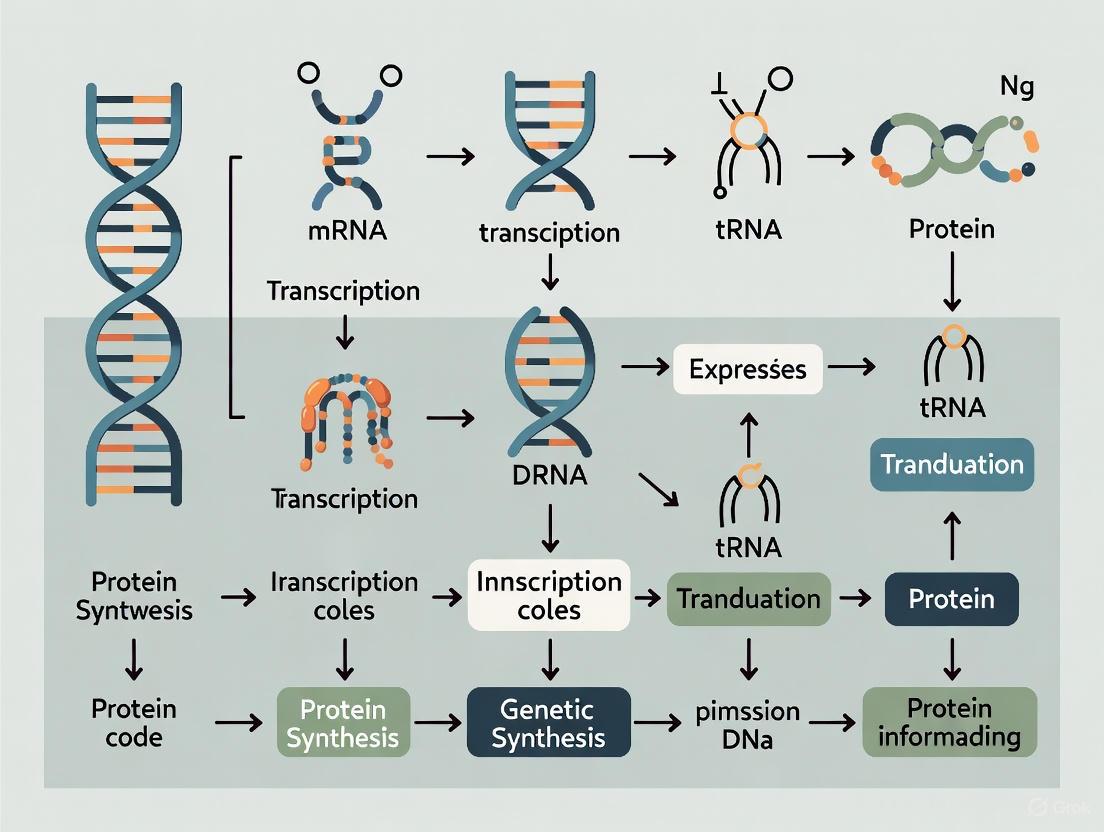
The central dogma of molecular biology is a fundamental theory stating that genetic information flows in a specific, unidirectional pathway: from DNA, to RNA, and then to protein [3]. First articulated by Francis Crick in 1958, this principle explains how the genetic code stored in DNA is used to create functional molecules within the cell [3] [1]. The process by which DNA is copied to RNA is called transcription, and that by which RNA is used to produce proteins is called translation [5]. A complementary process, DNA replication, ensures that this genetic information is faithfully copied for daughter cells during cell division [6]. These three processes—replication, transcription, and translation—form the core framework of molecular biology and provide the mechanistic basis for heredity and gene expression in living organisms [7] [5].
This information flow pathway is not merely a descriptive model but represents the actual biochemical operations performed by complex molecular machines. The precision of these operations enables the transmission of genetic traits across generations and the precise regulation of cellular functions in response to internal and external signals [8]. Modern quantitative biology continues to refine our understanding of these processes, investigating their dynamics in complex cellular environments such as the p53-mediated DNA damage response [9]. This technical guide examines the molecular mechanisms, key experimental elucidation, and research methodologies for studying these fundamental biological processes.
DNA Replication: The Semiconservative Mechanism
DNA replication is the biological process whereby a cell duplicates its entire DNA genome prior to cell division. This process occurs during the S-phase of the cell cycle and is essential for the faithful transmission of genetic information from parent to daughter cells [6]. The mechanism is termed semiconservative because each newly synthesized DNA double helix consists of one strand from the original parent molecule and one newly synthesized strand [6].
Molecular Mechanism of Replication
The replication process requires a coordinated series of steps facilitated by multiple enzymes and protein factors:
Initiation: Replication begins at specific genomic locations called origins of replication. The enzyme DNA helicase unwinds the double helix by breaking hydrogen bonds between base pairs, creating a replication fork characterized by Y-shaped structures [6]. This unwinding typically begins in adenine-thymine rich regions due to their weaker bonding (two hydrogen bonds versus three in guanine-cytosine pairs) [6]. Single-strand binding proteins stabilize the separated strands, while topoisomerase relieves torsional stress ahead of the replication fork [5].
Elongation: The enzyme DNA polymerase catalyzes the addition of nucleotides to the growing DNA chain, but requires a short RNA primer synthesized by primase to begin synthesis [6]. DNA synthesis always proceeds in the 5' to 3' direction, which creates an inherent asymmetry between the two template strands [5]. The leading strand is synthesized continuously toward the replication fork, while the lagging strand is synthesized discontinuously away from the fork in short segments called Okazaki fragments [5] [6].
Termination: On the lagging strand, the RNA primers are removed by flap endonuclease 1 (FEN1) and RNase H, and the resulting gaps are filled by DNA polymerase. DNA ligase then joins the Okazaki fragments by creating phosphodiester bonds, completing the new DNA strand [6]. In eukaryotic cells, the ends of chromosomes (telomeres) are extended by the enzyme telomerase to prevent progressive shortening with each replication cycle [6].
Key Experiments: Meselson-Stahl Experiment
The Meselson-Stahl experiment (1958) provided definitive evidence for the semiconservative model of DNA replication [8]. By growing E. coli bacteria in a medium containing the heavy nitrogen isotope ^15^N and then transferring them to a light ^14^N medium, the researchers could track parental and newly synthesized DNA strands through density gradient centrifugation [8]. After one generation, all DNA molecules exhibited intermediate density, ruling out conservative replication. After two generations, both intermediate and light DNA molecules were present, exactly as predicted by the semiconservative model [8].
Table 1: Key Enzymes in DNA Replication and Their Functions
| Enzyme/Protein | Function |
|---|---|
| DNA Helicase | Unwinds the DNA double helix by breaking hydrogen bonds |
| DNA Polymerase | Synthesizes new DNA strands by adding nucleotides; possesses proofreading activity |
| Primase | Synthesizes short RNA primers to initiate DNA synthesis |
| DNA Ligase | Joins Okazaki fragments on lagging strand by forming phosphodiester bonds |
| Topoisomerase | Relieves torsional stress ahead of replication fork |
| Single-Strand Binding Proteins | Stabilize separated DNA strands |
| Telomerase | Adds telomeric repeats to chromosome ends |
Transcription: DNA to RNA Conversion
Transcription is the process by which a specific DNA sequence is copied into a complementary RNA molecule by RNA polymerase enzymes [6]. This process represents the first step of gene expression, where genetic information encoded in DNA is converted into a messenger RNA (mRNA) template for protein synthesis [5].
Mechanism of Transcription
Transcription occurs in three main stages and involves different molecular components in prokaryotic and eukaryotic cells:
Initiation: RNA polymerase binds to specific DNA sequences called promoter regions, typically characterized by TATA box sequences (TATAAT in prokaryotes, TATA(A/T)A in eukaryotes) [6]. In eukaryotes, transcription factors help recruit and position RNA polymerase at the transcription start site. Unlike DNA polymerase, RNA polymerase can initiate RNA synthesis without a primer [6].
Elongation: RNA polymerase moves along the DNA template in the 3' to 5' direction, synthesizing a complementary RNA strand in the 5' to 3' direction [5] [6]. The DNA double helix temporarily unwinds, creating a transcription bubble of approximately 14 base pairs. Nucleotide triphosphates (ATP, GTP, CTP, UTP) align with the template strand through Watson-Crick base pairing, with uracil (U) pairing with adenine instead of thymine [5] [6].
Termination: Transcription concludes when RNA polymerase encounters a termination sequence in the DNA. In prokaryotes, this often involves a hairpin loop structure in the newly synthesized RNA that causes the polymerase to dissociate [6]. In eukaryotes, termination mechanisms are more complex and involve additional protein factors.
RNA Processing in Eukaryotes
Eukaryotic mRNA undergoes extensive post-transcriptional processing before export to the cytoplasm:
- 5' Capping: A 7-methylguanosine cap is added to the 5' end of the pre-mRNA, which protects from degradation and facilitates ribosome binding [5] [6].
- 3' Polyadenylation: A poly-A tail (150-200 adenine nucleotides) is added to the 3' end, which enhances stability and facilitates nuclear export [5].
- RNA Splicing: Non-coding sequences (introns) are removed, and coding sequences (exons) are joined together by spliceosome complexes [5]. Alternative splicing allows a single gene to produce multiple protein isoforms by including or excluding different exons, significantly expanding proteomic diversity [5].
Table 2: Types of RNA and Their Functions in Gene Expression
| RNA Type | Function | Synthesized By |
|---|---|---|
| Messenger RNA (mRNA) | Carries genetic code from DNA to ribosomes for translation | RNA Polymerase II |
| Transfer RNA (tRNA) | Brings amino acids to ribosomes during translation | RNA Polymerase III |
| Ribosomal RNA (rRNA) | Structural and catalytic component of ribosomes | RNA Polymerase I |
| MicroRNA (miRNA) | Regulates gene expression by binding to target mRNAs | RNA Polymerase II |
Translation: RNA to Protein Synthesis
Translation is the process by which the genetic code carried by mRNA is decoded to synthesize a specific protein [5]. This complex process occurs on ribosomes and involves multiple forms of RNA, including transfer RNA (tRNA) and ribosomal RNA (rRNA) [5].
The Genetic Code
The genetic code is a set of rules by which the nucleotide sequence of mRNA is translated into the amino acid sequence of proteins [5]. Key features include:
- Triplet Code: Three consecutive nucleotides (codon) specify one amino acid [5].
- Universality: The code is nearly universal across organisms, with minor variations in mitochondria and some protists [5].
- Degeneracy: Most amino acids are encoded by multiple codons (e.g., Arg and Ser each have 6 codons) [5].
- Non-overlapping: Codons are read sequentially without overlapping [5].
- Start and Stop Signals: AUG (encoding methionine) serves as the initiation codon, while UAA, UAG, and UGA serve as termination codons [5].
Mechanism of Translation
Translation occurs in three main stages through the coordinated action of ribosomes, tRNAs, and various protein factors:
Initiation: The small ribosomal subunit binds to the 5' end of mRNA and scans until it encounters the AUG start codon. The initiation complex is formed with the help of initiation factors, and the large ribosomal subunit joins to form the complete ribosome [5] [1].
Elongation: Aminoacyl-tRNAs carrying specific amino acids enter the ribosome's A site, where the anticodon on the tRNA base-pairs with the complementary codon on the mRNA. The ribosome catalyzes peptide bond formation between the growing polypeptide chain and the new amino acid. The ribosome then translocates to the next codon, moving the tRNAs through the P and E sites before releasing them [5] [1].
Termination: When a stop codon (UAA, UAG, or UGA) enters the A site, release factors bind and catalyze the hydrolysis of the completed polypeptide from the final tRNA. The ribosome dissociates from the mRNA, and the components are recycled for further rounds of translation [5] [1].
Following translation, proteins often undergo post-translational modifications (folding, cleavage, cross-linking, chemical group additions) to achieve their functional forms [1]. Molecular chaperones assist in proper protein folding, ensuring biological activity [1].
Experimental Methods and Research Tools
The study of replication, transcription, and translation relies on sophisticated experimental techniques that allow researchers to visualize, manipulate, and quantify these molecular processes.
Key Experimental Techniques
Polymerase Chain Reaction (PCR): This technique allows exponential amplification of specific DNA sequences through repeated cycles of denaturation, annealing, and extension [6]. PCR is fundamental to modern molecular biology, with applications in cloning, mutation detection, forensics, and diagnostics [6].
DNA Sequencing: Methods to determine the exact nucleotide sequence of DNA molecules provide crucial information for investigating gene function and identifying mutations [6]. Next-generation sequencing technologies now enable rapid, high-throughput analysis of entire genomes [8].
Southern Blotting: This technique detects specific DNA sequences in a sample through electrophoretic separation, transfer to a membrane, and hybridization with labeled complementary probes [6].
Live Single-Cell Imaging: Advanced microscopy techniques enable real-time visualization of transcription and translation dynamics in living cells, such as tracking p53 and its target genes in response to DNA damage [9].
Research Reagent Solutions
Table 3: Essential Research Reagents for Studying Central Dogma Processes
| Reagent/Technique | Application | Key Features |
|---|---|---|
| Restriction Enzymes | DNA manipulation; genetic engineering | Recognize and cut specific DNA sequences |
| Reverse Transcriptase | cDNA synthesis; RT-PCR | Converts RNA to complementary DNA (cDNA) |
| Taq Polymerase | PCR amplification | Thermostable DNA polymerase for PCR |
| Plasmid Vectors | Molecular cloning; protein expression | Extrachromosomal DNA for gene insertion and amplification |
| CRISPR-Cas9 Systems | Gene editing; functional genomics | RNA-guided genome editing technology |
| RNA Interference (RNAi) | Gene silencing; functional studies | Sequence-specific degradation of target mRNA |
| Nucleoside Analogs (e.g., Acyclovir, AZT) | Antiviral/anticancer therapy; replication studies | Inhibit DNA replication by chain termination |
Visualization of Molecular Processes
Diagram 1: Central Dogma Information Flow
Diagram 2: DNA Replication Process
Diagram 3: Transcription and RNA Processing
The coordinated processes of replication, transcription, and translation represent the fundamental mechanisms by which genetic information is preserved, expressed, and utilized within biological systems. The central dogma provides a robust framework for understanding how information flows from DNA sequence to functional protein, with numerous regulatory checkpoints ensuring fidelity at each step [3] [1]. Current research continues to expand our understanding of these processes, particularly through quantitative approaches that examine their dynamic regulation in complex cellular environments such as stress responses and disease states [9].
Modern molecular biology techniques, from CRISPR-based genome editing to single-cell omics technologies, build upon this foundational knowledge [8]. The integration of quantitative measurements with mathematical modeling promises to further elucidate the intricate relationships between molecular components, advancing both basic science and therapeutic applications in areas such as cancer research, genetic engineering, and drug development [7] [9]. As research progresses, our understanding of these core processes continues to refine, revealing new layers of complexity in the flow of genetic information.
The genetic code is the universal set of rules used by living cells to translate the information encoded within genetic material into functional proteins [10]. This process of translation is a critical step in the central dogma of molecular biology, which describes the directional flow of genetic information within biological systems [3]. The central dogma, first articulated by Francis Crick in 1958, fundamentally states that genetic information flows from DNA to RNA to protein, and that once information has passed into protein, it cannot flow back to nucleic acids [1]. This framework establishes the context in which the genetic code operates - as the essential cipher that enables the translation of nucleic acid sequences into the amino acid sequences that determine protein structure and function.
The genetic code achieves this translation through a system of nucleotide triplets called codons, which specify which amino acid will be added next during protein biosynthesis [10]. With few exceptions, each three-nucleotide codon in a nucleic acid sequence specifies a single amino acid, creating a standardized biological language that is highly conserved across virtually all organisms [10] [11]. The elucidation of this code represented a landmark achievement in molecular biology, revealing how the four-letter alphabet of nucleic acids (A, C, G, T/U) could specify the 20-letter alphabet of amino acids that build proteins [12].
Core Concepts and Quantitative Structure of the Genetic Code
Fundamental Properties of the Code
The genetic code possesses several defining characteristics that enable its function in protein synthesis:
Triplet Nature: Each amino acid is encoded by a sequence of three nucleotides [11]. This triplet system provides 64 (4³) possible codons, which is more than sufficient to encode the 20 standard amino acids [10].
Degeneracy: The code is degenerate, meaning that most amino acids are encoded by more than one codon [13] [11]. This redundancy provides a buffer against harmful mutations and allows for nuanced regulation of gene expression.
Universality: With minor exceptions (such as in mitochondria), the genetic code is shared across almost all organisms, providing powerful evidence for the common origin of all life on Earth [11].
Non-overlapping and Commaless: The code is read in sequential, non-overlapping triplets from a fixed start point, without punctuation between codons [10].
Codon Assignments and Amino Acid Specifications
Table 1: The Standard Genetic Code Table Showing Codon-Amino Acid Assignments
| Codon | Amino Acid | Codon | Amino Acid | Codon | Amino Acid | Codon | Amino Acid |
|---|---|---|---|---|---|---|---|
| UUU | Phe | UCU | Ser | UAU | Tyr | UGU | Cys |
| UUC | Phe | UCC | Ser | UAC | Tyr | UGC | Cys |
| UUA | Leu | UCA | Ser | UAA | Stop | UGA | Stop |
| UUG | Leu | UCG | Ser | UAG | Stop | UGG | Trp |
| CUU | Leu | CCU | Pro | CAU | His | CGU | Arg |
| CUC | Leu | CCC | Pro | CAC | His | CGC | Arg |
| CUA | Leu | CCA | Pro | CAA | Gln | CGA | Arg |
| CUG | Leu | CCG | Pro | CAG | Gln | CGG | Arg |
| AUU | Ile | ACU | Thr | AAU | Asn | AGU | Ser |
| AUC | Ile | ACC | Thr | AAC | Asn | AGC | Ser |
| AUA | Ile | ACA | Thr | AAA | Lys | AGA | Arg |
| AUG | Met (Start) | ACG | Thr | AAG | Lys | AGG | Arg |
| GUU | Val | GCU | Ala | GAU | Asp | GGU | Gly |
| GUC | Val | GCC | Ala | GAC | Asp | GGC | Gly |
| GUA | Val | GCA | Ala | GAA | Glu | GGA | Gly |
| GUG | Val | GCG | Ala | GAG | Glu | GGG | Gly |
The table illustrates several key features: the start codon (AUG) initiates translation and also codes for methionine; the three stop codons (UAA, UAG, UGA) terminate protein synthesis; and most amino acids are specified by multiple codons, with degeneracy particularly evident in the third nucleotide position of many codons [10] [11].
Reading Frames and Frameshift Mutations
The reading frame is established by the initial triplet from which translation begins, setting the frame for a run of successive, non-overlapping codons known as an open reading frame (ORF) [10]. Any sequence can be read in three possible reading frames in the 5'→3' direction, each potentially producing a different amino acid sequence. In double-stranded DNA, six possible reading frames exist - three forward and three reverse on the complementary strand [10].
Mutations that disrupt the reading frame by insertions or deletions of a non-multiple of 3 nucleotide bases are known as frameshift mutations [10]. These mutations completely alter the translational reading frame, typically resulting in a nonfunctional protein and often introducing a premature stop codon. The devastating effects of frameshift mutations underscore the critical importance of maintaining the correct reading frame for protein synthesis.
The Genetic Code in the Central Dogma Framework
Information Flow from DNA to Protein
The central dogma describes the sequential flow of genetic information from DNA to RNA to protein [14]. This process involves two major steps:
Transcription: The process by which information in a section of DNA is copied into a newly assembled piece of messenger RNA (mRNA) [1]. In eukaryotic cells, the primary transcript (pre-mRNA) undergoes processing including 5' capping, polyadenylation, and splicing to produce mature mRNA.
Translation: The process by which the mRNA sequence is decoded by ribosomes to synthesize proteins [1]. Transfer RNA (tRNA) molecules serve as adaptors that match codons in the mRNA to their corresponding amino acids, facilitating the assembly of the polypeptide chain.
The following diagram illustrates this sequential information flow:
Key Molecular Players in Protein Synthesis
Table 2: Essential Components of the Translation Machinery
| Component | Role in Protein Synthesis | Key Features |
|---|---|---|
| Messenger RNA (mRNA) | Carries genetic code from DNA to ribosomes | Contains codons that specify amino acid sequence; modified with 5' cap and poly-A tail in eukaryotes |
| Transfer RNA (tRNA) | Adaptor molecule that links codons to amino acids | Contains anticodon complementary to mRNA codon; carries corresponding amino acid |
| Ribosome | Catalytic machinery for protein synthesis | Composed of rRNA and proteins; has A, P, and E sites for tRNA binding |
| Aminoacyl-tRNA Synthetases | Enzymes that charge tRNAs with correct amino acids | Ensure fidelity of translation; one synthetase exists for each amino acid |
The ribosome reads the mRNA triplet codons, usually beginning with an AUG start codon, and complexes of initiation and elongation factors bring aminoacylated tRNAs into the ribosome-mRNA complex [1]. This matching of codon to anticodon ensures the accurate translation of the genetic message into a polypeptide chain with the specified amino acid sequence.
Historical Experimental Elucidation of the Genetic Code
Key Methodology: The Poly-U Experiment (Nirenberg and Matthaei, 1961)
The first breakthrough in deciphering the genetic code came from Marshall Nirenberg and J. Heinrich Matthaei in 1961 [10]. Their experimental protocol involved:
Materials and Methods:
- A cell-free system derived from E. coli bacteria containing ribosomes, tRNAs, amino acids, and energy sources
- Synthetic RNA homopolymer poly-uracil (poly-U) containing only uracil bases
- Radioactive labeling to detect protein synthesis
- A system to identify the specific amino acid incorporated into the polypeptide chain
Experimental Workflow:
- The cell-free system was incubated with poly-U mRNA template
- The resulting polypeptide was analyzed for amino acid composition
- Researchers discovered the synthesized polypeptide consisted solely of phenylalanine
Conclusion: The codon UUU specifies the amino acid phenylalanine [10]. This represented the first specific codon assignment and demonstrated that synthetic mRNAs could be used to decipher the genetic code.
Subsequent Codon Elucidation Experiments
Following this discovery, Severo Ochoa's laboratory extended this approach using different synthetic mRNAs [10]:
- Poly-adenine (poly-A) RNA coded for poly-lysine, identifying AAA as the codon for lysine
- Poly-cytosine (poly-C) RNA coded for poly-proline, identifying CCC as the codon for proline
Har Gobind Khorana subsequently used more complex copolymers with defined repeating sequences to determine most of the remaining codons [10]. Meanwhile, Robert W. Holley determined the structure of tRNA, the adapter molecule that facilitates translation [10]. The combined work of Nirenberg, Khorana, and Holley was recognized with the Nobel Prize in Physiology or Medicine in 1968.
The following diagram summarizes the key historical experiments:
Research Reagent Solutions for Genetic Code Studies
Table 3: Essential Research Reagents for Genetic Code and Protein Synthesis Studies
| Reagent/Material | Function in Experimental Research |
|---|---|
| Cell-Free Translation Systems | In vitro protein synthesis without intact cells; allows controlled manipulation of components |
| Synthetic mRNA Templates | Defined sequences to test specific codon assignments and translation efficiency |
| Radioactive Amino Acids | Tracing and quantifying amino acid incorporation into newly synthesized proteins |
| Ribosome Isolation Kits | Purification of functional ribosomes for structural and mechanistic studies |
| tRNA Purification Systems | Isolation of specific tRNAs for charging and binding studies |
| Aminoacyl-tRNA Synthetase Assays | Measuring enzyme activity in charging tRNAs with correct amino acids |
Contemporary Applications: Codon Usage and Optimization
Codon Usage Bias and Its Biological Significance
While the genetic code is universal, organisms exhibit codon usage bias - preferential use of certain synonymous codons over others [13]. This bias reflects evolutionary adaptation to various factors including:
- Cellular tRNA abundance and availability
- Translation efficiency and speed
- Protein folding requirements
- GC content of genomic DNA
Codon usage bias varies significantly across species and even between different genes within the same organism [13]. Highly expressed genes often show stronger codon bias, preferentially using codons that match abundant tRNAs for optimal translation efficiency.
AI-Driven Codon Optimization in Synthetic Biology
Recent advances in machine learning have revolutionized codon optimization for synthetic biology and biotechnology applications. Deep learning models like CodonTransformer demonstrate how AI can design host-specific DNA sequences with natural-like codon distribution profiles [13]. Key features of these approaches include:
- Training on massive datasets (e.g., ~1 million DNA-protein pairs from 164 organisms)
- Context-awareness through Transformer architectures
- Species-specific token representation combining organism, amino acid, and codon encodings
- Generation of DNA sequences that minimize negative cis-regulatory elements
Similarly, DeepCodon represents another deep learning tool focused on preserving functionally important rare codon clusters while enhancing overall protein expression [15]. These AI models address the combinatorial challenge of codon optimization, where for a typical 300-amino acid protein, approximately 10¹⁵⁰ possible synonymous DNA sequences exist [13].
Practical Applications in Biotechnology and Medicine
The strategic optimization of codon usage has significant practical applications:
Heterologous Protein Expression: Optimizing codons to match the host organism's preference is crucial for efficient production of recombinant proteins in biomanufacturing [13] [15].
Vaccine Development: Understanding viral codon usage patterns, as seen in SARS-CoV-2 evolution where the Omicron variant showed increased adaptation to human hosts, informs vaccine design strategies [16].
Gene Therapy: Codon optimization of therapeutic transgenes can enhance protein expression in target tissues while minimizing immune responses.
Synthetic Genomics: Recent achievements include creating bacterial strains with fully synthetic recoded genomes, such as the E. coli "Syn61" strain with a refactored genome that removes the use of three codons completely [10].
The genetic code represents one of biology's most fundamental concepts, providing the critical link between genetic information stored in nucleic acids and functional protein products. Its triplet, degenerate nature allows the four-letter alphabet of nucleotides to specify the 20-amino acid alphabet of proteins with remarkable fidelity. Operating within the framework of the central dogma, the genetic code enables the directional flow of genetic information from DNA to RNA to protein.
Contemporary research continues to reveal new dimensions of this ancient biological code, from its role in regulating gene expression through codon usage bias to its manipulation through AI-driven optimization for synthetic biology applications. The continued elucidation of how codons specify protein sequences remains essential for advancing fields ranging from basic molecular biology to drug development and genetic engineering.
The Central Dogma of Molecular Biology represents a foundational principle for understanding genetic information flow. However, a significant discrepancy exists between Francis Crick's original sophisticated conceptualization and James Watson's simplified DNA→RNA→protein pathway that permeates scientific education and discourse. This analysis examines the historical context, conceptual framework, and biochemical evidence distinguishing these two versions, demonstrating that Crick's hypothesis specifically forbids information transfer from protein to nucleic acids, while Watson's reductive model fails to capture this essential constraint. The clarification of this distinction has profound implications for accurate scientific communication and interpretation of molecular genetic phenomena.
The Central Dogma of Molecular Biology originated from Francis Crick's 1957 lecture to the Society for Experimental Biology and was formally published in 1958 [17]. Crick's conceptual framework emerged during a period of significant uncertainty in molecular biology, when the mechanisms linking nucleic acids to protein synthesis remained largely undefined [17]. In his own words, Crick acknowledged the speculative nature of his hypothesis, stating that "the psychological drive behind this hypothesis is at the moment independent of such evidence" [17]. This historical context is crucial for understanding the Dogma's original intent as a guiding principle rather than an established fact.
Crick's Central Dogma was fundamentally concerned with the directionality of information flow at the molecular level, specifically positing that "once 'information' has passed into protein it cannot get out again" [1]. The term "information" here precisely meant "the precise determination of sequence, either of bases in the nucleic acid or of amino acid residues in the protein" [1]. This negative formulation—specifying what cannot happen—represented the core of Crick's conceptual insight, which was far more nuanced than subsequent simplified versions would suggest.
Crick's Original Conceptualization: A Detailed Analysis
The Sequence Hypothesis and Information Flow
Crick's thinking was underpinned by what he termed the "sequence hypothesis," which proposed that the DNA sequence determines the protein sequence through an informational RNA intermediate [17]. This hypothesis boldly claimed that three-dimensional protein folding was "simply a function of the order of the amino acids," an idea that remains essentially correct today despite the recognized role of molecular chaperones [17]. Crick introduced the novel concept of "information flow" as distinct from mere chemical transformations, adding this conceptual framework to the established biological flows of matter and energy.
Crick's original 1956 notes contained a diagram illustrating permitted and forbidden information transfers, which he later reproduced in his 1970 Nature paper [18]. This schema categorized information transfers into three distinct classes:
- General transfers: Those believed to occur in all cellular organisms (DNA→DNA, DNA→RNA, RNA→protein)
- Special transfers: Those that occur but only in specific circumstances (RNA→DNA, RNA→RNA, DNA→protein)
- Unknown transfers: Those considered impossible (protein→protein, protein→DNA, protein→RNA)
Table 1: Crick's Original Classification of Information Transfers
| Transfer Type | Direction | Status in Crick's Schema | Known Mechanisms |
|---|---|---|---|
| General | DNA → DNA | Possible | DNA replication |
| General | DNA → RNA | Possible | Transcription |
| General | RNA → Protein | Possible | Translation |
| Special | RNA → RNA | Possible | RNA virus replication |
| Special | RNA → DNA | Possible | Reverse transcription |
| Special | DNA → Protein | Theoretically possible | No known natural mechanism |
| Unknown | Protein → Protein | Impossible | - |
| Unknown | Protein → DNA | Impossible | - |
| Unknown | Protein → RNA | Impossible | - |
The Crucial Negative Statement
The most significant aspect of Crick's hypothesis was its negative formulation—the explicit prohibition of certain information transfers [18]. Crick repeatedly emphasized that "once information has passed into protein it cannot get out again" [1] [17]. This specific constraint carried profound implications for understanding cellular function and evolutionary mechanisms, as it established that acquired characteristics could not become genetically encoded—a molecular reaffirmation of August Weismann's barrier between germline and somatic cells [1].
Crick himself acknowledged that his use of the term "dogma" was problematic, noting in his autobiography that Jacques Monod pointed out he "did not appear to understand the correct use of the word dogma, which is a belief that cannot be doubted" [1]. Crick explained that he used the term differently, applying it "to a grand hypothesis that, however plausible, had little direct experimental support" [1]. This admission highlights the hypothetical nature of the Central Dogma in its original formulation, contrary to how the term "dogma" is typically understood in scientific contexts.
Watson's Simplification: The DNA→RNA→Protein Pathway
Origins and Dissemination of the Simplified Version
James Watson introduced the simplified DNA→RNA→protein version of the Central Dogma in the first edition of his influential 1965 textbook, The Molecular Biology of the Gene [1]. This formulation presented the Dogma as a sequential, two-step process of information transfer: DNA to RNA (transcription) followed by RNA to protein (translation). Watson's version differed fundamentally from Crick's original by omitting the crucial negative statement about the impossibility of reverse information flow [1] [18].
Watson's simplification gained rapid traction in biological education due to several factors:
- Pedagogical accessibility: The linear pathway was easier to teach and comprehend
- Textbook authority: Watson's stature as co-discoverer of DNA structure lent credibility to his formulation
- Experimental focus: Early molecular biology emphasized protein-coding genes, making the simplified version seemingly sufficient
Conceptual Consequences of Simplification
The reductive DNA→RNA→protein model fundamentally altered the conceptual meaning of the Central Dogma in several critical ways:
- Transformation of a constraint into a pathway: Crick's prohibition against reverse information flow became merely a descriptive sequence of information transfer
- Elimination of theoretical framework: The simplified version discarded Crick's systematic classification of possible and impossible information transfers
- Vulnerability to disproof: By presenting a positive description rather than a negative constraint, Watson's version became susceptible to apparent exceptions
Table 2: Comparative Analysis of Crick's vs. Watson's Formulations
| Aspect | Crick's Original Concept | Watson's Simplified Version |
|---|---|---|
| Core Statement | "Once information has passed into protein it cannot get out again" | "DNA makes RNA, and RNA makes protein" |
| Primary Emphasis | Directionality constraints on information flow | Sequential steps of gene expression |
| Theoretical Scope | Comprehensive classification of all possible information transfers | Limited to protein-coding genes |
| Key Omission | - | Reverse information flow prohibitions |
| Conceptual Type | Negative constraint (specifies impossibilities) | Positive pathway (describes process) |
| Vulnerability | Resistant to exceptions from new transfer discoveries | Vulnerable to apparent exceptions |
Experimental Validation and Protocol Analysis
Foundational Experiments in Information Transfer
The validation of different information transfers required diverse methodological approaches across multiple experimental systems:
DNA → DNA (DNA Replication)
- Protocol: Meselson-Stahl density gradient centrifugation (1958)
- Methodology: E. coli grown in ¹⁵N-heavy medium, transferred to ¹⁴N-light medium
- Analysis: CsCl equilibrium density gradient centrifugation
- Key Reagents: ¹⁵NH₄Cl (heavy nitrogen isotope), CsCl (density gradient medium)
- Outcome: Demonstrated semi-conservative replication through intermediate density bands
DNA → RNA (Transcription)
- Protocol: In vitro transcription systems with radioactive labeling
- Methodology: Isolation of RNA polymerase, DNA templates with specific promoters, ³²P-UTP
- Analysis: Gel electrophoresis, autoradiography
- Key Reagents: ³²P-UTP (radiolabeled nucleotide), α-amanitin (RNA polymerase inhibitor)
- Outcome: Confirmed DNA-dependent RNA synthesis with sequence complementarity
RNA → Protein (Translation)
- Protocol: Cell-free translation systems
- Methodology: Reticulocyte lysates or wheat germ extracts, synthetic mRNA templates, ³⁵S-methionine
- Analysis: SDS-PAGE, immunoprecipitation, scintillation counting
- Key Reagents: ³⁵S-methionine (radiolabeled amino acid), cycloheximide (translation inhibitor)
- Outcome: Demonstrated sequence-specific protein synthesis directed by mRNA
Special Transfers: Exceptions to the Simple Linear Pathway
RNA → DNA (Reverse Transcription)
- Protocol: Temin-Mizutani and Baltimore experiments (1970)
- Methodology: Incubation of retroviral virions with dNTPs including ³H-TTP
- Analysis: Velocity sedimentation, DNase/RNase sensitivity assays
- Key Reagents: ³H-TTP (tritiated thymidine triphosphate), actinomycin D (DNA-dependent DNA synthesis inhibitor)
- Outcome: Identified RNA-dependent DNA polymerase (reverse transcriptase)
RNA → RNA (RNA Replication)
- Protocol: RNA virus replication in enucleated cells
- Methodology: Infection with purified RNA viruses, metabolic inhibitors of DNA-dependent RNA synthesis
- Analysis: Northern blotting, plaque assays
- Key Reagents: Actinomycin D (DNA-dependent RNA synthesis inhibitor), α-amanitin (RNA polymerase II inhibitor)
- Outcome: Demonstrated RNA-dependent RNA polymerase activity
Apparent Exceptions and Their Resolution
Prion-Mediated Information Transfer
Prions represent one of the most frequently cited challenges to the Central Dogma. These infectious proteins, associated with diseases such as Creutzfeldt-Jakob disease, propagate by inducing conformational changes in normal cellular proteins [3] [18]. However, detailed analysis reveals that prion replication does not violate Crick's original formulation.
The critical distinction lies in the definition of "information." As Crick specified, information means "the precise determination of sequence" [1]. Prions transmit a pathological conformation without altering the amino acid sequence of the recipient protein [18]. As researcher Rosalind Ridley noted, "The prion hypothesis is not heretical to the central dogma of molecular biology... because it does not claim that proteins replicate" [1]. Rather, prions propagate structural information through protein-mediated template-directed misfolding, which does not constitute sequence information transfer from protein to protein.
Epigenetic Inheritance
Epigenetic mechanisms, including DNA methylation and histone modification, enable the transmission of gene expression patterns across cell divisions and sometimes generations. While these phenomena expand our understanding of inheritance, they do not violate Crick's Central Dogma.
Epigenetic information is ultimately encoded in the chemical modifications of nucleic acids or chromatin proteins, not in protein sequences [18]. The machinery establishing and maintaining epigenetic marks—including DNA methyltransferases and histone modifiers—are themselves proteins encoded by genomic DNA sequences. As Crick acknowledged in 1970, "I do not subscribe to the view that all 'information' is necessarily located in nucleic acid" [18], recognizing that cellular context defines genetic expression without contradicting the core principle that sequence information cannot flow backward from protein to nucleic acid.
Diagram 1: Crick's original conception of information flow. Solid arrows represent general transfers, dashed arrows represent special transfers, and red dashed arrows represent forbidden transfers according to the Central Dogma.
Essential Research Reagents and Methodologies
Table 3: Key Research Reagents for Studying Information Transfer Processes
| Reagent/Category | Specific Examples | Research Application | Mechanism of Action |
|---|---|---|---|
| Nucleotide Analogs | ³²P-dNTPs, ³²P-NTPs, BrdU, EdU | Nucleic acid labeling and detection | Incorporates into nascent DNA/RNA for detection |
| Translation Inhibitors | Cycloheximide, Puromycin, Anisomycin | Protein synthesis studies | Blocks ribosomal function at different stages |
| Transcription Inhibitors | Actinomycin D, α-Amanitin, Rifampicin | RNA synthesis analysis | Inhibits DNA-dependent RNA polymerases |
| Reverse Transcriptase Inhibitors | AZT, Nevirapine, Efavirenz | Retroviral research and therapeutics | Blocks RNA-dependent DNA synthesis |
| Molecular Enzymes | Restriction enzymes, Ligases, Polymerases | Recombinant DNA technology | Specific DNA cleavage, joining, and synthesis |
| Antibiotics (Selection) | Ampicillin, Kanamycin, Tetracycline | Plasmid selection and maintenance | Inhibits bacterial growth for transformant selection |
Implications for Research and Therapeutic Development
The distinction between Crick's original concept and Watson's simplification has significant implications for contemporary biological research and drug development. Understanding the precise constraints on information flow guides appropriate experimental design and interpretation across multiple domains:
Genetic Engineering and Gene Therapy
The impossibility of protein-to-nucleic acid information transfer necessitates nucleic acid-based approaches for permanent genetic modification. This understanding underpins the development of:
- Gene therapy vectors: Viral and non-viral delivery systems for therapeutic genes
- Gene editing technologies: CRISPR-Cas systems that target DNA sequences directly
- mRNA therapeutics: Transient protein production without genomic integration
Antiviral Drug Development
Recognition of special information transfers, particularly RNA→DNA reverse transcription, enabled targeted development of:
- Reverse transcriptase inhibitors: Nucleoside analogs and non-nucleoside inhibitors for HIV treatment
- RNA-dependent RNA polymerase inhibitors: Antivirals targeting RNA virus replication
- Integration inhibitors: Blocking cDNA integration into host genomes
Diagnostic Applications
The central dogma framework informs molecular diagnostic approaches, including:
- PCR-based detection: DNA amplification for pathogen identification
- RNA expression profiling: Transcriptomic analysis of gene regulation
- Protein biomarker detection: Immunoassays for disease diagnosis and monitoring
The historical divergence between Crick's sophisticated conceptual framework and Watson's simplified pedagogical version has created persistent confusion in molecular biology. Crick's Central Dogma was fundamentally a hypothesis about constraints—specifically prohibiting the flow of sequence information from proteins back to nucleic acids. In contrast, Watson's DNA→RNA→protein formulation described a common biological pathway without the crucial theoretical constraints.
Reclaiming Crick's original framework provides several advantages for contemporary research:
- Conceptual clarity: Distinguishing between possible and impossible information transfers prevents misinterpretation of biological phenomena
- Experimental guidance: Understanding informational constraints directs appropriate research strategies for genetic manipulation
- Theoretical robustness: Crick's formulation has withstood six decades of scientific discovery, while Watson's simplified version requires continual qualification
As Crick himself emphasized late in his life, "As far as I know there are no exceptions to the Central Dogma. However, there are to Jim Watson's incorrect version of it" [18]. For researchers, educators, and drug development professionals, returning to Crick's original conception provides a more accurate and productive framework for understanding and manipulating the flow of genetic information.
The classical central molecular biology dogma, formulated by Francis Crick, established a unidirectional flow of genetic information from DNA to RNA to protein [19] [20]. This framework primarily focused on the approximately 2% of the human genome that codes for proteins, leaving the remaining 98% historically dismissed as "junk DNA" [20]. However, post-genomic era research has fundamentally overturned this view, revealing that the vast non-coding regions constitute an essential regulatory genome [21] [20].
We are now in the RNA revolution, propelled by the realization that over 95% of the genome, initially considered junk DNA between protein-coding genes, encodes essential, functionally diverse non-protein-coding RNAs (ncRNAs) [20]. This expanded understanding reveals that RNA diversity underlies most intra- and interspecies biological diversity, far exceeding diversity associated with DNA structural and functional complexities [20]. The regulatory genome operates through a complex network of ncRNAs that control epigenetic trajectories, chromatin remodeling, and gene expression at multiple levels, fundamentally updating our understanding of the central dogma to include multidirectional information flow with RNA as a primary determinant of cellular functional diversity [19] [20].
The Non-Coding RNA Universe: From Junk to Functional Repertoire
Non-coding RNAs represent a diverse class of RNA molecules that function without being translated into proteins. They are now recognized as essential regulators of diverse biological processes that drive development, cellular identity, and disease pathogenesis [22] [23]. The ncRNA landscape encompasses multiple RNA families with distinct functional mechanisms.
Table 1: Major Classes of Non-Coding RNAs and Their Functions
| ncRNA Class | Size Range | Key Functions | Mechanistic Roles |
|---|---|---|---|
| miRNA (microRNA) | 20-25 nt | Gene silencing, post-transcriptional regulation | Binds to target mRNAs leading to degradation or translational repression [23] |
| lncRNA (long non-coding RNA) | >200 nt | Chromatin remodeling, transcriptional regulation, genomic architecture | Guides enhancers to chromosomal sites; forms ribonucleoprotein complexes [22] [20] |
| circRNA (circular RNA) | Variable | miRNA sponging, protein decoys, biomarkers | Competes with endogenous RNAs; regulates transcription and splicing [23] |
| piRNA (Piwi-interacting RNA) | 26-31 nt | Transposon silencing, germline development | Binds Piwi proteins for transcriptional and post-transcriptional silencing [19] |
| snoRNA (small nucleolar RNA) | 60-300 nt | rRNA modification, guiding chemical modifications | Directs methylation and pseudouridylation of ribosomal RNAs [20] |
The functional significance of ncRNAs is underscored by their prevalence in disease pathways. Approximately 95% of disease-associated mutations occur in non-coding regions, including 5' and 3' untranslated regions (UTRs) that play crucial roles in post-transcriptional regulation by controlling RNA stability, cellular localization, and translation efficiency [24]. Notably, variants with strong effects on translation in oncogenes and tumor suppressors are often catalogued as somatic variants in the Catalogue of Somatic Mutations in Cancer (COSMIC), highlighting the crucial role of 5'UTR variants in cancer biology [24].
Quantitative Landscape of ncRNA Functional Networks
Advanced computational frameworks have enabled the systematic mapping of ncRNA functional networks. The ncFN framework, a comprehensive tool for ncRNA function annotation, illustrates the scale and complexity of ncRNA interactions through a Global Interaction Network (GIN) that integrates diverse molecular relationships [23].
Table 2: Quantitative Composition of the Global ncRNA Interaction Network (ncFN)
| Network Component | Count | Data Sources | Validation Criteria |
|---|---|---|---|
| PCG-PCG Interactions | 462,943 interactions | KEGG, Reactome, NetPath, PANTHER, PID, INOH, HumanCyc | High-confidence PPIs reported in ≥2 independent databases [23] |
| ncRNA-PCG Interactions | 53,619 interactions | starBase, LncRNA2Target, mirTarBase, TransmiR | Experimental validation (CLIP, degradome, low-throughput) [23] |
| ncRNA-ncRNA Interactions | 49,920 interactions | LncBase, starBase, LncRNA2Target | Simultaneous CLIP and degradome validation [23] |
| Total Network Edges | 565,482 edges | Integrated from multiple databases | Largest connected component analysis [23] |
| Network Nodes | 29,676 molecules (17,060 PCGs + 12,616 ncRNAs) | Standardized identifiers | Entrez Gene IDs, Ensembl IDs, miRBase accessions [23] |
This quantitative framework demonstrates that ncRNAs participate in extensive regulatory networks, with the association strengths between ncRNAs and protein-coding genes quantified using Random Walk with Restart (RWR) algorithms to predict functional relationships [23]. The network topology reveals that ncRNAs exert their functions by regulating highly associated protein-coding genes within the global interaction network.
Methodologies for ncRNA Functional Characterization
High-Throughput Experimental Assays
Cutting-edge technologies have enabled systematic functional characterization of ncRNA variants and their mechanisms:
NaP-TRAP (Nascent Peptide-Translating Ribosome Affinity Purification): This novel massively parallel reporter assay quantifies translational consequences of 5'UTR variants. The method enables sensitive measurements of protein output by capturing mRNAs associated with actively translating ribosomes through immunocapture-based techniques [24]. Researchers applied this approach to quantify the effects of over one million 5'UTR variants identified across approximately 17,000 genes from UK Biobank and gnomAD [24]. By integrating NaP-TRAP with machine learning, the researchers identified critical 5'UTR regulatory features that modulate protein output, including functional effects of variants altering sequence motifs and novel 5'UTR structures extending beyond well-characterized elements like upstream open reading frames (uORFs) [24].
Single-Cell Transcriptomics for lncRNA Characterization: In studies of the lncRNA Evf2 during brain development in mouse embryos, single-cell transcriptomics revealed that Evf2 "guides" an enhancer to chromosomal sites that influence gene expression [22]. This approach uncovered a sophisticated system of gene regulation that both activates and represses genes linked to seizure susceptibility and adult brain function, revealing a potentially novel chromosome organizing principle where Evf2 RNA binding patterns across each chromosome are distinct [22].
Computational Framework for ncRNA Functional Annotation
The ncFN framework employs a systematic computational approach for annotating ncRNA functions:
Random Walk with Restart (RWR) Algorithm: The mathematical formulation of the RWR algorithm is represented as: Pt+1 = (1-r)WPt + rP0 where P0 represents the initial probability vector (with value 1 for the seed ncRNA node and 0 for others), Pt denotes the probability distribution vector at iteration step t, and W is the column-normalized adjacency matrix of the network [23]. The restart coefficient r balances local exploration and global diffusion within the heterogeneous network.
Functional Enrichment Analysis: Association strengths between ncRNAs and protein-coding genes calculated by RWR are used as input for Gene Set Enrichment Analysis (GSEA) against collections of functional gene sets (e.g., 299 KEGG pathways) to annotate ncRNA functions [23]. This approach leverages the global network topology rather than focusing solely on direct connections, enhancing annotation accuracy and revealing previously overlooked functional relationships.
Research Reagent Solutions for ncRNA Studies
Table 3: Essential Research Reagents and Resources for ncRNA Investigation
| Reagent/Resource | Function/Application | Key Features & Examples |
|---|---|---|
| Massively Parallel Reporter Assays | Functional screening of non-coding variants | NaP-TRAP for translational quantification; captures ribosome-associated mRNAs [24] |
| Single-Cell RNA Sequencing Kits | Cell-type-specific ncRNA expression profiling | Enables discovery of uncharacterized cell types and transient regulatory states [21] [22] |
| Crosslinking Immunoprecipitation | Mapping RNA-protein interactions | Identifies binding sites of RBPs on ncRNAs; validated protocols from starBase [23] |
| Long-Read Sequencing Technologies | Characterization of full-length RNA isoforms | Reveals alternative splicing and transcript diversity; illuminates repetitive regions [21] |
| Computational Frameworks | Functional annotation and network analysis | ncFN for comprehensive annotation; integrates heterogeneous interactions [23] |
| Genome-Scale Databases | Variant interpretation and functional prediction | gnomAD (15,708 genomes; 125,748 exomes); COSMIC for somatic mutations [24] |
Signaling Pathways and Regulatory Networks
Non-coding RNAs participate in complex regulatory networks that control gene expression through multiple mechanisms. The following diagram illustrates key ncRNA regulatory pathways and their interactions:
Clinical Implications and Therapeutic Applications
The functional impact of non-coding RNAs extends significantly to human disease and therapeutic development. Several key areas demonstrate particular promise:
Cancer Biology and Somatic Mutations: Research has revealed that variants with strong effects on translation in oncogenes and tumor suppressors are frequently catalogued as somatic variants in COSMIC [24]. The 5'UTR represents a crucial regulatory region where mutations can disrupt translational control mechanisms, contributing to oncogenesis. Mapping the translational impact of non-coding variants across disease-related genes highlights candidate variants for further clinical studies [24].
Neurological Disorders and Brain Development: Studies of lncRNAs like Evf2 have uncovered regulation of networks of seizure-related genes in the embryonic brain that influence adult circuitry and seizure susceptibility [22]. The complex co- and post-transcriptional regulation in the human brain, including extensive alternative splicing affecting over 90% of multiexon genes, creates substantial transcript diversity that influences differential brain region development, function, and plasticity [20].
RNA-Based Therapeutics: The success of RNA-based coronavirus vaccines demonstrates the transformative potential of RNA technology in medicine [20]. As with recombinant DNA technology in the 1980s, RNA therapeutics represent a new frontier for addressing diseases through direct manipulation of regulatory networks.
Pharmacogenomics and Personalized Medicine: Large-scale eQTL studies leveraging biobank-scale resources enable detection of rare variants with finer resolution of tissue-specific and context-dependent regulatory effects [21]. These data contribute to personalized therapies based on genomic information, potentially explaining individual variations in drug response and disease susceptibility through non-coding regulatory variants.
The classical central molecular biology dogma requires expansion to incorporate the essential regulatory functions of the non-coding genome. RNA is now recognized as the primary determinant of cellular to populational functional diversity, disease-linked and biomolecular structural variations, and cell function regulation [20]. The regulatory genome, operating through complex networks of non-coding RNAs, represents a sophisticated control system that orchestrates developmental trajectories, cellular identity, and physiological responses.
Future research directions will focus on elucidating the complete regulatory network topology, understanding the dynamics of ribonucleoprotein complexes in response to cellular needs and environmental conditions, and translating these insights into targeted therapeutic interventions [22] [20]. As technological advances in single-cell sequencing, long-read transcriptomics, and artificial intelligence continue to accelerate, our understanding of the regulatory genome will yield increasingly sophisticated insights into evolution, development, and disease mechanisms [21].
The central dogma of molecular biology represents the foundational framework describing the flow of genetic information within biological systems. First articulated by Francis Crick in 1958, the principle originally emphasized that sequence information can be transferred between nucleic acids or from nucleic acids to proteins, but once information has passed into protein, it cannot flow back to nucleic acids [1]. While popularly simplified to "DNA makes RNA makes protein," this simplistic DNA → RNA → protein pathway differs significantly from Crick's more nuanced conception, which focused on the irreversible nature of information transfer once it reaches protein form [1] [3].
Contemporary research has revealed that biological systems employ sophisticated control mechanisms regulating each step of this information flow, with recent quantitative studies demonstrating that transcriptional control predominates in bacterial systems, while eukaryotic systems exhibit more complex layers of regulation [25] [26]. This whitepaper examines the core principles of the central dogma, explores emerging exceptions and paradigm-challenging processes, and details experimental approaches for quantifying information flow, with particular relevance for drug discovery and therapeutic development.
Fundamental Principles of Information Flow
The central dogma encompasses three primary information transfers: replication, transcription, and translation. Each process maintains the fidelity of genetic information through precise molecular recognition.
DNA Replication: Preserving Genetic Information
DNA replication represents the fundamental transfer of genetic information from parent DNA to daughter DNA, providing the molecular basis for inheritance [1]. A complex group of proteins called the replisome performs this replication, ensuring accurate copying of information from the parent strand to the complementary daughter strand [1]. This process maintains information stability through:
- Complementary base pairing (A-T, G-C)
- Semi-conservative mechanism where each new DNA molecule contains one original and one new strand
- Proofreading and repair systems that correct incorporation errors
Transcription: DNA to RNA Information Transfer
Transcription transfers information from DNA to messenger RNA (mRNA), creating a temporary copy of the gene sequence [1] [27]. In eukaryotic cells, this process occurs in the nucleus and involves several key steps:
- Initiation: RNA polymerase binds to promoter regions, signaling the DNA to unwind [27]
- Elongation: RNA polymerase adds nucleotides to the growing mRNA chain using complementary base pairing (A-U, T-A, G-C) [27]
- Termination: Transcription ends when RNA polymerase encounters a termination sequence [27]
Eukaryotic cells employ three specialized RNA polymerase enzymes [27]:
- RNA polymerase I: Transcribes ribosomal RNA (rRNA) in the nucleolus
- RNA polymerase II: Synthesizes all protein-coding nuclear pre-mRNAs
- RNA polymerase III: Transcribes transfer pre-RNAs (pre-tRNAs) and small nuclear RNAs
In eukaryotes, the initial pre-mRNA transcript undergoes extensive processing including 5' capping, 3' polyadenylation, and splicing to remove introns and join exons, producing mature mRNA [1] [28].
Translation: RNA to Protein Information Transfer
Translation converts the genetic code carried by mRNA into functional polypeptide chains [29]. This complex process occurs on ribosomes and involves multiple components:
- mRNA template containing the protein-coding information
- Transfer RNAs (tRNAs) that serve as adaptor molecules, recognizing codons on mRNA and carrying corresponding amino acids [29]
- Ribosomes composed of ribosomal RNAs (rRNAs) and proteins that catalyze peptide bond formation [29]
The genetic code uses nucleotide triplets called codons to specify amino acids [30]. Key features include:
- Degeneracy: Most amino acids are encoded by multiple codons [30]
- Universality: Nearly all organisms use the same genetic code [30]
- Start and stop signals: AUG serves as the initiation codon, while UAA, UAG, and UGA function as termination signals [29] [30]
The translation process occurs in three phases [27]:
- Initiation: The small ribosomal subunit with initiator tRNA binds the mRNA start codon, followed by large subunit joining
- Elongation: Aminoacyl-tRNAs enter the ribosome's A site, peptide bonds form, and the ribosome translocates along the mRNA
- Termination: Release factors recognize stop codons, triggering polypeptide release and ribosome dissociation
Central Dogma Information Flow
Quantitative Principles of Gene Expression Control
Recent quantitative studies have revealed fundamental design principles governing information flow in gene expression. Research in E. coli has demonstrated that protein concentration is determined primarily by promoter activity, with surprisingly uniform translational characteristics across most mRNAs [25].
Mathematical Framework for Gene Expression
In exponentially growing bacteria where protein degradation is negligible, protein concentrations are determined by the balance between synthesis and dilution [25]. The steady-state relationship between mRNA and protein concentrations follows:
[Pᵢ] = (αₚᵢ × [mRᵢ]) / λ [25]
Where:
- [Pᵢ] = concentration of protein i
- αₚᵢ = translation initiation rate of mRNA i
- [mRᵢ] = concentration of mRNA i
- λ = growth rate
Summing over all genes yields the total protein synthesis flux: ᾱₚ[mR] = λ[P] [25]
Where ᾱₚ represents the average translation initiation rate across all mRNAs.
Experimental Quantification of Expression Parameters
Genome-wide measurements of absolute mRNA and protein concentrations in E. coli across multiple growth conditions revealed that mRNA and protein fractional abundances are approximately equal (ψₘ,ᵢ ≈ ψₚ,ᵢ) for most genes [25]. This relationship implies that translation initiation rates are similar across most mRNAs, enabling the average translational initiation rate (ᾱₚ) to represent the majority of mRNAs.
Table 1: Quantitative Parameters of Bacterial Gene Expression
| Parameter | Symbol | Experimental Value | Condition |
|---|---|---|---|
| Average ribosome spacing | - | ~200 nucleotides | Various growth conditions [25] |
| Physical packing limit | - | ~40 nt/ribosome | Maximum ribosome density [25] |
| Protein number fractions | ψₚ,ᵢ | 10⁻² to 10⁻⁶ | Glucose minimal medium [25] |
| mRNA-protein correlation | r | 0.80 | E. coli K-12 [25] |
| Growth rate range | λ | 0.3/h to 0.9/h | Carbon limitation conditions [25] |
Coordination Between Transcription and Translation
Quantitative analysis reveals sophisticated coordination between transcriptional and translational machineries [25]:
- Total mRNA abundance matches translational capacity across growth conditions
- Ribosome spacing remains constant at approximately 200 nucleotides per ribosome across different mRNAs and nutrient conditions
- RNA polymerase activity is fine-tuned to match translational output
- This coordination makes regulation insulated from concentrations of shared machineries
These design principles enable bacteria to allocate their proteome according to functional needs while complying with cellular constraints, with transcriptional control primarily setting protein concentrations [25].
Gene Expression Control Principles
Exceptions and Emerging Paradigms
While the central dogma provides a robust framework for understanding information flow, several significant exceptions challenge and expand this model, with important implications for biological function and therapeutic development.
Reverse Transcription: RNA to DNA Transfer
Reverse transcription transfers information from RNA to DNA, reversing the normal transcription pathway [1]. This process occurs in:
- Retroviruses (such as HIV) that replicate their RNA genomes through a DNA intermediate
- Eukaryotic retrotransposons that move within genomes via RNA intermediates
- Telomere maintenance in certain cell types The reverse transcriptase enzyme family catalyzes this information transfer, enabling RNA sequences to be copied into DNA [1].
RNA Replication: Direct RNA to RNA Transfer
RNA replication involves direct copying from RNA to RNA without DNA intermediates [1]. This occurs in:
- Many viruses (including SARS-CoV-2) that replicate their RNA genomes directly
- Eukaryotic RNA silencing pathways that employ RNA-dependent RNA polymerases
- RNA editing systems where guide RNAs direct sequence alterations in target RNAs
Protein-Based Information Transfer
Several mechanisms enable protein-to-protein information transfer, challenging the strictest interpretation of the central dogma:
Prions: Infectious proteins that propagate by inducing conformational changes in normally-folded proteins of identical amino acid sequence [1]. While prion replication does not alter nucleic acid sequences, it represents a form of protein-based information transfer that can affect biological function and cause diseases like Creutzfeldt-Jakob disease [1] [3].
Inteins: "Parasitic" protein segments that excise themselves from nascent polypeptide chains and rejoin the flanking regions with a peptide bond [1]. Some inteins contain homing endonuclease domains that enable them to mediate insertion of their DNA sequence into intein-free genes, representing protein-directed DNA sequence editing [1].
Nonribosomal peptide synthesis: Large protein complexes called nonribosomal peptide synthetases assemble peptides without mRNA templates, producing compounds like some antibiotics that often contain non-proteinogenic amino acids and cyclic structures [1].
Table 2: Exceptions to the Central Dogma
| Exception | Information Flow | Biological Example | Molecular Mechanism |
|---|---|---|---|
| Reverse transcription | RNA → DNA | Retroviruses (HIV) | Reverse transcriptase enzyme [1] |
| RNA replication | RNA → RNA | RNA viruses | RNA-dependent RNA polymerase [1] |
| Prions | Protein → Protein | Infectious prion proteins | Conformational change induction [1] |
| Inteins | Protein → DNA | Protein-splicing elements | Homing endonuclease activity [1] |
| Nonribosomal peptide synthesis | Protein → Protein | Antibiotic synthesis | Nonribosomal peptide synthetases [1] |
Experimental Approaches and Methodologies
Quantitative analysis of information flow requires sophisticated experimental designs that measure both concentrations and fluxes of mRNAs and proteins across different biological conditions.
Genome-Wide Quantification of Expression Parameters
A comprehensive approach to quantifying gene expression involves multiple complementary techniques [25]:
Proteomics Workflow:
- Data-independent acquisition (DIA) mass spectrometry for protein identification and quantification
- Ribosome profiling for accurate normalization and translation rate determination
- Enables quantification of protein number fractions (ψₚ,ᵢ = [Pᵢ]/[P]) for >1900 proteins
Transcriptomics Analysis:
- RNA-sequencing to determine mRNA number fractions (ψₘ,ᵢ = [mRᵢ]/[mR])
- High-reproducibility measurements across growth conditions
Total mRNA Quantification:
- ³H-uracil labeled RNA hybridization to genomic DNA
- Quantitative Northern blotting for validation
- Enables determination of total mRNA concentration [mR]
Ribosome Activity Measurements:
- Determination of actively translating ribosome concentration ([Rb]ₐcₜ)
- Translation elongation rate (ε) quantification
- Calculation of ribosome spacing on mRNAs
Research Reagent Solutions
Table 3: Essential Research Reagents for Central Dogma Studies
| Reagent / Method | Function | Application Example |
|---|---|---|
| RNA polymerase | Catalyzes DNA-directed RNA synthesis | In vitro transcription studies [27] |
| Reverse transcriptase | Synthesizes DNA from RNA templates | cDNA synthesis for RNA viruses [1] |
| RNA-dependent RNA polymerase | Replicates RNA templates | RNA virus replication studies [1] |
| Data-independent acquisition (DIA) MS | Protein identification and quantification | Absolute proteome quantification [25] |
| Ribosome profiling | Maps ribosome positions on mRNAs | Translation initiation rate measurement [25] |
| ³H-uracil labeling | Metabolic RNA labeling | Total cellular RNA quantification [25] |
| Quantitative Northern blotting | RNA detection and quantification | mRNA concentration validation [25] |
Gene Expression Quantification Workflow
Implications for Therapeutic Development
The expanding understanding of information flow in biological systems has profound implications for drug discovery and therapeutic development, enabling new approaches that leverage genetic programmability.
Expanding the Central Dogma to Small Molecule Therapeutics
The same principles underlying biologics development can be extended to small molecule therapeutics through genetic chemistry approaches [31]. This paradigm involves:
- Mining microbial DNA from the human microbiome for biosynthetic gene clusters
- Programming engineered bacteria to produce therapeutic small molecules
- Leveraging evolutionary optimization through molecules tested across human history
This approach combines the benefits of small molecules (oral availability, tissue penetration, manufacturing scalability) with the programmability and human relevance of biologics [31].
AI and Computational Approaches in Drug Development
Artificial intelligence and machine learning methods are being applied to multiple aspects of drug development [26]:
- Target identification and validation through analysis of complex biological data
- Drug candidate design and optimization using structural and chemical information
- Clinical trial design and patient stratification based on multidimensional profiling
- Digital twin technology for simulating drug effects and disease progression
However, these approaches remain primarily correlative rather than causal, and have yet to produce FDA-approved drugs developed solely using AI methods [26].
Challenges in Therapeutic Translation
Successful therapeutic development requires addressing fundamental complexities in biological information processing [26]:
- Disease as a process: Diseases represent evolving processes rather than static states, requiring temporal understanding of pathology
- Accurate patient stratification: Heterogeneous disease manifestations necessitate precise subtyping for targeted therapies
- Real-world data limitations: Electronic health records and claims data contain inherent biases and incompletions
- Pathway complexity: Biological pathways function in four-dimensional space with dynamic component interactions
- Patient diversity: Real-world patients present with complex comorbidities, polypharmacy, and diverse environmental exposures
The central dogma of molecular biology continues to provide a powerful framework for understanding information flow in biological systems, while evolving to incorporate newly discovered exceptions and quantitative principles. The emerging paradigm recognizes the primacy of transcriptional control in setting protein concentrations, coordinated with translational capacity through elegant design principles [25]. Furthermore, the expansion of the central dogma to include reverse transcription, RNA replication, and protein-based information transfer provides a more comprehensive understanding of genetic information processing.
These insights are driving innovative therapeutic approaches, including genetic chemistry platforms that leverage the programmability of genetic information for small molecule drug discovery [31]. As quantitative methods improve and computational approaches mature, our ability to precisely measure and manipulate biological information flow will continue to advance, enabling more effective targeting of disease processes and development of novel therapeutics with enhanced precision and efficacy.
The continuing evolution of our understanding of the central dogma underscores the dynamic nature of biological information processing and its fundamental importance for both basic research and therapeutic innovation.
Harnessing the Central Dogma: From CRISPR to Synthetic Biology and Therapeutic Design
CRISPR-Cas as a Programmable Tool for Genome Editing and Regulation
The advent of CRISPR-Cas systems has revolutionized molecular biology by providing an unprecedented ability to interrogate and manipulate the flow of genetic information. This technical guide explores CRISPR-Cas technology as a programmable toolkit for genome editing and regulation, contextualized within the central dogma of molecular biology. We examine molecular mechanisms, experimental applications, and recent advances—including AI-designed editors—while providing detailed methodologies and analytical frameworks for research scientists and drug development professionals. The content emphasizes practical implementation while considering the broader implications of intervening in genetic information transfer processes.
The central dogma of molecular biology describes the fundamental flow of genetic information from DNA to RNA to protein [3]. CRISPR-Cas systems represent a paradigm-shifting technology that enables precise intervention at each stage of this information transfer process. Originally discovered as an adaptive immune system in bacteria and archaea that protects against invading viruses and mobile genetic elements [32] [33], CRISPR-Cas has been repurposed as a highly programmable molecular toolkit for targeted genome manipulation.
CRISPR systems contain two key components: CRISPR-associated (Cas) proteins that perform enzymatic functions, and CRISPR RNA (crRNA) that provides targeting specificity through complementary base pairing [33]. The simplicity of programming these systems by designing short RNA guides has fundamentally transformed genetic engineering approaches, overcoming limitations of earlier technologies like zinc-finger nucleases (ZFNs) and transcription activator-like effector nucleases (TALENs) that required complex protein engineering for each new target [34]. This programmability positions CRISPR-Cas as a powerful tool for investigating and manipulating the central dogma with unprecedented precision and efficiency.
Molecular Mechanisms of CRISPR-Cas Systems
Classification and Functional Diversity
CRISPR-Cas systems exhibit significant diversity across prokaryotic organisms and are categorized into two major classes based on their effector complex architecture [33]:
- Class 1 Systems (Types I, III, and IV) utilize multi-protein effector complexes for target recognition and cleavage
- Class 2 Systems (Types II, V, and VI) employ single-protein effectors, making them more suitable for genome engineering applications
Table 1: Major CRISPR-Cas Systems and Their Characteristics
| System Type | Example Effectors | Target | PAM Requirement | Key Features |
|---|---|---|---|---|
| Type II (Class 2) | Cas9 (SpCas9) | dsDNA | 5'-NGG-3' (SpCas9) | First engineered for genome editing; uses HNH and RuvC nuclease domains |
| Type V (Class 2) | Cas12a (Cpf1) | dsDNA | 5'-TTTV-3' | Single RuvC domain; creates staggered ends; processes its own crRNAs |
| Type VI (Class 2) | Cas13a | ssRNA | None | RNA-targeting; exhibits collateral cleavage activity |
DNA Recognition and Cleavage Mechanisms
The core functionality of DNA-targeting CRISPR-Cas systems involves a sequence-specific recognition and cleavage process. For the well-characterized Cas9 system, this occurs through several defined steps [32]:
Guide RNA Formation: In native systems, two RNA molecules - CRISPR RNA (crRNA) and trans-activating CRISPR RNA (tracrRNA) - form a complex that guides Cas9 to its target. For experimental applications, these are typically combined into a single guide RNA (sgRNA) [32].
PAM Recognition: The Cas9 protein first identifies a short protospacer adjacent motif (PAM) sequence adjacent to the target site. For Streptococcus pyogenes Cas9 (SpCas9), this is typically 5'-NGG-3' [32] [33].
Target Binding: Once the PAM is recognized, the Cas9 protein unwinds the adjacent DNA, allowing the guide RNA to form base pairs with the target DNA strand.
DNA Cleavage: If the target DNA sequence matches the guide RNA, Cas9 activates its two nuclease domains: the HNH domain cleaves the target DNA strand complementary to the guide RNA, while the RuvC domain cleaves the non-target strand [32]. This creates a precise double-strand break (DSB) approximately 3-4 nucleotides upstream of the PAM sequence.
Following DNA cleavage, cellular repair mechanisms are engaged to repair the damage, primarily through two pathways [32] [33]:
- Non-Homologous End Joining (NHEJ): An error-prone repair pathway that often results in small insertions or deletions (indels) at the cleavage site, potentially leading to gene knockouts.
- Homology-Directed Repair (HDR): A precise repair mechanism that uses a DNA template to repair the break, allowing for specific genetic modifications when an exogenous donor template is provided.
Current Advances in CRISPR-Cas Technology
Expanding the CRISPR Toolbox
The fundamental CRISPR-Cas9 system has been extensively engineered to overcome limitations and expand functionality:
High-Fidelity Variants: Engineered Cas9 variants like SpCas9-HF1 and eSpCas9 demonstrate reduced off-target effects by modulating protein-DNA interaction dynamics, incorporating mutations that decrease non-specific binding while maintaining on-target activity [33].
PAM Expansion: Wild-type SpCas9 requires a 5'-NGG-3' PAM sequence, restricting targetable genomic sites. Engineered variants such as xCas9 and SpCas9-NG recognize alternative PAM sequences (e.g., NG, GAA), significantly expanding the targetable genome space [33].
CRISPR Nickases: By mutating one nuclease domain (either HNH or RuvC), CRISPR nickases create single-strand breaks rather than double-strand breaks. When used in pairs targeting opposite strands, nickases can create DSB-like edits with significantly reduced off-target effects [33].
AI-Designed CRISPR Systems
Recent breakthroughs have demonstrated the application of artificial intelligence to design novel CRISPR systems with enhanced properties. In a landmark 2025 study, researchers curated a dataset of more than 1 million CRISPR operons through systematic mining of 26 terabases of assembled genomes and metagenomes to create the "CRISPR-Cas Atlas" [35].
Using large language models (LMs) trained on this biological diversity, the team successfully generated 4.8 times the number of protein clusters across CRISPR-Cas families found in nature. The AI-generated editors showed comparable or improved activity and specificity relative to SpCas9, despite being "400 mutations away in sequence" from any known natural protein [35]. One AI-designed editor, OpenCRISPR-1, demonstrated compatibility with base editing applications and has been released to facilitate broad ethical use across research and commercial applications.
Table 2: Comparison of Natural and AI-Designed CRISPR Systems
| Property | Natural Cas9 (SpCas9) | AI-Designed Editors (OpenCRISPR-1) |
|---|---|---|
| Sequence origin | Streptococcus pyogenes | AI-generated based on natural diversity |
| Diversity | Limited to natural sequences | 4.8× expansion of protein clusters |
| Sequence similarity | Reference standard | ~56.8% identity to nearest natural sequence |
| Specificity | Baseline | Comparable or improved |
| PAM flexibility | NGG-dependent | Varies by design |
| Experimental validation | Extensive | Demonstrates functionality in human cells |
Experimental Applications and Methodologies
CRISPR-Based Genome Editing Workflow
A standard workflow for CRISPR-based genome engineering involves several key steps, from target selection to validation:
Quantitative Evaluation of Editing Efficiency
Accurate assessment of CRISPR editing efficiency is critical for experimental success. The qEva-CRISPR method provides a quantitative approach that overcomes limitations of traditional assays [36].
Principle: qEva-CRISPR is a ligation-based, dosage-sensitive method that adapts the multiplex ligation-based probe amplification (MLPA) assay design. It utilizes short oligonucleotide probes that can be chemically synthesized for any target of interest [36].
Advantages Over Traditional Methods:
- Detects all mutation types (point mutations and large deletions)
- Enables multiplex analysis of multiple targets or off-target sites
- Functions effectively in "difficult" genomic regions, including areas flanking microsatellite repeats
- Distinguishes between NHEJ and HDR repair outcomes
- Not affected by common SNPs that hamper mismatch detection assays
Protocol Overview:
- Design target-specific probes: Each probe consists of two oligonucleotides that hybridize to adjacent target sequences.
- Hybridization: Probes hybridize to the target DNA sequence.
- Ligation: Adjacent hybridized probes are joined by DNA ligase.
- PCR Amplification: Amplification with fluorescently labeled primers.
- Capillary Electrophoresis: Separation and quantification of amplification products.
- Data Analysis: Comparison of peak areas between treated and control samples quantifies editing efficiency.
This method has been successfully applied to evaluate editing at multiple genomic loci (TP53, VEGFA, CCR5, EMX1, HTT) across different cell lines and experimental conditions [36].
Analysis of CRISPR Editing with ICE
The Inference of CRISPR Edits (ICE) tool provides a robust computational method for analyzing CRISPR editing results using Sanger sequencing data [37].
Key Features:
- Delivers NGS-quality analysis from Sanger sequencing data at substantially reduced cost
- Calculates overall editing efficiency and characterizes specific edit profiles
- Compatible with multiple nucleases (SpCas9, hfCas12Max, Cas12a, MAD7)
- Analyzes edits from single or multiple gRNAs
- Provides both Knockout Score (proportion of cells with frameshift or 21+ bp indels) and Knock-in Score (proportion with desired knock-in edit)
Implementation Workflow:
- Sample Preparation: Extract genomic DNA and perform PCR amplification of the target region.
- Sanger Sequencing: Sequence the amplified products.
- Data Upload: Submit sequencing files to the ICE platform with gRNA sequence and nuclease information.
- Analysis: ICE algorithm compares edited traces to control traces to determine indel percentage and characterize specific mutations.
- Interpretation: Assess editing efficiency through multiple parameters including Indel Percentage, Model Fit (R²) Score, and Knockout/Knock-in Scores [37].
Table 3: Key Research Reagent Solutions for CRISPR Experiments
| Reagent Category | Specific Examples | Function | Considerations |
|---|---|---|---|
| CRISPR Nucleases | SpCas9, NmeCas9, GeoCas9, Cas12a | DNA cleavage effector proteins | Size, PAM requirement, specificity, temperature stability |
| Delivery Systems | Lentiviral vectors, AAV, Electroporation, Lipofection | Introduce CRISPR components into cells | Efficiency, cargo size, cell type compatibility, safety |
| gRNA Design Tools | CRISPRscan, ChopChop, Synthego Design Tool | Predict gRNA efficiency and specificity | On-target score, off-target predictions, genomic context |
| Analysis Software | ICE (Inference of CRISPR Edits), TIDE, CRISPResso | Quantify editing efficiency and characterize mutations | Sequencing method compatibility, accuracy, ease of use |
| Control Reagents | Non-targeting gRNAs, GFP reporters, Selection markers | Experimental controls and enrichment | Validation of specificity, tracking efficiency, selecting edited cells |
Regulatory Applications and Therapeutic Translation
CRISPR-Based Transcriptional and Epigenetic Regulation
Beyond DNA cleavage, CRISPR technology has been adapted for programmable regulation of gene expression and epigenetic modifications:
Catalytically Inactive Cas9 (dCas9): By mutating the nuclease domains of Cas9 while retaining DNA-binding capability, researchers have created a programmable DNA-binding platform that can be fused to various effector domains [34].
Transcriptional Regulation: dCas9 fused to transcriptional activation domains (e.g., VP64, p65) creates CRISPRa systems for gene activation, while fusions to repressive domains (e.g., KRAB) create CRISPRi systems for gene silencing [34].
Epigenetic Editing: dCas9 fused to epigenetic modifiers (e.g., DNA methyltransferases, histone acetyltransferases/deacetylases) enables targeted modification of epigenetic marks, potentially creating stable changes in gene expression states [33].
Clinical Applications and Approved Therapies
CRISPR-based therapies have rapidly advanced from concept to clinical reality:
Casgevy (exagamglogene autotemcel): In 2023, this became the first CRISPR-based therapy to receive FDA approval for treating sickle cell anemia and beta thalassemia [32]. The therapy involves ex vivo editing of patients' hematopoietic stem cells to reactivate fetal hemoglobin production.
In Vivo Clinical Trials: Intellia Therapeutics demonstrated the first successful in vivo CRISPR gene editing in humans for treating transthyretin amyloid cardiomyopathy, while Editas Medicine and Allergan have partnered on a trial for LCA10, a form of blindness [32].
Cancer Immunotherapy: CRISPR is being extensively used to engineer chimeric antigen receptor (CAR) T-cells with enhanced anti-tumor activity and persistence [33].
CRISPR-Cas systems have fundamentally transformed our ability to interrogate and manipulate the central dogma of molecular biology. From basic research to therapeutic applications, these programmable tools provide unprecedented control over genetic information flow. The field continues to evolve rapidly, with recent advances in AI-designed editors [35] and precision editing tools expanding the capabilities and applications of CRISPR technology.
Future directions include enhancing specificity and efficiency, developing more sophisticated delivery systems for clinical applications, and establishing ethical frameworks for responsible development. As these technologies mature, CRISPR-based approaches will continue to drive innovations in basic research, therapeutic development, and our fundamental understanding of genetic information processing in biological systems.
The central dogma of molecular biology, which describes the flow of genetic information from DNA to RNA to protein, provides the fundamental operating system for biological systems [1]. In synthetic biology, this paradigm is transformed from a descriptive model to an engineering framework, enabling the programming of cellular machinery for pharmaceutical production. Cellular factories are living systems—typically microorganisms like E. coli or Chinese Hamster Ovary (CHO) cells—that have been engineered to function as miniature production facilities for complex therapeutic molecules [38]. This approach has evolved from early applications like recombinant insulin production to sophisticated platforms capable of manufacturing monoclonal antibodies, bispecifics, viral vectors, and other emerging therapeutic modalities [38]. The engineering process involves precisely reprogramming each stage of the central dogma—transcription, translation, and post-translational modification—to optimize the cell's native assembly line for industrial-scale protein production.
Core Principles: Reprogramming the Central Dogma
Transcription Engineering
At the transcription level, synthetic biology employs promoter engineering and genetic circuit design to control the timing and magnitude of gene expression. Advanced tools include transposon-based systems for stable gene integration and inducible promoters that respond to specific environmental triggers [38]. Research using chromoproteins (CPs) as visual markers has demonstrated how codon optimization of eukaryotic genes for bacterial expression is critical for high-level functional expression, enabling instrument-free detection of successful transformation and gene expression in E. coli [39] [40].
Translation and Secretory Pathway Optimization
The translation process and subsequent protein handling represent critical bottlenecks in cellular factories. The endoplasmic reticulum (ER) functions as the primary quality control station where polypeptide chains fold and initial glycosylation occurs, while the Golgi apparatus further refines glycan patterns [38]. Engineering solutions must address ER overload, which can trigger the unfolded protein response (UPR) when incoming translation rates exceed the ER's processing capacity, potentially reducing yields [38]. Balancing high productivity with cellular viability remains a central challenge, as evidenced by plasma cell models that achieve massive antibody secretion at the cost of limited lifespan [38].
Quantitative Dynamics of Gene Expression
Recent quantitative studies of the p53-mediated DNA damage response have revealed the complex temporal relationships between transcription and translation, demonstrating that mRNA and protein levels often show poor correlation due to transcriptional bursting, delayed protein synthesis, and differing degradation rates [9]. These insights inform the engineering of cellular factories, highlighting the need to optimize not only synthesis rates but also degradation rates to achieve desired protein output. Live single-cell imaging and omics approaches have been instrumental in uncovering these dynamics, enabling more predictive engineering of gene expression systems [9].
Experimental Results and Quantitative Analysis
Chromoprotein Performance in Bacterial Systems
The engineering of 14 eukaryotic chromoproteins for expression in E. coli provides valuable insights into the practical constraints of heterologous protein production. Table 1 summarizes the performance characteristics of selected chromoproteins, highlighting the trade-offs between color intensity, maturation time, and fitness cost.
Table 1: Performance Characteristics of Engineered Chromoproteins in E. coli
| Chromoprotein | Color | Maturation Time | Fitness Cost | Expression Stability |
|---|---|---|---|---|
| aeBlue | Blue | Moderate (t₁/₂ ~24 min) | High | Unstable (loss-of-function mutations) |
| amilCP | Purple | Moderate (t₁/₂ ~54 min) | Medium | Stable in chromosomal integration |
| meffRed | Red | Slow | High | Unstable in high-copy plasmids |
| asPink | Pink | Fast | Low | Stable |
| eforRed | Red | Fast | Low | Stable |
The variation in cellular fitness costs was particularly striking, with some high-copy-plasmid-borne CPs leading to selection pressure for loss-of-expression mutations during overnight liquid cultures [39] [40]. This phenomenon was solved through chromosomal integration of CP genes, highlighting the importance of expression context on genetic stability.
Advanced Cellular Engineering Strategies
More sophisticated engineering approaches have focused on balancing growth and productivity in CHO cells, the industry standard for therapeutic protein production. Table 2 compares key parameters for different engineering strategies.
Table 2: Comparison of Cellular Engineering Strategies for Protein Production
| Engineering Strategy | Typical Titer Increase | Development Timeline | Key Challenges | Best Applications |
|---|---|---|---|---|
| Plasma cell-inspired transcription factors | 2-3 fold | Medium (6-12 months) | Reduced cell viability, apoptosis activation | Short-term, high-yield production |
| Secretory pathway engineering | 1.5-2 fold | Long (12-24 months) | ER stress, unbalanced glycosylation | Complex proteins requiring precise modification |
| Continuous bioprocessing | 3-5 fold (productivity) | Medium (12-18 months) | Process control, contamination risk | Established platforms with high demand |
| Synthetic genetic circuits | 2-4 fold | Variable (6-18 months) | Circuit stability, metabolic burden | Dynamic control of expression timing |
Engineering transcription factors inspired by plasma cell differentiation can dramatically increase secretion but often at the cost of cellular lifespan, as these modifications may activate apoptosis pathways [38]. Successful implementation requires fine-tuned regulation and careful screening for subclones that maintain viability while boosting production.
Methodologies: Protocols for Engineering and Analysis
Protocol: Chromoprotein Engineering and Assessment
This protocol adapts methodologies from successful chromoprotein engineering in E. coli for assessing gene expression components in cellular factories [39] [40].
Gene Synthesis and Codon Optimization:
- Utilize proprietary codon optimization programs to recode eukaryotic genes for bacterial expression.
- Remove restrictive enzyme sites ("illegal" BioBrick sites) for compatibility with standardized assembly systems.
- Synthesize genes commercially or amplify via PCR from existing templates.
Vector Assembly and Transformation:
- Ligate optimized genes into medium- (15-20 copies/cell) and high-copy (100-300 copies/cell) BioBrick plasmids under constitutive promoters.
- Transform into appropriate E. coli strains (MG1655 preferred for uniform colony size).
Functional Expression Assessment:
- Plate transformed cells on LB agar and incubate at 28-37°C for 24-72 hours.
- Assess color development under ambient lighting daily.
- For quantitative analysis, image colonies and analyze color intensity using software such as ImageJ.
Maturation Time Quantification:
- Grow cultures anaerobically overnight to prevent chromophore formation.
- Expose to air and monitor color development over time.
- Calculate t₁/₂ (time to half-maximal color intensity).
Fitness Cost Evaluation:
- Perform serial dilution and growth competitions between colored and non-colored variants.
- Monitor culture color stability through repeated subculturing.
- Sequence CP genes from both colored and non-colored colonies to identify loss-of-function mutations.
Protocol: Multi-Omics Analysis of Cellular Factories
This protocol outlines an integrated omics approach for analyzing transcription-translation relationships in engineered cells, based on methodologies used to study p53 dynamics [9].
Sample Preparation:
- Culture engineered cells under production conditions.
- Collect samples at multiple time points (e.g., 0, 2, 6, 12, 24, 48 hours) post-induction.
- Process samples for transcriptomics, proteomics, and metabolomics analysis.
Transcriptomics Processing:
- Extract RNA using column-based purification kits.
- Prepare mRNA sequencing libraries using poly-A selection.
- Sequence on Illumina platform (minimum 30 million reads/sample).
- Map reads to reference genome and quantify gene expression levels.
Proteomics Analysis:
- Lyse cells in RIPA buffer with protease inhibitors.
- Digest proteins with trypsin and label with TMT reagents.
- Analyze by LC-MS/MS on Orbitrap mass spectrometer.
- Identify and quantify proteins using MaxQuant or similar software.
Data Integration:
- Normalize transcript and protein levels across time points.
- Calculate synthesis and degradation rates using mathematical modeling.
- Identify key bottleneck points in central dogma flow.
- Correlate omics data with productivity metrics.
Visualization of Cellular Factory Engineering
Central Dogma Engineering Workflow
The following diagram illustrates the comprehensive engineering approach for optimizing cellular factories across the central dogma pipeline.
Cellular Secretory Pathway Engineering
This diagram details the subcellular compartments and engineering targets in the protein secretion pathway of a typical cellular factory.
The Scientist's Toolkit: Essential Research Reagents
Table 3: Essential Research Reagents for Engineering Cellular Factories
| Reagent/Category | Function | Example Applications |
|---|---|---|
| BioBrick Plasmids | Standardized genetic parts for modular assembly | Chromoprotein expression, genetic circuit construction [39] |
| CHO Cell Lines | Mammalian host for complex protein production | Monoclonal antibody production, viral vector manufacturing [38] |
| Transposon Systems | Stable gene integration into host genome | Chromosomal integration to avoid plasmid loss [38] [39] |
| Codon Optimization Services | Algorithmic gene recoding for heterologous expression | Eukaryotic chromoprotein expression in E. coli [39] [40] |
| Synthetic Transcription Factors | Engineered regulators of gene expression | Plasma cell-inspired secretion enhancement [38] |
| Microfluidic Sorters | High-throughput single-cell screening | Isolation of high-producing clones from populations [38] |
| Multi-Omics Analysis Platforms | Integrated transcriptomic, proteomic, and metabolomic profiling | Analysis of transcription-translation dynamics [9] |
| Continuous Bioreactor Systems | Sustained protein production with feeding and harvesting | Fujifilm Diosynth's continuous manufacturing platform [38] |
The engineering of cellular factories represents a practical realization of the central dogma as an engineerable system rather than merely a biological concept. By applying synthetic biology principles to each step of the information flow from DNA to functional protein, researchers have developed increasingly sophisticated production platforms for pharmaceutical proteins. The integration of AI and machine learning with synthetic biology promises to further accelerate this field, enabling predictive design of genetic elements and host cell factories [38]. Future directions include the development of modular localized manufacturing facilities using continuous processing systems, expansion into next-generation therapeutics including RNA-based medicines and cell therapies, and improved educational resources to bridge the academic-to-industry gap [38]. As these technologies mature, the central dogma will continue to provide both the theoretical foundation and practical framework for reprogramming cellular machinery to meet humanity's evolving pharmaceutical needs.
In the landscape of precision medicine, the validation of therapeutic targets demands experimental models of the highest genetic fidelity. Isogenic cell lines—genetically identical cell pairs differing only at a specific locus of interest—have emerged as a cornerstone technology for this purpose. By engineering these controlled systems, researchers can directly attribute phenotypic changes, such as drug response, to specific genetic manipulations, thereby deconvoluting the complex molecular interactions that underlie disease. This technical guide details the methodology for deriving isogenic cell lines, frames their utility within the central dogma of molecular biology, and provides a toolkit for their application in robust, reproducible target validation.
A core challenge in molecular biology and drug development is distinguishing causal genetic drivers from passenger mutations. Isogenic cell line pairs provide an elegant solution to this problem. The fundamental premise involves creating two cell lines from the same genetic background: one with a disease-relevant genetic alteration (e.g., a driver oncogene mutation or tumor suppressor knockout) and a control where the wild-type allele is preserved or reintroduced. This model system allows for direct, isogenic comparison of how a specific genetic variant alters the flow of genetic information.
This process is intrinsically linked to the central dogma of molecular biology, which states that genetic information flows from DNA to RNA to protein [3] [1]. Isogenic cell line engineering intentionally perturbs the DNA sequence—the foundational layer of this dogma. The subsequent phenotypic consequences, observed through alterations in RNA transcription (e.g., transcriptomic profiles) and protein function (e.g., signaling pathway activation), can then be unequivocally attributed to the engineered genetic variant. This provides a powerful framework for validating that a drug target sits within a causal pathway driving a disease phenotype.
Methodological Framework: Deriving Isogenic Cell Lines
The generation of isogenic cell lines is a multi-stage process that requires careful planning and validation. The following workflow and detailed methodology outline the key steps.
Core Experimental Protocol for Isogenic Cell Line Generation
1. Parental Cell Line Selection and Culture
- Principle: Begin with a well-characterized cell line relevant to the disease of interest. Authentication and confirmation of a stable karyotype are critical first steps.
- Protocol:
- Source and Authenticate: Obtain cell lines from reputable repositories (e.g., ATCC). Authenticate using short tandem repeat (STR) DNA fingerprinting [41].
- Culture and Maintain: Grow cells under standard conditions (e.g., 37°C, 5% CO₂). Routinely test for and eliminate Mycoplasma species and interspecies contaminants to ensure genetic purity [41].
- Baseline Genotyping: Perform comprehensive genomic profiling (e.g., whole-exome or targeted sequencing) on the parental line to establish its baseline genetic landscape.
2. Genetic Manipulation via Genome Engineering
- Principle: Introduce a specific, desired genetic alteration (e.g., a point mutation, gene knockout, or small insertion) using CRISPR-Cas9 or similar technologies.
- Protocol (CRISPR-Cas9 Mediated Knock-in):
- Design gRNAs and Donor Template: Design single-guide RNAs (sgRNAs) targeting the genomic locus of interest. Synthesize a single-stranded oligodeoxynucleotide (ssODN) donor template containing the desired mutation and homologous arms.
- Transfection: Co-transfect cells with plasmids encoding Cas9 nuclease, the sgRNA, and the donor template using a method appropriate for the cell type (e.g., lipofection, electroporation).
- Selection: Apply an appropriate antibiotic selection (e.g., puromycin) for 48-72 hours if a selection marker is co-delivered.
3. Clonal Isolation and Expansion
- Principle: Isolate single cells to ensure the genetic uniformity of the resulting cell line.
- Protocol (Limiting Dilution):
- After transfection and recovery, seed cells at a very low density into 96-well plates, aiming for a statistical probability of <1 cell per well.
- Monitor wells daily under a microscope and flag those containing a single cell.
- Allow single cells to proliferate into clonal populations over 2-4 weeks, expanding sequentially to larger culture vessels.
4. Genotypic Validation of Clones
- Principle: Confirm the presence of the intended genetic modification and the absence of off-target events.
- Protocol:
- Extract genomic DNA from expanded clonal populations.
- Perform PCR amplification of the targeted genomic region.
- Confirm the edit via Sanger sequencing. For critical applications, use next-generation sequencing (NGS) to analyze the on-target locus and a panel of predicted off-target sites for unintended mutations.
5. Phenotypic Validation and Control Line Complementation
- Principle: Confirm that the genetic alteration produces the expected molecular phenotype and generate a matched control.
- Protocol:
- Phenotypic Assay: For a knockout, perform Western blotting to confirm the absence of the target protein. For a pathway activation, use phospho-specific antibodies to detect changes in signaling.
- Control Line Generation (Rescue): To create the isogenic control, reintroduce the wild-type gene into the genetically engineered clone. This can be achieved via lentiviral transduction [42] or targeted insertion into a genomic "safe harbor" locus, such as the SHS231 site on chromosome 4 [41]. This "complementation" control confirms that observed phenotypes are due to the specific genetic loss.
The Scientist's Toolkit: Essential Reagents for Isogenic Line Generation
The following table catalogs the essential materials required for the successful derivation and validation of isogenic cell line pairs.
Table 1: Research Reagent Solutions for Isogenic Cell Line Generation
| Research Reagent | Function & Application in Workflow |
|---|---|
| Authenticated Parental Cell Lines | The genetically defined starting material; ensures experimental reproducibility and relevance to the human disease being modeled [41]. |
| CRISPR-Cas9 System | Enables precise genomic edits (knockout, knock-in) via targeted DNA double-strand breaks; the core technology for introducing the genetic variant of interest. |
| Lentiviral / Retroviral Vectors | Used for stable delivery of transgenes, such as for the complementation (rescue) of a knocked-out gene to create the isogenic control pair [42] [41]. |
| Selection Antibiotics (e.g., Puromycin) | Allows for the enrichment of cells that have successfully incorporated engineered constructs containing resistance markers. |
| Short Tandem Repeat (STR) Profiling | A standardized method for authenticating cell lines and confirming their unique genetic identity, preventing cross-contamination [41]. |
| Sanger Sequencing / NGS Kits | Critical for genotypic validation; confirms the presence of the intended edit and screens for potential off-target effects in engineered clones. |
Application in Target Validation: A Case Study in Fanconi Anemia
The power of isogenic cell lines is exemplified by their use in studying rare cancers and therapy-resistant diseases. The Fanconi Anemia Cancer Cell Line Resource (FA-CCLR) was developed to address the clinical challenges of Fanconi anemia (FA), a DNA repair disorder that confers a high risk of squamous cell carcinomas [41].
Experimental Approach:
- Model System: The resource comprises ten isogenic head and neck squamous cell carcinoma (HNSCC) cell line pairs. Five were derived from FA patients, and five were engineered from sporadic HNSCC lines by knocking out the dominant FANCA gene [41].
- Validation: The FANCA-deficient sporadic lines were subsequently complemented with a wild-type FANCA transgene, creating a perfectly matched isogenic pair from a non-FA genetic background [41].
- Utility in Target Validation: These pairs allow researchers to:
- Identify HNSCC-associated phenotypes specifically driven by the loss of Fanconi pathway function.
- Screen for compounds that selectively kill FA-pathway deficient cells while sparing the complemented controls, a classic example of synthetic lethality.
- Understand the molecular rewiring that occurs due to constitutional genomic instability, informing new therapeutic strategies for a patient population that cannot tolerate standard DNA-damaging therapies.
Table 2: Exemplar Isogenic Cell Line Pairs from the FA-CCLR
| ICLAC Systematic Name | Abbreviation | Origin / Genotype | Genetic Complementation Method |
|---|---|---|---|
CCH-SCC-FA1d |
FA1 | FA Patient-derived (FANCA) | Lentiviral Transduction |
OHSU-SCC-974f |
974 | FA Patient-derived (FANCA) | Safe Harbor / Lentiviral |
JHU-SCC-FaDuh |
FaDu | Sporadic HNSCC (FANCA KO) | Safe Harbor / Lentiviral |
CAL-SCC-27i |
CAL27 | Sporadic HNSCC (FANCA KO) | Retroviral Transduction |
Visualizing the Molecular Workflow from Gene to Phenotype
The following diagram maps the experimental logic of using isogenic cell lines to dissect a molecular pathway, contextualized within the central dogma. This approach directly tests how a perturbation at the DNA level impacts the flow of information to produce a measurable, drug-gable phenotype.
The central dogma of molecular biology, a fundamental theory stating that genetic information flows sequentially from DNA to RNA to protein, provides the essential framework for understanding how biological systems store and execute their instructional code [3]. This flow of information is not merely a descriptive biological concept; it is the very engine that drives modern, innovative medical treatments. Advanced cell therapies, particularly Chimeric Antigen Receptor (CAR) T-cell therapy, represent the central dogma in actionable therapeutic form. This approach involves the deliberate genetic reprogramming of a patient's own T-cells to combat cancer, translating the core principles of molecular biology into a powerful clinical application [43] [44].
The process of creating CAR-T cells is a direct manifestation of the central dogma. It begins with the isolation of T-cells from a patient, after which scientists introduce a new genetic code in the form of DNA that instructs the cell to produce a custom CAR protein. This DNA is transcribed into messenger RNA (mRNA), which is then translated into the functional CAR protein. This engineered receptor is expressed on the T-cell's surface, enabling it to recognize and eliminate cancer cells with high specificity [43] [45]. This therapy has revolutionized the treatment landscape for certain relapsed or refractory B-cell malignancies, demonstrating the profound potential of harnessing the body's own cellular machinery through genetic redirection [44].
The Core Principle: From Genetic Code to Cellular Therapy
The Central Dogma as a Foundational Workflow
The creation of a CAR-T cell product is a direct application of the central dogma's sequential information transfer. The following workflow outlines the key steps, from accessing the genetic code to generating a therapeutic "living drug":
CAR-T Cell Structure and Generational Evolution
The chimeric antigen receptor is a synthetic protein that is deliberately designed to redirect T-cell specificity. Its structure intelligently combines the antigen-recognition domain of an antibody with the potent signaling machinery of a T-cell [43] [45]. The table below summarizes the components of a typical second-generation CAR, which forms the basis of all currently approved therapies [45].
Table 1: Core Structural Components of a Second-Generation CAR
| Component | Description | Function |
|---|---|---|
| Extracellular Domain | Single-chain variable fragment (scFv) derived from a monoclonal antibody [45]. | Provides antigen recognition and binding specificity. |
| Hinge/Spacer | A flexible structural region (e.g., derived from CD8 or IgG) [45]. | Provides flexibility, allowing the scFv access to the target antigen. |
| Transmembrane Domain | A hydrophobic alpha-helix (e.g., from CD8 or CD28) [45]. | Anchors the CAR structure within the T-cell membrane. |
| Intracellular Signaling Domains | Combination of a costimulatory domain (e.g., CD28 or 4-1BB) and the CD3ζ chain [45]. | Transduces activation signals upon antigen binding, initiating T-cell effector functions. |
CAR-T cells have undergone significant evolution since their inception, categorized into "generations" based on the complexity of their intracellular signaling domains.
This progression from first to fifth-generation constructs illustrates the field's focus on enhancing CAR-T cell persistence, potency, and control [45]. The sixth approved CAR-T cell products are all second-generation, utilizing either a CD28 or 4-1BB costimulatory domain, which have been shown to impact the T-cells' metabolic profile and long-term durability in patients [45].
Clinical Translation and Quantitative Outcomes
Approved CAR-T Cell Therapies and Their Efficacy
The successful translation of CAR-T cell therapy from a laboratory concept to a clinical reality is evidenced by multiple FDA approvals. These therapies have shown remarkable efficacy in treating hematological malignancies that were previously considered incurable. The table below summarizes key approved products and their documented clinical performance.
Table 2: Clinical Efficacy of Selected FDA-Approved CAR-T Cell Therapies
| CAR-T Product (Generic Name) | Target Antigen | Approved Indication(s) | Key Clinical Trial Efficacy Data |
|---|---|---|---|
| Tisagenlecleucel (Kymriah) [44] | CD19 | Relapsed/Refractory (R/R) B-cell ALL in children and young adults [44]. | Eliminated leukemia in most children with R/R ALL; many achieved long-term survival without cancer recurrence [44]. |
| Axicabtagene ciloleucel (Yescarta) [44] | CD19 | R/R Follicular Lymphoma; R/R Large B-cell Lymphoma [44]. | Eliminated cancer in nearly 80% of patients with advanced follicular lymphoma; many remained cancer-free at 3 years [44]. |
| Brexucabtagene autoleucel (Tecartus) [44] | CD19 | R/R B-cell ALL in adults; Mantle cell lymphoma [44]. | A standard and recommended treatment for adults with R/R ALL [44]. |
| Ciltacabtagene autoleucel (Carvykti) [45] | BCMA | R/R Multiple Myeloma [44]. | Notable for using a camelid binding domain instead of a murine scFv [45]. |
Detailed Protocol: Manufacturing and Administration
The journey from patient leukapheresis to CAR-T cell infusion is a complex, multi-step process that directly applies molecular biology techniques. The following protocol details the standard methodology for creating and implementing autologous CAR-T cell therapy [44]:
- Leukapheresis and T-cell Isolation: Peripheral blood is collected from the patient via leukapheresis. Mononuclear cells are isolated, and T-cells are purified using density gradient centrifugation or magnetic-activated cell sorting (MACS).
- T-cell Activation: Isolated T-cells are stimulated ex vivo using anti-CD3/CD28 antibodies or magnetic beads, initiating cell cycle progression and making the cells receptive to genetic modification.
- Genetic Engineering (Transduction): The activated T-cells are transduced with a viral vector—most commonly a gamma-retrovirus or lentivirus—that carries the DNA sequence encoding the CAR. This step is the critical application of the central dogma, introducing new genetic instructions into the cell [43] [45].
- Ex Vivo Expansion: The successfully transduced CAR-T cells are cultured in bioreactors with supportive media containing cytokines like IL-2 for approximately 7-10 days. This expansion phase generates a sufficient therapeutic dose of hundreds of millions to billions of cells.
- Lymphodepleting Chemotherapy: Prior to infusion, the patient typically undergoes lymphodepleting chemotherapy (e.g., with cyclophosphamide and fludarabine). This creates a favorable immunologic environment by depleting endogenous lymphocytes, which enhances the expansion and persistence of the infused CAR-T cells.
- CAR-T Cell Infusion: The final cryopreserved CAR-T cell product is thawed and administered to the patient via a single intravenous infusion.
- Post-Infusion Monitoring: Patients are closely monitored for efficacy and for the management of acute toxicities, primarily Cytokine Release Syndrome (CRS) and Immune Effector Cell-Associated Neurotoxicity Syndrome (ICANS) [44].
Current Challenges and Innovative Solutions
Limitations in Solid Tumors and Antigen Escape
Despite their success in blood cancers, CAR-T therapies face significant challenges, particularly in solid tumors. The major barriers include:
- Target Antigen Heterogeneity: Solid tumors often display a non-uniform mix of antigens. Tumor cells may lack the target antigen entirely, allowing them to escape CAR-T cell recognition through a process called "antigen escape" [45] [46].
- On-Target/Off-Tumor Toxicity: The absence of truly tumor-specific antigens means that many target antigens are also expressed at low levels on healthy tissues. CAR-T cell attack of these healthy cells can lead to severe, sometimes life-threatening, toxicities [45].
- Hostile Tumor Microenvironment (TME): Solid tumors create an immunosuppressive TME that can directly inhibit CAR-T cell function through metabolic restrictions, immune checkpoint molecules, and suppressive cytokines, leading to T-cell exhaustion [43] [45].
- Poor Tumor Infiltration: Physical and chemical barriers within solid tumors can prevent CAR-T cells from trafficking to and efficiently penetrating the tumor mass [45].
The Scientist's Toolkit: Key Reagents for CAR-T Cell Research
The development and production of CAR-T cells rely on a sophisticated set of research reagents and materials. The following table outlines essential components and their functions in the experimental and manufacturing process.
Table 3: Essential Research Reagents and Materials for CAR-T Cell Development
| Research Reagent / Material | Function in CAR-T Cell Workflow |
|---|---|
| Viral Vectors (Lentivirus, Retrovirus) [43] [45] | Delivery system for the stable genomic integration of the CAR gene into the host T-cell DNA. |
| mRNA for Transfection | Enables transient CAR expression for preliminary testing or in next-generation platforms, avoiding genomic integration. |
| Cytokines (e.g., IL-2) | Used in T-cell culture media to promote T-cell activation, survival, and expansion ex vivo. |
| Anti-CD3/CD28 Antibodies/Antibody-coated Beads | Used for T-cell activation, a critical step that primes T-cells for successful genetic transduction. |
| Fab Fragments [46] | In novel "split" CAR systems, these serve as the modular, interchangeable antigen-recognition component. |
| Selection Markers (e.g., EGFRt) | Allows for the purification and tracking of successfully transduced CAR-T cells post-manufacturing. |
Next-Generation Strategies and the GA1CAR Platform
To overcome existing limitations, the field is rapidly advancing next-generation CAR designs. One innovative approach is the GA1CAR platform, a "plug-and-play" system developed at the University of Chicago [46]. This technology represents a significant departure from conventional CARs:
- Split Design: The GA1CAR system separates the antigen-recognition element (a Fab fragment) from the T-cell signaling machinery (an engineered protein G variant, GA1, fused to signaling domains) [46].
- Enhanced Safety and Control: The connection between the Fab and the GA1 CAR is strong but reversible. The Fab has a short half-life (~2 days), acting as an "on-off" switch. If severe side effects occur, Fab administration can be stopped to pause the therapy [46].
- Multi-Targeting Flexibility: Clinicians can redirect the same universal GA1CAR-T cell product to different tumor antigens simply by administering a different Fab fragment. This allows for rapid adaptation if a tumor develops resistance by losing the initial target antigen [46].
In preclinical models of breast and ovarian cancer, GA1CAR-T cells demonstrated equal or superior efficacy compared to conventional CAR-T cells and could be reactivated weeks later with a fresh Fab dose, enabling tunable, repeatable therapy [46].
CAR-T cell therapy stands as a powerful validation of the central dogma of molecular biology, demonstrating how the deliberate redirection of genetic information flow—from DNA to RNA to a therapeutic protein—can be harnessed to create a transformative "living drug." The journey from basic molecular principles to clinically approved products for hematologic malignancies marks a monumental achievement in biomedical science.
The future of the field lies in overcoming the challenges of solid tumors and improving the safety and accessibility of these therapies. Key future directions include the development of "off-the-shelf" allogeneic CAR-T products from healthy donors to eliminate the need for custom manufacturing [44], the integration of CAR-T therapy with other treatment modalities like radiation [46], and the application of advanced gene editing tools like CRISPR to create more potent and persistent cells [45]. As research continues, the synergy between fundamental molecular biology and innovative clinical application promises to unlock the full potential of cellular immunotherapy for a broader range of diseases.
The Central Dogma of Molecular Biology, first articulated by Francis Crick in 1957, establishes the fundamental flow of genetic information within a biological system: from DNA to RNA to protein [3] [17]. This principle defines how the sequence of nucleotides in DNA is transcribed into messenger RNA (mRNA), which is then translated into the amino acid sequence of a protein, ultimately determining cellular structure and function [1] [28]. For decades, a significant challenge in molecular biology has been moving beyond simply identifying gene sequences (as enabled by the Human Genome Project) to understanding the specific functions of these genes and their roles in health and disease.
Functional genomics addresses this challenge by aiming to systematically assign functions to genetic elements [47]. In this context, CRISPR screening has emerged as a powerful perturbomics approach—a method for annotating gene function based on phenotypic changes resulting from targeted gene perturbations [47]. By creating precise, targeted disruptions in the DNA sequence, CRISPR screens directly intervene in the initial step of the Central Dogma, enabling researchers to systematically investigate the consequences of losing gene function on downstream cellular and molecular phenotypes. This approach provides a direct method for establishing causal links between genes and their biological functions, thereby expanding our understanding of the Central Dogma from a descriptive theory to a manipulable framework for biological discovery.
The Core Principles of CRISPR Screening
CRISPR screening is a large-scale experimental approach that enables the systematic perturbation of thousands of genes in parallel to identify those influencing specific biological phenotypes [48] [49]. Its power lies in its ability to connect genotypic alterations to phenotypic outcomes in an unbiased, genome-wide manner.
From RNAi to CRISPR: The Evolution of Genetic Screening
Before CRISPR, RNA interference (RNAi) was the predominant technology for loss-of-function screens. However, RNAi has several limitations, including off-target effects due to unintended mRNA degradation and incomplete gene knockdown, which can lead to false positives and false negatives [47] [49].
The CRISPR-Cas9 system, derived from a bacterial adaptive immune system, revolutionized functional genomics by enabling more precise and complete gene disruption [50]. The system consists of two key components: the Cas9 nuclease, which acts as "molecular scissors" to create double-strand breaks in DNA, and a guide RNA (gRNA), which directs Cas9 to a specific genomic locus via base-pair complementarity [48]. The cell's repair of these breaks often introduces insertion or deletion (InDel) mutations that disrupt the gene's reading frame, leading to effective gene knockout [47]. Compared to RNAi, CRISPR-Cas9 screens offer greater specificity, more consistent results, and permanent protein ablation, which often produces stronger phenotypic signals [49].
The Basic Workflow of a CRISPR Screen
The fundamental steps in a CRISPR screen are as follows:
- Library Design: A pooled library is constructed containing tens of thousands of unique gRNAs designed to target protein-coding genes across the genome or within a specific gene set of interest [51] [48].
- Library Delivery: The gRNA library is packaged into viral vectors (typically lentivirus) and transduced into a population of Cas9-expressing cells at a low multiplicity of infection (MOI) to ensure most cells receive only a single gRNA [47] [52].
- Selection and Phenotyping: The genetically diverse population of cells is subjected to a selective pressure (e.g., drug treatment, nutrient deprivation) or sorted based on a specific marker (e.g., cell survival, fluorescence) [47] [49].
- Sequencing and Hit Identification: Genomic DNA is extracted from selected cells, and the integrated gRNAs are amplified and sequenced using next-generation sequencing (NGS) [48]. gRNAs that are significantly enriched or depleted in the selected population compared to the starting library identify genes that confer sensitivity or resistance to the applied condition, implicating them in the phenotype of interest [47].
The diagram below illustrates the logical sequence of a typical pooled CRISPR screen workflow.
Experimental Design and Protocols
Successful execution of a CRISPR screen requires careful planning and optimization at each step. Below is a detailed guide to the key methodologies.
Selecting the CRISPR Tool and Designing the gRNA Library
The first critical decision is choosing the appropriate CRISPR tool based on the experimental goal.
- CRISPR Knockout (CRISPRn): Uses active Cas9 nuclease to create double-strand breaks, leading to gene knockout. Ideal for determining the effect of complete gene loss on cell phenotype [48].
- CRISPR Interference (CRISPRi): Uses a catalytically dead Cas9 (dCas9) fused to a transcriptional repressor domain (e.g., KRAB) to block transcription without cutting DNA. Useful for studying essential genes and non-coding elements like lncRNAs, and reduces off-target effects related to DNA damage [47].
- CRISPR Activation (CRISPRa): Uses dCas9 fused to transcriptional activators (e.g., VP64, VPR) to enhance gene expression, enabling gain-of-function screens [47].
- Base Editing: Uses Cas9 nickase fused to a deaminase enzyme to directly convert one base pair to another (e.g., C to T) without causing double-strand breaks, allowing for precise single-nucleotide editing [47].
gRNA Design Protocol:
- Target Selection: For knockout screens, target early exons of the protein-coding sequence to maximize the probability of generating a frameshift and a non-functional protein [49].
- Specificity Check: Use bioinformatics tools like CRISPOR or CHOPCHOP to design gRNA sequences (typically 18-23 bases) and scan the genome for potential off-target sites with sequence similarity [48].
- Efficiency Optimization: Select gRNAs with a GC content between 40-60% to ensure stable binding without complex secondary structures [48].
- Library Synthesis: Chemically synthesize the oligonucleotide pool for the selected gRNAs, clone them into a lentiviral vector, and amplify the library in E. coli before viral packaging [48].
Library Delivery and Cell Line Selection
Cell Line Selection Criteria:
- Growth Characteristics: Choose robust, easily cultured cell lines (e.g., HeLa, 293T) to obtain sufficient cell numbers [48].
- Biological Relevance: Use disease-relevant models, including immortalized lines, primary cells, or organoids, depending on the research question [47].
- Viral Susceptibility: Ensure the cell line is efficiently transducible by the chosen viral vector (e.g., lentivirus) [48].
Delivery Protocol:
- Stable Cas9 Expression: Generate a cell line that stably expresses the Cas9 nuclease (or dCas9 for CRISPRi/a) to ensure consistent editing activity.
- Viral Transduction: Package the gRNA library plasmid into lentiviral particles by co-transfecting with packaging plasmids into 293T cells. Harvest the virus-containing supernatant [48].
- Titer Determination: Perform a pilot transduction to determine the viral titer.
- Library Transduction: Transduce the Cas9-expressing cell population at a low MOI (typically ~0.3) to ensure most cells receive only a single gRNA. Use a high library coverage (e.g., >500 cells per gRNA) to maintain library diversity [48] [49].
- Selection: Apply antibiotics (e.g., puromycin) for several days to select for cells that have successfully integrated the gRNA vector.
Phenotypic Assays and Selection Methods
The choice of phenotypic assay is dictated by the biological question. The table below summarizes common assay types used in CRISPR screens.
Table 1: Phenotypic Assays for CRISPR Screening
| Assay Type | Description | Readout Method | Example Application |
|---|---|---|---|
| Viability/Robust Growth | Measures cell survival or proliferation under selective pressure (e.g., drug treatment). | Bulk sequencing to identify gRNAs depleted in the surviving population. | Identifying genes essential for cell survival or those that confer drug sensitivity [47] [49]. |
| FACS-Based Sorting | Uses fluorescence-activated cell sorting to isolate cells based on protein marker expression. | Bulk sequencing of gRNAs from sorted cell populations. | Uncovering genes regulating cell surface markers, cell cycle stages, or apoptosis [47] [49]. |
| Single-Cell RNA Seq | Measures the transcriptomic profile of thousands of individual cells. | Single-cell sequencing to link each gRNA to a full transcriptome. | Providing deep mechanistic insight into the effect of a gene knockout on cellular pathways [47] [52]. |
| Imaging-Based Assays | Quantifies morphological features, protein localization, or other visual traits. | High-content microscopy and image analysis. | Discovering genes involved in organelle morphology, cell migration, or synapse formation [49]. |
Advanced Screening Modalities and Data Analysis
Pooled vs. Arrayed Screening Formats
CRISPR screens can be conducted in two primary formats, each with distinct advantages and limitations.
Table 2: Comparison of Pooled vs. Arrayed CRISPR Screens
| Feature | Pooled Screen | Arrayed Screen |
|---|---|---|
| Format | Mixed population of gRNAs in a single vessel. | One gene target per well in a multiwell plate. |
| Library Delivery | Lentiviral transduction. | Transfection or transduction in separate wells. |
| Phenotypic Assay | Limited to bulk or FACS-based readouts. | Compatible with any assay, including high-content imaging and multiplexed readouts. |
| Data Analysis | Requires NGS and deconvolution of gRNA abundance. | Simpler; phenotype is directly linked to the known gRNA in each well. |
| Throughput | High; suitable for genome-wide screens. | Lower throughput due to well-by-well processing. |
| Cost and Labor | Lower cost and labor for large libraries. | Higher cost and labor, requires automation [49]. |
| Primary Use | Primary, unbiased discovery screens. | Secondary validation and focused screens with complex phenotypes [49]. |
Single-Cell CRISPR Screening
A major technological advancement is the integration of CRISPR screening with single-cell RNA sequencing (scRNA-seq), in techniques such as Perturb-seq or CROP-seq [47] [52]. This approach addresses a key limitation of pooled screens: while traditional pooled screens reveal which genes are important for a phenotype, they do not explain why [52].
In single-cell CRISPR screens, the gRNA is captured alongside the full transcriptome from each individual cell. This allows researchers to not only identify hits based on gRNA enrichment/depletion but also to observe the specific transcriptional changes caused by each gene perturbation [52]. This direct genotype-to-phenotype correlation provides immediate mechanistic insight, significantly shortening the target validation timeline and reducing false positives [52]. The following diagram illustrates this integrated workflow.
Data Analysis and Hit Validation
After NGS, the raw gRNA counts are processed using specialized computational tools [47]. The basic steps include:
- Alignment: Mapping sequenced reads to the reference gRNA library.
- Normalization: Adjusting counts for sequencing depth and other technical biases.
- Statistical Testing: Using algorithms (e.g., MAGeCK, DESeq2) to identify gRNAs and target genes that are significantly enriched or depleted under selection compared to a control (e.g., the initial plasmid library or a non-selected sample) [47] [48].
Hit Validation: Genes identified in the primary screen must be rigorously validated. This typically involves:
- Orthogonal Validation: Using independent gRNAs targeting the same gene or an alternative technology (e.g., RNAi) to confirm the phenotype [49].
- Individual Assays: Performing knockout and phenotypic assays on individual candidate genes in a non-pooled format.
- Mechanistic Studies: Investigating the role of the validated gene in relevant biological pathways and disease models to understand its function and therapeutic potential [47].
The Scientist's Toolkit: Essential Research Reagents
Table 3: Key Reagents for CRISPR Screening
| Reagent / Solution | Function | Key Considerations |
|---|---|---|
| CRISPR Library | A pooled collection of thousands of plasmid vectors, each encoding a specific gRNA. | Choose between genome-wide or focused libraries. Quality control is critical to ensure full representation and accurate sequence [48] [49]. |
| Lentiviral Packaging System | A set of plasmids (e.g., psPAX2, pMD2.G) used to produce replication-incompetent lentiviral particles that deliver the gRNA library into cells. | Essential for high-efficiency delivery, especially in hard-to-transfect cells. Requires biosafety level 2 (BSL-2) containment [48]. |
| Cas9-Expressing Cell Line | A cell line that stably expresses the Cas9 nuclease (or dCas9 for CRISPRi/a). | Ensures consistent editing activity across the entire cell population. Can be generated in-house or purchased commercially. |
| Selection Antibiotics | Antibiotics (e.g., Puromycin) used to select for cells that have successfully integrated the gRNA vector after transduction. | The concentration and duration of selection must be optimized for each cell line [48]. |
| Next-Generation Sequencing (NGS) | Platform (e.g., Illumina) and reagents for high-throughput sequencing of gRNA amplicons from genomic DNA of selected cells. | Required for deconvoluting the results of a pooled screen and determining gRNA abundance [47] [48]. |
Applications in Drug Discovery and Therapeutics
CRISPR screening has become an indispensable tool in the drug development pipeline, particularly in oncology [51] [50] [52].
- Target Identification and Validation: CRISPR screens can systematically identify genes essential for cancer cell survival or those that, when knocked out, mimic the therapeutic effect of a drug, thereby nominating novel drug targets [49]. The joint AstraZeneca–Cancer Research Horizons Functional Genomics Centre uses pooled CRISPR screening specifically to identify new targets for cancer drug discovery [52].
- Unraveling Drug Resistance Mechanisms: A key application is identifying genes that confer resistance or sensitivity to existing therapies. For example, screens have successfully identified genes that cause resistance to BRAF inhibitors in cancer, providing insights for designing combination therapies to overcome resistance [47] [49].
- Optimizing Immunotherapy: CRISPR screens are used to identify genes in cancer cells that modulate sensitivity to T-cell killing, leading to the discovery of novel targets for improving cancer immunotherapy [51].
- Personalized Medicine: By performing screens in diverse cellular models, including patient-derived organoids, researchers can identify genetic dependencies specific to cancer subtypes, paving the way for more personalized treatment strategies [47].
CRISPR screening technology has fundamentally transformed functional genomics by providing a direct, scalable, and precise method for interrogating gene function at the level of the genome. By creating targeted perturbations in DNA—the foundational repository of genetic information in the Central Dogma—this approach allows for the systematic establishment of causal relationships between genes and phenotypic outcomes. As the field advances with innovations like single-cell transcriptomic readouts and advanced base editing, CRISPR screens are poised to yield even deeper insights into the complex wiring of biological systems. Their integration into the drug discovery process is already accelerating the identification and validation of novel therapeutic targets, ultimately bridging the gap between the information encoded in our DNA and the development of life-saving medicines.
The central dogma of molecular biology, which describes the unidirectional flow of genetic information from DNA to RNA to protein, provides a fundamental framework for understanding cellular function [3] [1]. However, this flow is not a simple linear pathway but is intricately regulated at multiple steps. The tumor suppressor protein p53 serves as a critical nexus in the DNA damage response (DDR), orchestrating gene expression programs that determine cell fate decisions. Recent advances in quantitative live-cell imaging have enabled researchers to move beyond static population-level observations to capture the dynamic behaviors of transcription factors like p53 in single cells. These studies reveal that p53 dynamics—its oscillatory patterns and concentration changes over time—are not mere epiphenomena but are functionally significant in regulating the transcription and translation of target genes, thereby shaping cellular outcomes such as cell cycle arrest, DNA repair, or apoptosis [9] [53]. This whitepaper details the quantitative methodologies, analytical frameworks, and practical considerations for investigating transcription factor dynamics, using the p53-mediated DNA damage response as a central paradigm.
The classical view of the central dogma outlines the transfer of sequential information from nucleic acids to proteins [1]. While this principle remains foundational, modern cell biology has uncovered immense complexity in its execution. Information flow is regulated by dynamic signaling systems, with the p53 network presenting a premier example. In response to genotoxic stress, p53 activates the transcription of target genes such as:
- MDM2: Encoding a negative regulator that feeds back to control p53 stability.
- p21: Encoding a key cyclin-dependent kinase inhibitor that mediates cell cycle arrest [9].
The relationship between p53 dynamics and the downstream steps of the central dogma (transcription and translation) is complex and non-linear. Quantitative single-cell analysis has been crucial in demonstrating that the temporal pattern of p53 signaling (e.g., sustained vs. oscillatory) can determine the expression levels of its target mRNAs and proteins, ultimately influencing the cell's fate [53]. This technical guide outlines the experimental and computational tools required to dissect these relationships.
Core Quantitative Imaging Methodologies
Live-Cell Fluorescence Microscopy
The cornerstone of dynamic TF analysis is live-cell fluorescence microscopy, which allows for the non-invasive monitoring of protein location and concentration in real time.
Experimental Protocol for Live-Cell Imaging of p53 Dynamics:
- Cell Line Engineering:
- Construct Design: Generate a fusion of p53 with a fluorescent protein (e.g., GFP). It is critical to use the native p53 promoter within the transgene to preserve its natural stimulus-dependent regulation, rather than a constitutive promoter like CMV [54].
- Expression Level Validation: The fluorescent fusion protein must be expressed at near-physiological levels to avoid perturbing the endogenous network. This can be confirmed via snapshot comparisons (e.g., immunofluorescence, western blot) between the transgene and the endogenous p53 in wild-type cells [54].
- Functional Validation: The fusion protein's function must be confirmed by comparing its subcellular localization, degradation kinetics, and protein-protein interactions to those of the endogenous, unlabeled p53 [54].
Microscopy Setup and Image Acquisition:
- Microscope Platform: Wide-field microscopy is often preferred over confocal for long-term imaging due to reduced photo-toxicity [54].
- Spatial Resolution: Balance the need for sufficient spatial resolution with the imperative to minimize photo-damage. Use the lowest resolution that allows for accurate cell segmentation and tracking [54].
- Temporal Resolution: The sampling interval (e.g., every 15-30 minutes) must be frequent enough to capture the expected dynamics of the biological process (p53 oscillations can occur over hours) [54] [55].
- Environmental Control: Maintain cells at 37°C, 5% CO₂, and high humidity throughout imaging to ensure physiological health [54] [55].
- Autofocus: Implement a reliable hardware-based autofocus system to maintain focus over extended durations (hours to days) without inducing excessive photo-damage [54].
Data Extraction:
- Cell Segmentation and Tracking: Use automated software (e.g., CellProfiler) to identify cell boundaries and track the same cell over multiple frames [55].
- Signal Quantification: For each cell and time point, measure the mean or total fluorescence intensity of p53 in the nucleus to create a dynamic trajectory.
Complementary Omics and Label-Free Approaches
Imaging data is powerfully complemented by other quantitative approaches:
- Omics Approaches: Techniques like single-cell RNA-seq provide comprehensive, quantitative insights into mRNA changes of p53 target genes following DNA damage, allowing for the broad surveying and clustering of transcriptional responses [9].
- Label-Free Imaging: Quantitative Phase Imaging (QPI) is a label-free method that measures cellular dry mass and biomass by detecting optical path differences. It is ideal for non-invasively monitoring long-term cell growth, death, and mitosis in response to stimuli like DNA damage [55]. The integrated dry mass of a cell population correlates well with cell number and provides a robust metric for physiological status without fluorescent labels.
Quantitative Analysis of p53 Dynamics
Characterizing Dynamic Behaviors
Single-cell live imaging of p53 in response to DNA damage has revealed distinct dynamic behaviors, which can be quantified and classified.
Table 1: Quantified Dynamic Behaviors of p53 and Correlated Cellular Outcomes
| Dynamic Behavior | Quantitative Description | Key Target Genes | Correlated Cell Fate |
|---|---|---|---|
| Sustained Oscillations | Repeated pulses with a period of several hours [9] [53]. | MDM2, p21 | Transient Cell Cycle Arrest [9] |
| Damped Oscillations | Pulse amplitude decreases over time. | MDM2, p21 | Variable Outcome |
| Single Pulse | One sharp increase followed by a return to baseline. | p21, PUMA | Senescence or Apoptosis |
Mathematical Modeling
Mathematical models are essential for connecting observed p53 dynamics with the regulation of its target genes. These models often take the form of ordinary differential equations that capture the core interactions within the p53 network.
- Core p53-MDM2 Negative Feedback Loop: The model incorporates the synthesis of p53, its activation of MDM2 transcription, the translation of MDM2 protein, and the subsequent degradation of p53 by MDM2. This simple negative feedback is sufficient to generate oscillations under certain conditions [9].
- Integrating with Central Dogma Steps: More sophisticated models explicitly represent the transcription of p53 target genes (DNA→RNA) and the translation of the corresponding proteins (RNA→protein), allowing researchers to simulate and predict how p53 dynamics are decoded into distinct patterns of gene expression [9].
The Scientist's Toolkit: Research Reagent Solutions
Successful quantitative imaging requires a carefully selected suite of reagents and tools.
Table 2: Essential Research Reagents and Materials for Live-Cell Analysis of TF Dynamics
| Reagent/Material | Function/Description | Key Considerations |
|---|---|---|
| Fluorescent Protein Fusion Construct | Visualizes the transcription factor (e.g., p53) in live cells. | Use BAC-based constructs or endogenous promoter-driven knock-ins for physiological regulation [54]. |
| DNA Damage Agents | Induces p53 pathway activation (e.g., Etoposide, Doxorubicin, Ionizing Radiation). | Different agents and doses can elicit distinct p53 dynamics [9]. |
| Small Molecule Inhibitors | Perturbs specific network nodes (e.g., Nutlin-3 inhibits MDM2-p53 interaction). | Essential for testing model predictions and establishing causality [9]. |
| Genome-Editing Tools (CRISPR/Cas9) | For knock-in of fluorescent tags at endogenous loci [54]. | Ensures native regulation and avoids overexpression artifacts. |
| Environmental Chamber | Maintains cells at 37°C, 5% CO₂ during live imaging. | Critical for long-term cell health and physiological relevance [54] [55]. |
Signaling Pathways and Experimental Workflows
The following diagrams, generated with Graphviz using the specified color palette, illustrate the core signaling network and a generalized experimental workflow.
Diagram 1: p53 Signaling Network Logic
Diagram 2: Quantitative Imaging Workflow
Quantitative live-cell imaging has fundamentally transformed our understanding of transcription factor dynamics, revealing a layer of temporal control that is deeply integrated with the core principles of the central dogma. The p53 system exemplifies how the dynamic behavior of a regulatory protein can directly influence the rates and outcomes of transcription and translation, thereby determining cell fate. The integration of rigorous live-cell imaging, omics technologies, and mathematical modeling provides a powerful, multidisciplinary framework for deciphering the complexity of cellular information processing. This approach not only deepens our fundamental biological knowledge but also holds great promise for identifying novel therapeutic strategies in diseases like cancer, where regulatory networks like p53 are frequently disrupted.
Navigating Complexity: Quantitative Relationships and Regulatory Hurdles in the Information Pathway
The central dogma of molecular biology, fundamentally describing the flow of genetic information from DNA to RNA to protein, provides a crucial framework for understanding gene expression [3] [1]. For decades, researchers operated under the assumption that mRNA transcript levels serve as reliable proxies for protein abundance, leading to widespread dependence on transcriptomic analyses in both basic research and drug development. However, accumulating evidence now reveals that the relationship between mRNA and protein expression is far more complex than this linear model suggests. The mRNA-protein disconnect represents a fundamental biological phenomenon with profound implications for interpreting genomic data and developing biological therapeutics, including mRNA vaccines and protein-targeting drugs.
While the central dogma correctly outlines the directional flow of genetic information, it does not fully capture the extensive regulatory complexity that occurs after mRNA transcription [56]. A typical cell contains only 1-6% of its total RNA as messenger RNA, with the remainder consisting of various non-coding RNAs that perform diverse functions [56]. The protein synthesis pathway involves multiple sophisticated steps beyond simple transcription, including RNA processing, nucleocytoplasmic transport, translation initiation and elongation, and extensive post-translational modifications [56] [1]. Each of these stages presents opportunities for regulation that can decouple mRNA levels from the resulting proteome, creating a significant challenge for researchers who rely on transcriptomic data to predict protein expression outcomes.
Quantitative Evidence of the mRNA-Protein Disconnect
Systematic Comparisons of mRNA and Protein Abundance
Recent technological advances enabling simultaneous quantification of mRNA and protein in single cells have revealed striking discrepancies between transcript and protein levels. A comprehensive 2020 study developed a CRISPR-based system for simultaneous quantification of mRNA and protein via dual fluorescent reporters in live yeast cells, mapping 86 trans-acting loci affecting the expression of ten genes [57]. Remarkantly, less than 20% of these loci had concordant effects on both mRNA and protein of the same gene, while most influenced protein without affecting mRNA levels [57]. This demonstrates that genetic variants can independently affect different layers of gene expression regulation, with profound implications for interpreting transcriptomic data.
Table 1: Concordance Between mRNA and Protein Quantitative Trait Loci (QTLs) Across Studies
| Organism | Sample Size | cis-QTL Concordance | trans-QTL Concordance | Key Findings | Reference |
|---|---|---|---|---|---|
| Yeast | Large populations | ~50% | <20% | Most trans-loci affect protein but not mRNA | [57] |
| Mouse | <200 individuals | Wide variation | Minimal overlap | trans-eQTLs and trans-pQTLs show little overlap | [57] |
| Human | Varying | ~50% of pQTLs | Variable between studies | Exact fraction varied between studies | [57] |
| Plants | - | Many buffered | Few protein-specific | Many trans-eQTLs buffered at protein level | [57] |
Technical and Biological Factors Contributing to Discrepancies
The observed discrepancies between mRNA and protein measurements stem from both technical limitations and biological reality. Methodologically, limited statistical power in many studies has inflated apparent discrepancies, as small-effect loci that genuinely influence both mRNA and protein may pass detection thresholds for one but not the other [57]. Additionally, experimental differences between studies conducted in separate laboratories under different conditions have further confounded comparisons, as environmental influences can drastically alter regulatory variant effects [57].
From a biological perspective, multiple mechanisms operate to buffer protein levels against variation in mRNA abundance. Research increasingly indicates that protein-specific effects often arise from variations in protein degradation rates, especially for proteins that form complexes, rather than from translational regulation [57]. The same study that found low concordance between mRNA and protein QTLs also discovered instances of 'discordant' trans-acting loci that affect both mRNA and protein of the same gene but in opposite directions [57], highlighting the sophisticated regulatory mechanisms that operate at multiple levels simultaneously.
Molecular Mechanisms Underlying the Disconnect
Post-Transcriptional Regulation
The journey from mRNA to functional protein involves numerous regulatory checkpoints that collectively determine the final protein output. After transcription, mRNA molecules undergo complex processing including 5' capping, splicing, and polyadenylation, each subject to regulation [56]. The cellular RNA content is dynamic, with rapid turnover mechanisms ensuring most mRNAs have short half-lives—from minutes in bacteria to hours in eukaryotes [56]. This rapid degradation, while energetically costly, enables rapid restructuring of the transcriptome in response to cellular signals.
Critical to the mRNA-protein disconnect is the regulation of RNA localization and local translation. Research on the survival of motor neuron (SMN) protein demonstrates that SMN deficiency severely disrupts local protein synthesis within neuronal growth cones without necessarily affecting overall mRNA levels [58]. This specific impairment of GAP43 mRNA localization and translation in spinal muscular atrophy illustrates how spatial regulation of translation can decouple local protein abundance from total cellular mRNA measurements [58].
Translation and Post-Translational Mechanisms
The translation process itself introduces multiple regulatory layers. While genetic effects on translation as measured by ribosome profiling were found to be similar to those on mRNA in both yeast and humans [57], this does not account for the protein-specific QTLs observed. Instead, research suggests that protein degradation dynamics, particularly for proteins participating in complexes, primarily drive the discordance between mRNA and protein measurements [57].
Table 2: Mechanisms Contributing to mRNA-Protein Disconnect
| Regulatory Level | Specific Mechanisms | Impact on Protein Output |
|---|---|---|
| Transcriptional | Promoter accessibility, Transcription factor availability | Determines initial mRNA levels but not final protein yield |
| Post-transcriptional | RNA processing, Nucleocytoplasmic transport, Localization | Affects which mRNAs reach translation machinery |
| Translational | Initiation efficiency, Ribosome stalling, miRNA regulation | Direct control of protein synthesis rates |
| Post-translational | Protein folding, Modifications, Degradation | Determines final functional protein concentration |
The following diagram illustrates the comprehensive pathway from DNA to functional protein, highlighting key regulatory points where discordance between mRNA and protein levels can occur:
Diagram 1: Gene Expression Pathway with Key Regulatory Points. Multiple regulatory mechanisms (dashed lines) at each step contribute to discordance between mRNA and protein levels.
Experimental Approaches for Simultaneous mRNA-Protein Analysis
Advanced Methodologies for Dual Quantification
Traditional approaches that measure mRNA and protein in separate experiments introduce significant confounding variables. To address this limitation, researchers have developed innovative systems for simultaneous quantification of mRNA and protein from the same gene in live single cells [57]. This approach utilizes dual fluorescent reporters to monitor both transcriptional and translational outputs in real time within genetically diverse populations, enabling direct comparison without technical artifacts introduced by separate processing.
The following workflow outlines a comprehensive experimental approach for investigating mRNA-protein relationships:
Diagram 2: Experimental Workflow for Simultaneous mRNA-Protein Analysis. This integrated approach minimizes technical artifacts and enables direct comparison of transcriptional and translational regulation.
Research Reagent Solutions for mRNA-Protein Studies
Table 3: Essential Research Reagents for mRNA-Protein Disconnect Investigations
| Reagent/Category | Specific Examples | Function/Application |
|---|---|---|
| Dual Reporter Systems | CRISPR-based dual fluorescent reporters | Simultaneous quantification of mRNA and protein in live cells |
| mRNA Labeling Tools | Molecular beacons, MS2-MCP system, SunTag | Real-time monitoring of mRNA localization and dynamics |
| Protein Detection Reagents | NanoLuc, GFP variants, HaloTag | Protein quantification and localization studies |
| Translation Inhibitors | Harringtonine, Lactimidomycin | Measuring translation initiation and elongation rates |
| Metabolic Labeling | AHA, HPG, SILAC, BONCAT | Monitoring nascent protein synthesis and degradation |
| RNA Sequencing Kits | SMART-seq, CEL-seq2, Drop-seq | Single-cell transcriptome analysis |
| Proteomics Reagents | TMT, iTRAQ, antibody-based proteomics | High-throughput protein quantification |
Implications for Pharmaceutical Development and mRNA Therapeutics
Challenges in mRNA Vaccine and Therapeutic Development
The mRNA-protein disconnect presents both challenges and opportunities for pharmaceutical development, particularly in the rapidly advancing field of mRNA therapeutics. While mRNA vaccines represent a breakthrough technology with advantages in safety, development cycle time, and production capacity [59], their effectiveness depends critically on predictable translation of administered mRNA into the target immunogen. The inherent instability of mRNA necessitates sophisticated optimization including nucleotide modification, sequence engineering, and advanced delivery systems to ensure adequate protein expression [60] [59].
A critical issue identified in COVID-19 mRNA vaccines involves frameshift events caused by modified nucleotides. Research has demonstrated that N1-methylpseudouridine, an artificial instruction inserted into mRNA vaccines to prevent degradation and enhance protein expression, causes ribosomal frameshifting in approximately 10% of translations [61]. This results in production of "off-target" proteins that can trigger unintended immune responses, highlighting how subtle molecular features can significantly impact therapeutic protein expression [61]. These findings emphasize the necessity of rigorous characterization of both intended and unintended protein products in mRNA therapeutic development.
Optimizing mRNA Constructs for Predictable Expression
The design of mRNA therapeutics requires careful optimization of multiple structural elements to balance expression efficiency with fidelity. Key modifications include:
- Nucleotide modifications: Substituting uridine with modified nucleotides like N1-methylpseudouridine and 5-methylcytosine reduces immunogenicity but may introduce translational errors [59] [61].
- Sequence optimization: Codon optimization, avoiding problematic sequence motifs, and optimizing untranslated regions (UTRs) can enhance translation fidelity and efficiency [59].
- Delivery system engineering: Lipid nanoparticles (LNPs) must protect mRNA and facilitate intracellular delivery while minimizing toxicity [60].
Research has demonstrated that novel mRNA sequences can be designed to significantly reduce frameshifting while maintaining intended protein expression [61], pointing toward next-generation mRNA therapeutics with improved predictability. Additionally, emerging platforms including self-amplifying mRNA (saRNA) and circular RNA (circRNA) offer alternative approaches with potentially superior stability and duration of expression [59].
The disconnect between mRNA transcript levels and protein abundance represents a fundamental consideration for both basic research and therapeutic development. While the central dogma correctly describes the directional flow of genetic information, the regulatory complexity intervening between transcription and functional protein production necessitates more sophisticated models of gene expression. The evidence from multiple organisms and experimental systems consistently demonstrates that mRNA levels alone are insufficient predictors of protein abundance, with genetic and environmental factors introducing substantial modulation at multiple regulatory layers.
Future research directions should prioritize the development of integrated experimental approaches that simultaneously capture information across multiple regulatory levels, particularly as single-cell multi-omics technologies continue to advance. For therapeutic development, particularly in the mRNA space, comprehensive characterization of both intended and unintended protein products will be essential for ensuring efficacy and safety. As our understanding of post-transcriptional regulatory mechanisms grows, so too does our ability to predict and manipulate the relationship between mRNA delivery and protein output—a crucial advancement for realizing the full potential of genetic medicine.
The scientific community must move beyond the oversimplified "DNA makes RNA makes protein" paradigm [56] toward a more nuanced understanding that acknowledges the sophisticated regulatory networks operating at each step of gene expression. Only through this more comprehensive framework can we accurately interpret functional genomics data and design effective biological therapeutics that reliably achieve their intended protein expression outcomes.
The central dogma of molecular biology, which outlines the flow of genetic information from DNA to RNA to protein, provides a foundational framework for understanding cellular function [1] [3]. However, the precise quantitative dynamics governing this flow—specifically the synthesis rates, decay rates, and their delicate balance—are what ultimately determine phenotypic outcomes and cellular fitness. This technical guide examines the key parameters that regulate gene expression during dynamic processes like cellular differentiation, where protein expression is predominantly controlled by changes in relative synthesis rates rather than degradation rates for the majority of proteins [62]. We explore the organizational principles of mRNA decay across functional classes, the coordination of synthesis and degradation in regulatory proteins like Arc, and provide methodologies for quantifying these parameters in biological systems. The insights presented herein are particularly relevant for researchers and drug development professionals seeking to manipulate gene expression patterns for therapeutic interventions.
The central dogma of molecular biology describes the transfer of sequential information from nucleic acids to proteins, specifically from DNA to RNA through transcription and from RNA to protein through translation [1] [63]. While this framework outlines the directional flow of genetic information, the quantitative dynamics—synthesis rates, decay rates, and their balance—determine the temporal and spatial concentrations of molecular species that drive cellular functions. External perturbations force cells to adapt to new environments through large-scale changes in gene expression, resulting in an altered proteome that improves cellular fitness [62]. Understanding these kinetic parameters provides the foundation for predictive models in systems biology and enables more precise interventions in drug development.
Quantitative biology approaches have revealed that the steady-state levels of any proteome depend on the intricate balance between transcription, transcript levels, translation, and protein degradation [62] [64]. The generally poor correlation observed between transcript and protein levels can be explained once protein synthesis and degradation rates are taken into account [62]. This whitepaper synthesizes current understanding of these key quantitative parameters, provides methodologies for their measurement, and illustrates their importance through case studies spanning simple model systems to complex regulatory networks.
Quantitative Dynamics of mRNA and Protein Expression
mRNA Decay Rates and Functional Organization
mRNA decay rates are a key determinant of steady-state concentration for any given mRNA species, with significant variation observed across functional classes [65] [63]. Genome-wide studies in human cell lines have revealed statistically significant organizational principles in the variation of decay rates among functional categories defined by the Gene Ontology hierarchy.
Table 1: mRNA Half-Lives Across Functional Classes
| Functional Class | Average Decay Rate (h⁻¹) | Average Half-Life (hours) | Percentage of Fast-Decaying mRNAs (Half-life < 2h) |
|---|---|---|---|
| Transcription Factors | 0.221 | ~3.1 | 13.1% |
| Biosynthetic Proteins | 0.085 | ~8.2 | 1.9% |
| All mRNAs | 0.127 | ~5.5 | ~5% |
The data reveal that transcription factor mRNAs have significantly increased average decay rates compared to other transcripts and are enriched in "fast-decaying" mRNAs [65]. This rapid turnover enables rapid adaptation to changing cellular conditions. In contrast, mRNAs for biosynthetic proteins have decreased average decay rates and are deficient in fast-decaying mRNAs, reflecting the stable requirements for housekeeping functions [65]. This functional organization of decay rates is conserved across eukaryotes, having been observed in both human cells and Saccharomyces cerevisiae [63].
The median half-life of mRNA in human cell lines is approximately 10 hours, though this varies significantly between functional classes [65]. This half-life scales roughly in proportion to the length of the cell cycle across organisms, with cell cycle lengths of 20, 90, and 3000 minutes corresponding to median mRNA half-lives of 5, 21, and 600 minutes for E. coli, S. cerevisiae, and human HepG2/Bud8 cells, respectively [65].
Sequence features also influence decay rates. mRNAs with 3′-UTR sequences longer than 1 kb decay at significantly faster rates than those with shorter 3′-UTRs [65]. While AU-rich elements (ARE) are known to correlate with increased decay, short mRNA motifs alone are poor predictors of decay rates, indicating that the regulation of mRNA decay involves complex cooperative binding of several RNA-binding proteins at different sites [63].
Protein Synthesis and Degradation Dynamics
During cellular differentiation, protein expression is largely controlled by changes in relative synthesis rate rather than relative degradation rate for the majority of proteins [62]. This suggests that synthesis rate is the predominant regulator of protein expression during this key biological process.
Table 2: Synthesis and Degradation Parameters for Specific Proteins
| Protein | Synthesis Regulation | Degradation Mechanism | Half-Life | Biological Context |
|---|---|---|---|---|
| Arc | Muscarinic cholinergic receptor stimulation triggers transcription and translation [66] | Ubiquitinated and targeted for proteasomal degradation [66] | ~37 minutes [66] | Synaptic plasticity, response to cholinergic signaling |
| General Protein Population | Predominant regulator during differentiation [62] | Majority show constant relative degradation rates during differentiation [62] | Varies by protein function | Cellular differentiation |
The balance between synthesis and degradation creates dynamic expression patterns that are crucial for regulatory functions. For Arc, a key regulator of synaptic plasticity, cholinergic activation induces transcription via ERK signaling and calcium release from IP3-sensitive stores, while translation requires ERK activation but not changes in intracellular calcium [66]. Concurrently, Arc mRNA is subject to rapid translation-dependent decay, while Arc protein is ubiquitinated and targeted for proteasomal degradation [66]. This coordinated regulation at multiple levels allows for precise control of Arc expression dynamics in response to cholinergic signaling.
For proteins in defined sub-structures of larger protein complexes, synthesis and degradation rates tend to be highly correlated, though this correlation does not necessarily extend to the holo-complex [62]. This suggests coordinated regulation for structural subunits but more individualized regulation for assembly factors or regulatory components.
Experimental Methodologies for Parameter Quantification
Measuring mRNA Decay Rates
Protocol: Genome-wide mRNA Decay Rate Measurement Using Actinomycin D
Cell Treatment: Apply the RNA polymerase inhibitor Actinomycin D to cells at a concentration sufficient to quantitatively halt RNA polymerases. For human hepatocellular carcinoma cell line HepG2 and primary fibroblast cell line Bud8, use 2-3 hours of treatment [65].
RNA Collection and Processing: Collect RNA from cells at multiple time points following inhibition (e.g., 0, 1, 2, 3 hours). Extract and purify total RNA using standard methodologies.
Microarray Analysis: Analyze RNA samples using high-density oligonucleotide arrays (e.g., Affymetrix U95Av2). Process using microarray analysis software (e.g., Affymetrix Microarray Suite 5.0) to quantify changes from untreated state.
Decay Rate Calculation: For each gene, estimate decay rates by combining data from all probe sets (including replicate probe sets on a single chip and across replicate decay experiments). Fit exponential decay curves to the time course data to calculate decay rates for each mRNA species.
Functional Analysis: Assign mRNAs to functional classes using Gene Ontology (GO) hierarchy of biological processes. Compare decay rate statistics between these classes using statistical tests such as decay rate inference (DRI) or percentage fast decay inference (PFDI) for categories containing more than 25 probe sets.
This approach has revealed the functional organization of mRNA decay rates, with transcription-related transcripts showing significantly faster decay compared to biosynthetic transcripts [65].
Parameter Identification Combining Qualitative and Quantitative Data
Systems biology models often benefit from incorporating both qualitative and quantitative data for parameter identification. The following protocol enables this integration:
Objective Function Formulation: Construct a single scalar objective function that accounts for both datasets:
where x is the vector of unknown model parameters [67].
Quantitative Data Term: Define the quantitative component as a standard sum of squares over all quantitative data points j:
Qualitative Data Term: Convert qualitative data into inequality constraints of the form g_i(x) < 0. Construct the qualitative component using a static penalty function:
where C_i is a problem-specific constant [67].
Optimization: Minimize f_tot(x) using optimization algorithms such as differential evolution or scatter search to identify parameter values that best fit both qualitative and quantitative data [67].
This approach has been successfully applied to parameterize a model of Raf activation and a more elaborate model characterizing cell cycle regulation in yeast, incorporating both quantitative time courses (561 data points) and qualitative phenotypes of 119 mutant yeast strains (1647 inequalities) to identify 153 model parameters [67].
Research Reagent Solutions
Table 3: Essential Research Reagents for Studying Synthesis and Decay Rates
| Reagent | Function | Example Application | Key Details |
|---|---|---|---|
| Actinomycin D | RNA polymerase inhibitor | Measuring mRNA decay rates [65] | Concentration: 5 μg/ml; Treatment duration: 2-3 hours |
| Carbachol (Cch) | Muscarinic cholinergic receptor agonist | Inducing Arc expression [66] | Concentration: 50 μM |
| U0126 | MEK/ERK pathway inhibitor | Blocking ERK-dependent transcription and translation [66] | Concentration: 10 μM |
| MG-132 | Proteasomal inhibitor | Studying proteasomal degradation [66] | Concentration: 10 μM |
| Anisomycin | Protein synthesis inhibitor | Measuring protein degradation rates [66] | Concentration: 50 μg/ml |
| Thapsigargin | Endoplasmic calcium-ATPase pump inhibitor | Studying calcium-dependent degradation [66] | Concentration: 1 μM |
| BAPTA-AM | Intracellular calcium chelator | Dissecting calcium-dependent signaling [66] | Concentration: 10 μM |
| Atropine | Muscarinic receptor antagonist | Blocking cholinergic signaling [66] | Concentration: 1 μg/ml |
Signaling Pathways and Regulatory Networks
The quantitative parameters of synthesis and decay rates are embedded within complex signaling pathways and regulatory networks. The following diagrams illustrate key relationships and experimental workflows using DOT language.
Cholinergic Regulation of Arc Expression
Experimental Workflow for mRNA Decay Analysis
Discussion and Research Implications
The quantitative parameters governing synthesis rates, decay rates, and their balance represent a critical layer of regulation beyond the sequential information transfer described by the central dogma. The observation that protein expression during cellular differentiation is primarily controlled by synthesis rates rather than degradation rates suggests a more efficient regulatory strategy focused on production rather than turnover for most proteins [62]. However, for key regulatory proteins like Arc, coordinated control of both synthesis and degradation enables dynamic responses to signaling events [66].
The functional organization of mRNA decay rates, with transcription factors displaying rapid turnover and biosynthetic proteins showing extended half-lives, reflects an evolutionary optimization for responsive regulation versus stable maintenance of cellular functions [65] [63]. This organization is conserved from yeast to humans, indicating its fundamental importance in eukaryotic biology.
From a drug development perspective, understanding these quantitative parameters provides multiple intervention points beyond simple target inhibition. Potential strategies include modulating mRNA decay rates through targeting RNA-binding proteins, influencing translation efficiency, or manipulating proteasomal degradation. The example of Arc regulation demonstrates how signaling epoch duration and pattern can dramatically influence expression dynamics, suggesting that chronotherapeutic approaches matching biological rhythms might optimize efficacy [66].
Future research directions should focus on multi-scale modeling that integrates quantitative parameters across transcriptional, translational, and degradative processes, leveraging both qualitative and quantitative data in parameter identification [67] [64]. The development of higher resolution measurement techniques, including single-molecule tracking in live cells [64], will further enhance our understanding of these fundamental biological parameters.
The central dogma of molecular biology, which outlines the flow of genetic information from DNA to RNA to protein, provides a fundamental framework for understanding biological systems. However, this flow is not the deterministic process once imagined. At the cellular level, gene expression is inherently stochastic, leading to significant heterogeneity in mRNA and protein levels among genetically identical cells under the same environmental conditions [68] [69]. This cell-to-cell heterogeneity drives phenotypic diversity and has profound implications for developmental processes, disease progression, and cellular responses to therapeutics.
Transcriptional bursting represents a fundamental molecular mechanism underlying this heterogeneity, where genes switch stochastically between transcriptionally active (ON) and inactive (OFF) states, resulting in the production of mRNA in sporadic, pulsatile events [70] [69]. Rather than a smooth, continuous process, gene transcription occurs in discontinuous bursts, creating substantial variability in transcriptional outputs across individual cells. This review explores the mechanisms, quantification, and biological implications of transcriptional bursting, situating our current understanding within the revised framework of the central dogma where stochasticity plays a functional role in cellular biology.
Molecular Mechanisms of Transcriptional Bursting
Promoter State Dynamics and Stochastic Switching
The core molecular mechanism driving transcriptional bursting involves stochastic transitions in promoter states. The simplest conceptual framework is the random telegraph (or two-state) model, which describes genes switching between transcriptionally active (ON) and inactive (OFF) states [68] [70]. In this model, the promoter switches from OFF to ON at rate α (burst frequency) and from ON to OFF at rate β. While in the ON state, transcription produces mRNA at rate ρ (burst size), and mRNA decays at rate γ [71]. These stochastic transitions create bursts of transcriptional activity interspersed with periods of silence.
In eukaryotes, the picture is complicated by chromatin structure and nuclear organization. The tight packaging of DNA into nucleosomes can lead to gene silencing, with genes progressing through multiple inactive states before achieving transcriptional competence [69]. Promoter architecture, transcription factor availability, and chromatin modification states collectively determine the kinetic parameters of bursting - the frequency with which bursts occur and the number of mRNA molecules produced per burst [68].
Extended Models and Regulatory Influences
While the two-state model provides a foundational framework, genome-wide studies have revealed the need for more complex models to explain observed transcriptional patterns. Multi-state promoter models incorporate additional intermediate states between fully active and completely silent promoters, reflecting the complexity of transcriptional initiation involving multiple rate-limiting steps [69]. These models can account for more complex burst arrival processes and waiting-time distributions that deviate from simple exponential distributions [70].
Feedback regulation further modulates bursting dynamics. In auto-negative feedback motifs, the protein product represses its own transcription, creating a regulatory circuit that can influence both burst frequency and size [72]. Such feedback loops can significantly alter the noise characteristics of gene expression and even induce oscillations in circumstances where deterministic systems would not oscillate [72].
Table 1: Key Metrics for Quantifying Transcriptional Bursting
| Parameter | Symbol | Biological Significance | Experimental Approach |
|---|---|---|---|
| Burst Frequency | α/β/λ | Rate of switching to active transcription; determines how often bursts occur | smFISH, MS2 tagging, scRNA-seq |
| Burst Size | ρ/σ | Mean number of mRNA molecules produced per burst; determines transcriptional output magnitude | smFISH, scRNA-seq inference |
| ON Time | 1/β | Duration of active transcription period | Live-cell imaging, MS2 system |
| OFF Time | 1/α | Duration between bursting events | Live-cell imaging, inference models |
| Burst Duration | Varies | Timescale of individual bursting events | Metabolic labeling, live imaging |
Quantitative Frameworks and Analytical Approaches
Mathematical Models of Bursting Dynamics
The quantitative analysis of transcriptional bursting employs diverse mathematical frameworks with varying degrees of complexity and computational tractability. The telegraph model represents the simplest approach, described by chemical master equations that define the probability distributions of mRNA counts in each promoter state [71]. For a gene with promoter switching rates α (OFF→ON) and β (ON→OFF), transcription rate ρ, and mRNA degradation rate γ, the master equations are:
dP₍G₎/dt = βP₍G*₎(m,t) + γ(m+1)P₍G₎(m+1,t) - (α + γm)P₍G₎(m,t)
dP₍G₎/dt = αP₍G₎(m,t) + ρP₍G₎(m-1,t) + γ(m+1)P₍G₎(m+1,t) - (β + ρ + γm)P₍G₎(m,t)
where P₍G₎(m,t) and P₍G*₎(m,t) represent the probability of having m mRNA molecules at time t when the promoter is OFF or ON, respectively [71].
For larger systems or those incorporating additional complexity, approximations like the chemical Langevin equation provide computational efficiency by representing discrete molecular events as continuous stochastic processes [72]. Recent extensions of this approach incorporate noise terms specifically representing transcriptional bursting, enabling analytical calculation of dynamic properties like power spectra while drastically reducing computation times [72].
Inference Challenges and Solutions
A fundamental challenge in quantifying bursting parameters lies in the inherent limitations of standard "snapshot" single-cell RNA sequencing data, which often cannot uniquely constrain parameters or discriminate between alternative models [71]. This structural unidentifiability means that different parameter combinations can generate statistically indistinguishable steady-state distributions, a phenomenon known as model mimicry [71].
Structured datasets with temporal, spatial, or multimodal features provide critical constraints to resolve these ambiguities. Metabolic labeling techniques (e.g., 4-thiouridine sequencing) that distinguish newly synthesized from pre-existing RNA enable direct estimation of absolute kinetic rates for RNA synthesis and degradation [71]. Similarly, integrating measurements of nascent and mature RNA, or combining RNA and protein measurements, provides additional constraints for model inference.
Advanced computational approaches, including simulation-based inference and machine learning techniques, are increasingly employed to extract bursting parameters from complex single-cell data [71] [73]. These methods can overcome limitations of classical inference approaches but require careful validation to ensure reliability.
Diagram 1: Analytical frameworks for transcriptional bursting
Experimental Methodologies and Technical Approaches
Single-Cell Technologies for Monitoring Bursting Dynamics
Advanced single-cell technologies have revolutionized our ability to observe and quantify transcriptional bursting dynamics. Single-molecule fluorescence in situ hybridization (smFISH) enables direct visualization and quantification of individual mRNA molecules within fixed cells, providing spatial information about transcript distribution [74] [69]. Live-cell imaging approaches using MS2 or PP7 stem-loop systems allow real-time monitoring of transcription by tagging nascent RNA with fluorescent proteins, enabling direct observation of bursting kinetics in living cells [69].
Single-cell RNA sequencing (scRNA-seq) provides comprehensive transcriptome-wide data but typically captures only steady-state snapshots [75] [76]. However, when combined with metabolic labeling techniques (e.g., scEU-seq, SLAM-seq), scRNA-seq can distinguish newly synthesized from pre-existing RNA, enabling inference of absolute transcriptional rates and degradation constants [71]. Mass cytometry and emerging multimodal technologies extend these capabilities to simultaneously measure RNA and protein, providing a more complete view of the central dogma flow [76].
Experimental Workflows for Burst Parameter Estimation
A typical workflow for investigating transcriptional bursting involves cell isolation using fluorescence-activated cell sorting (FACS) or microfluidics, followed by transcriptome analysis using scRNA-seq or targeted approaches [76]. For dynamic measurements, metabolic labeling with 4-thiouridine (4sU) can be incorporated prior to cell isolation, with sequencing protocols that distinguish labeled from unlabeled RNA [71]. Data analysis then employs specialized computational tools like Seurat or Scanpy for preprocessing, followed by mechanistic inference using custom models or specialized packages.
Diagram 2: Experimental workflow for bursting analysis
Table 2: Research Reagent Solutions for Transcriptional Bursting Studies
| Reagent/Technology | Function | Application Context |
|---|---|---|
| 4-thiouridine (4sU) | Metabolic RNA labeling | Distinguishing newly synthesized RNA in temporal studies |
| MS2/PP7 stem-loop system | RNA tagging for live imaging | Real-time visualization of transcription dynamics |
| smFISH probes | mRNA detection in fixed cells | Quantifying transcript numbers and spatial distribution |
| scRNA-seq reagents | Single-cell transcriptomics | Genome-wide expression profiling at single-cell resolution |
| Fluorescent proteins (GFP, RFP) | Reporter gene expression | Monitoring promoter activity in live cells |
| Chromatin modifiers | Epigenetic manipulation | Investigating chromatin effects on bursting parameters |
Biological Implications and Applications
Functional Consequences of Transcriptional Bursting
Transcriptional bursting is not merely molecular noise but has significant functional consequences across biological systems. In development, bursting dynamics contribute to cell fate decisions by creating heterogeneity that can be leveraged during differentiation [69]. In the nervous system, which is particularly enriched in regulatory RNAs, bursting contributes to cellular specialization and plasticity, allowing complex responses to environmental signals [77].
Bursting dynamics can also alter fundamental systems properties. In auto-negative feedback motifs, transcriptional bursting can induce oscillations when they would not otherwise be present in deterministic systems or magnify existing oscillations [72]. This phenomenon, known as stochastic amplification, demonstrates how noise can actively shape dynamical behaviors in gene regulatory networks.
Pathological Contexts and Therapeutic Implications
Altered bursting dynamics have been implicated in disease states, particularly in cancer, where tumor heterogeneity contributes to therapeutic resistance [69]. Variations in burst size, duration, and frequency can control how genes are expressed in the same cell nucleus, potentially driving the emergence of treatment-resistant subpopulations [69].
In viral infections such as HIV-1, transcriptional bursting of viral genes influences latency decisions, affecting whether infections remain dormant or progress to active replication [70]. Understanding these dynamics provides potential avenues for therapeutic intervention by modulating bursting parameters to steer cellular outcomes toward favorable states.
Advanced Analytical Techniques
Noise Analysis and Moment Calculations
Analytical approaches for characterizing bursting often focus on calculating moments of mRNA and protein distributions. For general stochastic models of gene expression with arbitrary burst arrival processes and burst size distributions, queueing theory provides a powerful analytical framework [70]. This approach enables derivation of exact expressions for steady-state moments, which can be used to derive "noise signatures" - conditions based on experimentally measurable quantities that determine if burst distributions deviate from geometric distributions or if burst arrival deviates from Poisson processes [70].
For the standard telegraph model, the steady-state distribution of mRNA counts follows a Poisson-Beta distribution, which reduces to the widely observed negative binomial distribution in the limit of short, infrequent transcriptional pulses (α ≪ γ, α ≪ β) [71]. The negative binomial distribution for observing k mRNA molecules is given by:
P(k | r, p) = [Γ(k + r)/(k! Γ(r))] × (1-p)^r p^k
where r is the dispersion parameter, p is the probability of success, and Γ is the gamma function [76].
Uncertainty Quantification in Model Inference
Accurate parameter estimation from single-cell data requires careful attention to uncertainty quantification. Bayesian inference approaches provide a natural framework for this challenge, allowing explicit representation of parameter uncertainties [73]. However, the nonlinearity and stochasticity of gene expression models create formidable computational challenges.
Synthetic likelihood approaches address these challenges by creating tractable coarse-grainings of complex models that are learned from simulations [73]. These methods can substantially outperform state-of-the-art approaches for uncertainty quantification in stochastic models of gene expression, providing accurate and computationally viable solutions for parameter estimation [73].
Diagram 3: Computational analysis pipeline
Future Directions and Concluding Perspectives
The study of transcriptional bursting continues to evolve with advancing technologies and analytical frameworks. Multi-omics approaches that simultaneously measure chromatin accessibility, transcription factor binding, and RNA expression are revealing how epigenetic features shape bursting parameters [69]. Live-cell imaging with improved spatial and temporal resolution is providing unprecedented views of single-molecule dynamics in real time [68].
Conceptually, the field is moving toward integrated models that incorporate transcriptional bursting within larger regulatory networks, acknowledging that bursting does not occur in isolation but is modulated by and modulates broader cellular states [77]. This integration is essential for understanding how stochasticity at the molecular level gives rise to robust or tunable responses at the cellular and tissue levels.
In conclusion, transcriptional bursting represents a fundamental mechanism reshaping our understanding of the central dogma. Rather than a perfectly deterministic process, the flow of genetic information is inherently stochastic, with functional consequences for cellular behavior, developmental processes, and disease mechanisms. Continued advances in single-cell technologies, combined with sophisticated mathematical modeling and inference approaches, promise to further elucidate how bursting dynamics contribute to biological function in health and disease.
The central dogma of molecular biology outlines the fundamental flow of genetic information from DNA to RNA to protein, a process that is safeguarded by intricate cellular surveillance systems [3]. Among these, the p53-mediated DNA damage response (DDR) represents a critical biological pathway that protects the integrity of the genome, the very blueprint of life. The tumor suppressor protein p53, often termed the "guardian of the genome," functions as a central hub in a complex network that detects DNA damage and coordinates appropriate cellular outcomes, including DNA repair, cell cycle arrest, and programmed cell death [78] [79]. When the DDR is compromised, genomic instability can occur, which is a recognized hallmark of cancer development [78] [80]. Studying this multifaceted system presents significant challenges due to its dynamic signaling, extensive post-translational regulation, and intricate crosstalk with other pathways. This whitepaper examines the core complexities of the p53-DDR network, details advanced methodologies for its study, and explores the therapeutic implications of this knowledge, providing a technical guide for researchers and drug development professionals.
Biological Foundations of the p53 Network
The Architecture and Function of p53
The p53 protein is a transcription factor whose structure is organized into several functional domains that dictate its activity. Its N-terminus contains the transactivation domain (TAD), which is subdivided into TAD1 and TAD2. These subdomains are critical for binding co-factors and mediating p53's transcriptional response to diverse stress signals, with TAD1 being particularly important for responses to acute DNA damage [79]. The central core of the protein houses the DNA-binding domain (DBD), which allows p53 to recognize and bind specific DNA sequences known as p53 response elements (p53 RE) within the genome [79]. The C-terminus contains the tetramerization domain (TD), which enables p4 p53 proteins to oligomerize into the active tetrameric form, and a regulatory domain that influences protein stability and function [79]. In non-stressed cells, p53 is kept at low levels through a continuous process of ubiquitination and proteasomal degradation mediated by its negative regulator, MDM2 [79] [81].
DNA Damage Response: A Framework for Genome Protection
The DNA damage response is a sophisticated network of pathways designed to detect and repair various types of DNA lesions, thereby maintaining genomic stability [78] [80]. The response can be broadly categorized into several specialized repair mechanisms, each handling specific types of DNA damage. Table 1 summarizes the key DNA repair pathways and their primary functions.
Table 1: Major DNA Damage Repair Pathways
| Repair Pathway | Type of Damage Repaired | Key Players |
|---|---|---|
| Base Excision Repair (BER) | Oxidized bases, single-strand breaks (SSBs) | DNA glycosylases, APE1, PARP1, POL β [78] [80] |
| Nucleotide Excision Repair (NER) | Helix-distorting lesions (e.g., pyrimidine dimers from UV light) | XPC, XPF-ERCC1, XPG, POL δ/ε [78] [80] |
| Mismatch Repair (MMR) | Replication errors, mispaired bases | MSH2:MSH6, MSH2:MSH3, EXO1 [80] |
| Homologous Recombination (HR) | DNA double-strand breaks (DSBs) during S/G2 phases | MRN complex, ATM, BRCA1, BRCA2, RAD51 [78] [80] |
| Non-Homologous End Joining (NHEJ) | DNA double-strand breaks (DSBs) across all cell cycles | Ku70/Ku80, DNA-PKcs, XRCC4, LIG4 [78] [80] |
The canonical response to the most threatening type of damage, DNA double-strand breaks (DSBs), begins with the MRN (MRE11-RAD50-NBS1) complex acting as a sensor that recruits and activates the ataxia telangiectasia mutated (ATM) kinase [78]. Activated ATM then phosphorylates numerous substrates, including the histone variant H2AX (forming γH2AX), which serves as a platform for the assembly of DNA repair proteins into visible foci and amplifies the damage signal [78] [82]. This signaling cascade ultimately activates effector proteins that control cell cycle checkpoints, DNA repair, and cell fate decisions.
p53 Activation and Downstream Consequences
In response to DNA damage, p53 is rapidly stabilized and activated primarily through post-translational modifications (PTMs), such as phosphorylation, which are orchestrated by upstream kinases like ATM and Chk2 [78] [81]. These modifications disrupt p53's interaction with MDM2, leading to p53 accumulation and nuclear translocation. Once activated, p53 functions as a sequence-specific transcription factor, binding to p53 response elements and regulating a vast network of target genes. The specific combination of genes activated determines the cellular outcome:
- Cell Cycle Arrest: p53 transactivates the gene encoding p21, a cyclin-dependent kinase inhibitor. p21 induction leads to halting the cell cycle, providing time for DNA repair before replication or mitosis proceeds [79] [83].
- DNA Repair: p53 directly and indirectly facilitates multiple DNA repair pathways, including nucleotide excision repair (NER) and base excision repair (BER) [83].
- Apoptosis: If the damage is irreparable, p53 induces the expression of pro-apoptotic genes, such as those encoding Bax, Puma, and Noxa, leading to programmed cell death [79].
The following diagram illustrates the core signaling pathway of p53 activation in response to DNA double-strand breaks.
Diagram 1: Core p53 activation pathway in response to DNA double-strand breaks (DSBs). The MRN complex senses DSBs and activates ATM, which phosphorylates p53. Stabilized p53 acts as a transcription factor, inducing target genes for cell fate decisions while also transactivating its negative regulator, MDM2, creating a feedback loop.
Key Challenges in p53-DDR Research
System Complexity and Dynamic Regulation
The p53-DDR network is not a simple linear pathway but a complex, dynamic system characterized by several challenging features:
- Feedback Loops and Pulsatile Dynamics: The p53-MDM2 interaction forms a critical negative feedback loop. In response to sustained DNA damage, this network can generate oscillatory pulses of p53 accumulation, with uniform amplitude and duration in single cells [81]. The timing of these pulses is asynchronous across a cell population due to intrinsic and extrinsic noise, making population-average measurements misleading and necessitating single-cell analysis [81].
- Context-Dependent Decision Making: The choice between p53-mediated outcomes (e.g., repair vs. apoptosis) is influenced by the cell type, the nature and extent of DNA damage, and the cellular microenvironment. The mechanisms that determine this fate decision are not fully understood but involve the specific pattern of p53 post-translational modifications and the differential regulation of distinct sets of target genes [79].
Extensive Crosstalk with Other Signaling Networks
p53 does not operate in isolation. It is embedded in a rich context of crosstalk with other major signaling pathways, which adds a layer of complexity to its study. A prominent example is its interaction with the NF-κB pathway, a key regulator of immunity and cell survival. Research indicates that inhibiting IKK2, a kinase in the NF-κB pathway, alters p53 dynamics in response to genotoxic stress. Computational modeling of single-cell data suggests that this crosstalk simultaneously affects multiple processes within the p53 network, including p53 activation, p53 degradation, and Mdm2 degradation [81]. This multifaceted interference makes it difficult to isolate the specific molecular mechanisms and outcomes of the crosstalk.
The Diversity of p53 Mutations and Gain-of-Function Phenotypes
In more than half of all human cancers, the TP53 gene is mutated, and a majority of these mutations are missense mutations that result in a full-length but dysfunctional p53 protein [84] [79]. These mutant p53 proteins not only lose their tumor-suppressive functions but can also acquire novel oncogenic activities, known as gain-of-function (GOF) phenotypes. Different TP53 mutations (e.g., contact mutations like R273H vs. structural mutations like Y220C) can have distinct biochemical and biological impacts, creating a heterogeneous landscape of p53 dysfunction in cancer that is difficult to target therapeutically [84]. Furthermore, mutant p53 proteins typically accumulate to very high levels within cancer cells because the negative feedback loop with MDM2 is broken, presenting a unique therapeutic opportunity [84].
Advanced Methodologies for Deconstructing Complexity
Systems-Level Mapping and Proteomics
To overcome the challenge of system complexity, researchers are employing comprehensive, unbiased systems biology approaches. A key methodology is the systematic mapping of protein assemblies. One such effort created the DNA Damage Response Assemblies Map (DDRAM), which integrated affinity purifications of 21 DDR factors with multi-omics data to organize 605 proteins into a hierarchy of 109 distinct assemblies [85]. This map captures known repair mechanisms and proposes new DDR-associated proteins, providing a global view of the network's organization. The workflow for such a study is outlined below.
Diagram 2: A proteomics-driven workflow for mapping DNA damage response protein assemblies. The process involves systematic purification of protein complexes, identification of components via mass spectrometry, integration with other data sources, computational network building, and finally, functional validation.
Single-Cell Dynamics and Computational Modeling
Understanding the dynamic and heterogeneous behavior of the p53 network requires moving beyond population-level studies. Single-cell time-lapse microscopy allows researchers to monitor p53 dynamics (e.g., pulsatility) in individual living cells over time [81]. The following protocol details this approach:
- Protocol: Analyzing p53 Crosstalk via Single-Cell Perturbation and Modeling
- Cell Line Engineering: Use a reporter cell line expressing a p53-fluorescent protein fusion (e.g., p53-mVenus) for live-cell imaging.
- Pathway Perturbation: Treat cells with a genotoxic agent (e.g., etoposide to induce DSBs) and a pharmacological inhibitor of a crosstalk pathway (e.g., an IKK2 inhibitor).
- Time-Lapse Microscopy: Acquire images of individual cells at regular intervals (e.g., every 20-30 minutes) over a period of 24-48 hours to track p53 levels and dynamics.
- Image Analysis: Extract quantitative p53 fluorescence intensity and nuclear localization data for each cell over time.
- Computational Modeling: Develop or adapt mathematical models of the p53-MDM2 network. Fit these models to the single-cell time-series data to infer how the perturbation alters specific kinetic parameters (e.g., p53 activation or degradation rates) [81].
Targeting Mutant p53 via Proximity-Inducing Modalities
The high-level accumulation of mutant p53 in cancer cells presents a unique therapeutic vulnerability. A novel strategy to exploit this uses proximity-inducing bifunctional molecules. As demonstrated in a 2025 study, these molecules are designed with one end that binds to a mutant p53 protein (e.g., the Y220C variant) and another end that binds to a critical, low-abundance cellular protein like PLK1 [84]. This forced proximity mislocalizes PLK1, inhibits its activity, and selectively kills TP53-mutant cells by concentrating the toxic effect in cells with high mutant p53 burden, sparing wild-type cells [84].
Table 2: Key Research Reagent Solutions for Studying the p53-DDR
| Reagent / Tool | Function / Application | Key Characteristics |
|---|---|---|
| p53 Fluorescent Reporters (e.g., p53-mVenus) | Live-cell imaging of p53 dynamics | Enables quantification of p53 levels and localization in single, living cells over time [81]. |
| CRISPR Dependency Maps (e.g., DepMap) | Genome-wide functional genomics | Identifies genetic vulnerabilities and synthetic lethal interactions in TP53-mutant vs. wild-type cells [84]. |
| Quantitative Proteomics (e.g., RPPA, LC-MS/MS) | Global protein abundance and PTM analysis | Measures protein levels and post-translational modifications (e.g., phosphorylation) across the DDR network [84] [85]. |
| Bifunctional Molecules (e.g., Halo-PEG2-BI2536) | Induced proximity and targeted protein modulation | Research tool used to validate the concept of concentrating toxins in p53-high cells [84]. |
Therapeutic Implications and Future Directions
The deep characterization of the p53-DDR network has direct translational implications, particularly in oncology. The concept of synthetic lethality, where a combination of two genetic defects leads to cell death while either defect alone is tolerable, has been successfully applied with PARP inhibitors for treating BRCA-deficient cancers [78] [80]. While no synthetic lethal partners have been consistently identified for TP53 mutation itself, the high abundance of mutant p53 protein is being leveraged as a direct target [84].
Future research directions will focus on translating our systems-level understanding into novel therapeutic strategies. This includes:
- Developing bifunctional proximity-inducing drugs that target various p53 mutants and recruit different essential effector proteins [84].
- Exploiting p53-independent apoptosis pathways, such as those mediated by SLFN11, which can be downregulated in cancers and linked to chemotherapy resistance, as potential biomarkers and therapeutic targets [86].
- Utilizing multi-scale maps of DDR assemblies to identify new druggable vulnerabilities within the network, especially for cancers with specific DNA repair deficiencies [85].
Overcoming the challenges of complexity in the p53-DDR system requires an integrated approach, combining high-resolution omics technologies, sophisticated computational models, and innovative chemical biology. By continuing to deconstruct this guardian's network, researchers can develop more precise and effective strategies to combat cancer and other diseases associated with genomic instability.
The Central Dogma of Molecular Biology establishes the fundamental flow of genetic information within a biological system, classically described as a transfer from DNA to RNA to protein [3]. This "detailed residue-by-residue transfer of sequential information" [1] provides the foundational logic for modern genetic engineering. CRISPR-Cas9 genome editing operates within this framework, intervening at the DNA level to create precise changes that then flow through transcription and translation to alter protein function and cellular phenotype. However, a significant technical challenge arises from the fact that the cell's native machinery for high-fidelity DNA repair is tightly coupled to the cell cycle, creating a major hurdle for therapeutic applications in non-dividing cells [87] [88].
This whitepaper examines the core technical hurdles in applying Homology-Directed Repair (HDR)-based genome editing to non-dividing cells and the advanced delivery systems designed to overcome them. As the field advances beyond research and into human therapeutics, mastering these challenges is critical for realizing the potential of CRISPR-based treatments for genetic disorders affecting tissues such as neurons and cardiomyocytes.
The Fundamental Challenge: HDR Inefficiency in Non-Dividing Cells
The DNA Repair Pathway Competition
Upon introducing a double-strand break (DSB) with CRISPR-Cas9, the cell engages one of several competing DNA repair pathways. The outcome of this competition determines the editing result [87].
- Non-Homologous End-Joining (NHEJ): This is the cell's rapid "first responder" to DSBs. The Ku70–Ku80 heterodimer recognizes and binds broken DNA ends, and the break is ligated by XRCC4 and DNA ligase IV. NHEJ is active throughout the cell cycle and is highly efficient in both proliferating and quiescent cells. However, it is an error-prone process that often results in small insertions or deletions (indels) [87].
- Homology-Directed Repair (HDR): This pathway provides a high-fidelity alternative by using a homologous donor template (such as a sister chromatid or an exogenously supplied DNA template) to direct error-free repair. The process involves end resection by the MRN complex, formation of 3' single-stranded overhangs, and a RAD51-mediated homology search. Critically, HDR is restricted to the S and G2 phases of the cell cycle, as it relies on the presence of a sister chromatid template [87].
- Alternative Pathways (MMEJ/SSA): Microhomology-Mediated End-Joining (MMEJ) and Single-Strand Annealing (SSA) are additional, highly mutagenic pathways that can generate larger deletions [87].
In non-dividing, or postmitotic, cells, this pathway balance is skewed. Recent research comparing human induced pluripotent stem cells (iPSCs) to isogenic iPSC-derived neurons reveals that neurons exhibit a much narrower distribution of editing outcomes, heavily biased toward the small indels characteristic of NHEJ, while dividing cells show a broader range of outcomes including larger deletions associated with MMEJ [88]. This fundamental difference in repair pathway utilization underscores the challenge of achieving precise HDR in therapeutically relevant non-dividing cells.
Kinetic and Mechanistic Hurdles in Postmitotic Cells
Beyond the simple restriction of HDR to certain cell cycle phases, studies in human neurons and cardiomyocytes reveal additional kinetic and mechanistic barriers [88]:
- Prolonged Indel Accumulation: Unlike in dividing cells, where Cas9-induced indels typically plateau within a few days, indels in neurons continue to accumulate for up to two weeks post-transduction. This suggests that DSB repair occurs over a fundamentally longer timescale in postmitotic cells [88].
- Upregulation of Non-Canonical Repair Factors: Neurons respond to Cas9-induced DNA damage by upregulating a distinct set of DNA repair genes compared to genetically identical dividing cells. Manipulating this unique repair response presents an opportunity to direct outcomes toward greater precision [88].
- Absence of Cell Cycle Pressures: Dividing cells face replication checkpoints that can favor mutagenic repair (like MMEJ) to avoid progressing through mitosis with unresolved DSBs. Postmitotic cells, lacking these pressures, may resolve breaks more slowly, but this also reduces the impetus to engage more efficient, though often less precise, repair mechanisms [88].
The following diagram illustrates the logical relationship between cell state, dominant DNA repair pathways, and resulting genomic outcomes.
Diagram: Logical flow from CRISPR-induced DNA damage to editing outcomes, highlighting pathway availability differences between dividing and non-dividing cells. HDR is inactive in postmitotic cells, leading to a dominance of NHEJ-mediated outcomes.
Quantitative Profiling of Editing Outcomes
Data from recent studies quantify the stark differences in how dividing and non-dividing cells resolve the same CRISPR-induced breaks. The table below summarizes key findings from a 2025 Nature Communications study that directly compared editing outcomes in iPSCs and iPSC-derived neurons [88].
Table 1: Comparative Analysis of CRISPR-Cas9 Editing Outcomes in Dividing vs. Non-Dividing Human Cells
| Parameter | Dividing Cells (iPSCs) | Non-Dividing Cells (Neurons) |
|---|---|---|
| Dominant Repair Pathway(s) | NHEJ, MMEJ, limited HDR (in S/G2) | Overwhelmingly NHEJ |
| Indel Kinetics | Plateaus within 2-4 days | Continues accumulating for up to 16 days |
| Indel Distribution | Broad range (small & large deletions) | Narrow range (predominantly small indels) |
| Insertion:Deletion Ratio | Lower | Significantly higher |
| Theoretical HDR Window | Narrow (dependent on S/G2 phase) | Effectively nonexistent via canonical HDR |
| Response to DSBs | Upregulation of canonical repair factors | Upregulation of non-canonical repair factors |
A critical finding is the prolonged timeline for achieving maximal editing in neurons. The slow resolution of DSBs, while potentially a challenge for efficiency, may open a longer therapeutic window for interventions aimed at biasing repair toward HDR.
Table 2: Kinetic Profile of Indel Accumulation Post-Cas9 Delivery [88]
| Time Post-Cas9 Delivery | Dividing Cells (iPSCs) | Non-Dividing Cells (Neurons) |
|---|---|---|
| 24-48 Hours | Initial indels detectable | Few to no indels detectable |
| 4 Days | Editing peaks or plateaus | Indels steadily increasing |
| 7 Days | Stable plateau | ~50-70% of maximum indel frequency |
| 14-16 Days | N/A | Editing peaks at maximum frequency |
Advanced Strategies to Enhance HDR and Precision
Chemical and Genetic Perturbations to Bias Repair
Given the natural inefficiency of HDR in non-dividing cells, researchers have developed strategies to manipulate the DNA repair machinery. These approaches primarily aim to suppress the dominant NHEJ pathway or enhance the residual capacity for homology-driven repair.
- Inhibition of Key NHEJ Factors: Transient suppression of core NHEJ proteins (e.g., Ku70/80, DNA-PKcs, 53BP1) using small-molecule inhibitors or RNA interference can reduce competing error-prone repair. For example, 53BP1 protects DNA ends from resection; its inhibition helps shift the balance toward resection-dependent pathways [87].
- Enhancing Resection and HDR Factors: Overexpression or activation of pro-resection factors (e.g., BRCA1, CtIP) can promote the initial steps required for both HDR and alternative end-joining pathways. However, in non-dividing cells, the absence of a sister chromatid template limits the effectiveness of this approach for canonical HDR [87].
- Exploiting Alternative Pathways: Since MMEJ utilizes microhomologies and can be more active than HDR in some contexts, it can be co-opted for precise editing by using donor templates designed with microhomology arms. This approach, known as microhomology-mediated end-joining-based integration, may be less cell-cycle-dependent than HDR [87] [88].
The following experimental workflow diagram outlines a protocol for testing HDR-enhancing chemical perturbations in non-dividing cells.
Diagram: Experimental workflow for evaluating HDR enhancement strategies in human iPSC-derived neurons.
Cutting-Edge Delivery Modalities for Non-Dividing Cells
Efficient delivery of CRISPR components to non-dividing cells remains a significant barrier. Standard transfection methods are often ineffective in postmitotic neurons or cardiomyocytes. Virus-like particles (VLPs) have emerged as a promising solution [88].
- VLP Engineering and Pseudotyping: VLPs are engineered to deliver Cas9 ribonucleoprotein (RNP) complexes, reducing off-target risks associated with prolonged Cas9 expression. Pseudotyping the VLP envelope with specific glycoproteins (e.g., VSVG) and engineered variants (e.g., BaEVRless/BRL) dramatically enhances transduction efficiency in human neurons, achieving up to 97% delivery in some studies [88].
- RNP Delivery Advantage: Delivering preassembled Cas9 RNP complexes, as opposed to plasmid DNA, leads to a rapid, transient editing activity. This is crucial for minimizing persistent off-target effects and immune responses, key considerations for therapeutic development [89] [88].
- Protocol for VLP-Mediated Delivery to Neurons:
- VLP Production: Produce VLPs (e.g., based on Friend murine leukemia virus (FMLV) or HIV) in packaging cell lines by co-transfecting plasmids for Gag-Pol, the engineered envelope (VSVG/BRL), and the Cas9-sgRNA RNP cargo.
- VLP Concentration: Concentrate and purify the supernatant containing VLPs via ultracentrifugation.
- Cell Transduction: Apply the VLP preparation to cultured human iPSC-derived neurons. Optimize the multiplicity of infection (MOI) for maximal delivery and cell viability.
- Validation: Confirm successful DSB induction via immunocytochemistry for markers like γH2AX and 53BP1 [88].
The Scientist's Toolkit: Essential Reagents and Materials
Table 3: Key Research Reagent Solutions for HDR in Non-Dividing Cells
| Reagent/Material | Function/Description | Example Use Case |
|---|---|---|
| iPSC-Derived Neurons | Clinically relevant, postmitotic human cell model. | Isogenic control for dividing iPSCs in repair studies [88]. |
| VSVG/BRL-Pseudotyped VLPs | High-efficiency delivery vehicle for Cas9 RNP. >95% transduction in human neurons [88]. | Acute, transient Cas9 delivery without viral genome integration. |
| NHEJ Inhibitors (e.g., DNA-PKcs inhibitors) | Small molecules that suppress the canonical NHEJ pathway. | Shifts repair balance toward resection-dependent pathways (HDR/MMEJ) [87]. |
| HDR Donor Template | Exogenous DNA (ssODN or dsDNA) with homologous arms. | Provides the correct sequence for precise repair of the Cas9-induced DSB [87]. |
| Pro-Resection Factors (e.g., BRCA1 expression vectors) | Genetic tools to promote end resection. | Enhances the initial step common to HDR and MMEJ [87]. |
| Anti-γH2AX & Anti-53BP1 | Antibodies for immunofluorescence detection of DSBs. | Validates and quantifies Cas9 cutting and repair kinetics [88]. |
| Next-Generation Sequencing (NGS) | High-throughput analysis of editing outcomes. | Quantifies HDR efficiency, indel spectrum, and off-target effects [89] [88]. |
Overcoming the technical hurdles of HDR in non-dividing cells requires a multi-faceted approach that integrates an understanding of cell-type-specific DNA repair mechanisms with advanced delivery technologies. The evidence now clearly shows that the rules governing CRISPR outcomes in standard dividing cell lines do not apply to postmitotic cells like neurons. The prolonged repair kinetics and unique repair factor expression in these cells, while presenting challenges, also offer new avenues for intervention.
Future progress will likely come from more refined manipulation of the native DNA repair response in non-dividing cells, further optimization of VLP and other RNP delivery platforms, and the development of novel editing techniques that bypass the inherent cell-cycle limitations of HDR altogether. As these tools mature, they will pave the way for precise genome editing therapies for a host of neurological and other genetic diseases that were previously considered intractable.
Addressing Off-Target Effects and Safety in Therapeutic Genome Editing
The central dogma of molecular biology, which describes the faithful, unidirectional flow of genetic information from DNA to RNA to protein, provides the fundamental theoretical framework for therapeutic genome editing [3] [1]. CRISPR-Cas systems represent a powerful technological embodiment of this principle, enabling researchers to make precise modifications to genomic DNA (the initial information repository) to create downstream functional changes in transcribed RNA and ultimately, translated proteins [1]. This intervention at the DNA level offers the potential for durable cures for genetic diseases by addressing their root cause.
However, a significant challenge impeding the clinical translation of these technologies is the occurrence of off-target effects—unintended, promiscuous editing at genomic sites other than the intended target [89] [90]. These effects pose substantial safety risks because an off-target edit in a protein-coding region could disrupt a critical gene, potentially leading to consequences such as oncogenesis [91]. This technical guide examines the genesis of off-target effects, details the methodologies for their prediction and detection, and outlines strategies to minimize their occurrence, thereby ensuring the development of safer therapeutic genome editing applications.
Understanding the Mechanisms of Off-Target Effects
Off-target effects primarily occur due to the inherent biochemical flexibility of the Cas nuclease-guide RNA (gRNA) complex. The ribonucleoprotein complex can tolerate mismatches—imperfect base pairing—between the gRNA spacer sequence and genomic DNA [90] [91]. For the commonly used Streptococcus pyogenes Cas9 (SpCas9), this tolerance can extend to 3-5 base pair mismatches, particularly if these mismatches are distributed in the distal region of the target sequence (farthest from the Protospacer Adjacent Motif or PAM) [91].
The risk is further modulated by cellular context. The chromatin landscape and local epigenetic modifications can influence accessibility, making some genomic regions with partial homology more susceptible to off-target cleavage than others [90]. The repair of these unintended double-strand breaks (DSBs) by the error-prone non-homologous end joining (NHEJ) pathway introduces small insertions or deletions (indels). When these indels occur within the coding sequence of a gene, they can cause frameshift mutations that lead to non-sense mediated decay of the mRNA or a truncated, non-functional protein, effectively silencing the gene [90].
The following diagram illustrates how off-target editing fits within the flow of genetic information, representing a deviation from the intended therapeutic path.
Predicting and Detecting Off-Target Effects
A multi-faceted approach is required to comprehensively nominate and validate potential off-target sites. This typically begins with in silico prediction, followed by experimental detection and confirmation.
In Silico Prediction Tools
Computational tools are the first line of defense against off-target effects. These algorithms scan the reference genome to identify sites with significant sequence homology to the gRNA. They can be broadly categorized as follows [90]:
- Alignment-based models (e.g., CasOT, Cas-OFFinder, FlashFry, Crisflash): These tools perform exhaustive searches for genomic sites that align to the gRNA sequence with a user-defined number of mismatches and bulges (small insertions/deletions). They are highly customizable regarding PAM sequence and mismatch tolerance.
- Scoring-based models (e.g., MIT score, CCTop, CROP-IT, CFD, DeepCRISPR): These tools not only identify potential off-target sites but also rank them based on the likelihood of cleavage. Scoring algorithms consider factors like the position and type of mismatch relative to the PAM sequence. More advanced tools like DeepCRISPR also incorporate epigenetic features to improve prediction accuracy.
Experimental Detection Methods
While in silico tools are essential for gRNA selection, they are insufficient alone, as they may miss off-target sites influenced by chromatin structure or other cellular factors. Experimental validation is therefore critical. The table below summarizes the key characteristics of major detection methodologies.
Table 1: Experimental Methods for Detecting CRISPR Off-Target Effects
| Method | Principle | Advantages | Disadvantages | Best For |
|---|---|---|---|---|
| GUIDE-seq [90] | Integrates double-stranded oligodeoxynucleotides (dsODNs) into DSBs in situ, followed by enrichment and sequencing. | High sensitivity; low false positive rate; cost-effective. | Limited by transfection efficiency of the dsODN. | Broad profiling in cell culture models. |
| CIRCLE-seq [90] | Circularizes sheared genomic DNA, incubates with Cas9 RNP in vitro, and sequences linearized fragments. | Ultra-sensitive; works on purified DNA; no transfection needed. | Cell-free system may not reflect intracellular chromatin state. | Comprehensive, unbiased in vitro profiling. |
| DISCOVER-seq [90] | Utilizes the DNA repair protein MRE11 as bait to perform ChIP-seq on sites of Cas9-induced DSBs. | Highly sensitive and precise in cells; leverages endogenous repair machinery. | Can have false positives; requires specific antibodies. | Detecting off-targets in a more native cellular context. |
| Digenome-seq [90] | Digests purified genomic DNA with Cas9 RNP and performs whole-genome sequencing (WGS). | Highly sensitive; uses WGS for broad detection. | Expensive; requires high sequencing coverage; needs a reference genome. | Unbiased detection when budget allows. |
| Whole Genome Sequencing (WGS) [90] [91] | Sequences the entire genome of edited and control cells to identify all mutations. | Most comprehensive; detects chromosomal rearrangements and single-nucleotide variants (SNVs). | Very expensive; low sensitivity for rare edits without deep sequencing. | Final safety assessment of clinical candidate cells. |
The typical workflow for a comprehensive off-target assessment integrates both prediction and detection, as shown below.
Quantifying Editing Efficiency
Accurately quantifying both on-target and off-target editing efficiencies is crucial for assessing the specificity of a CRISPR system. Multiple techniques exist, each with its own trade-offs in accuracy, sensitivity, and cost. Targeted amplicon sequencing (AmpSeq) is widely considered the "gold standard" for quantifying editing frequency due to its high sensitivity and accuracy [92].
Table 2: Methods for Quantifying Genome Editing Efficiency
| Method | Principle | Accuracy & Sensitivity | Throughput & Cost |
|---|---|---|---|
| Targeted Amplicon Sequencing (AmpSeq) [92] | High-throughput sequencing of PCR-amplified target loci. | High accuracy and sensitivity (can detect edits <0.1%). | High throughput; moderate to high cost. |
| Droplet Digital PCR (ddPCR) [92] | Partitions sample into thousands of droplets for absolute quantification of edited vs. wild-type alleles. | Highly accurate and sensitive; benchmarked closely to AmpSeq. | Medium throughput; requires specialized equipment. |
| T7 Endonuclease I (T7E1) Assay [92] | Detects heteroduplex DNA formed by mixing wild-type and edited sequences, which are cleaved by the enzyme. | Low sensitivity; poor accuracy for low-frequency edits. | Low cost; simple and fast. |
| PCR-Capillary Electrophoresis (PCR-CE/IDAA) [92] | Separates PCR amplicons by size using capillary electrophoresis to resolve small indels. | Accurate when benchmarked to AmpSeq. | Medium throughput; medium cost. |
| Sanger Sequencing + Deconvolution [92] | Sanger sequences a mixed population and uses algorithms (ICE, TIDE) to infer the spectrum of edits. | Sensitivity depends on base-caller and algorithm; lower than AmpSeq for rare edits. | Low throughput; low cost. |
Strategies to Minimize Off-Target Effects
Several sophisticated strategies have been developed to enhance the precision of CRISPR-based genome editing, mitigating the risk of off-target effects.
High-Fidelity Cas Variants and Alternative Nucleases
Protein engineering has yielded high-fidelity variants of SpCas9, such as eSpCas9 and SpCas9-HF1, which contain mutations that reduce non-specific interactions with the DNA backbone, thereby increasing specificity without completely sacrificing on-target activity [91]. Furthermore, exploring natural orthologs or engineering novel Cas nucleases with different PAM requirements can expand the targeting space and reduce the likelihood of off-target activity. For instance, Staphylococcus aureus Cas9 (SaCas9) has a longer PAM, which inherently reduces the number of potential off-target sites in the genome.
Optimized gRNA Design and Delivery
The design and formulation of the gRNA itself are critical levers for controlling specificity.
- Chemical Modifications: Adding chemical modifications like 2'-O-methyl analogs (2'-O-Me) and 3' phosphorothioate bonds (PS) to synthetic gRNAs can increase stability and reduce off-target binding while potentially enhancing on-target efficiency [91].
- Truncated gRNAs: Using gRNAs shorter than the standard 20 nucleotides can reduce off-target activity by making the complex less tolerant to mismatches [91].
- Optimal Cargo and Delivery: The choice of cargo (e.g., mRNA or ribonucleoprotein (RNP) complexes versus plasmid DNA) significantly impacts off-target risk. RNP delivery leads to a rapid, sharp peak of nuclease activity that quickly dissipates, minimizing the window for off-target cleavage. Plasmid DNA, which leads to prolonged nuclease expression, is associated with higher off-target effects [91].
Advanced Editing Modalities
Moving beyond standard nuclease-based editing can virtually eliminate certain classes of off-target effects.
- Base Editing: This technology uses a catalytically impaired Cas nuclease (nCas9) fused to a deaminase enzyme to directly convert one base pair to another without creating a DSB. The absence of a DSB dramatically reduces the incidence of indels at both on-target and off-target sites [91].
- Prime Editing: This system utilizes an nCas9 fused to a reverse transcriptase and is programmed with a prime editing guide RNA (pegRNA). It can mediate all 12 possible base-to-base conversions, as well as small insertions and deletions, without requiring DSBs or donor DNA templates, offering exceptional precision and a lower risk of off-target effects [92] [91].
Table 3: The Scientist's Toolkit: Key Reagents for Safe Genome Editing
| Reagent / Solution | Function | Key Considerations |
|---|---|---|
| High-Fidelity Cas Nuclease | Engineered nuclease with reduced off-target activity. | Balance between specificity and on-target efficiency is crucial. |
| Chemically Modified Synthetic gRNA | Enhanced stability and specificity; reduced immune stimulation. | 2'-O-Me and PS modifications are common. |
| Ribonucleoprotein (RNP) Complex | Pre-complexed Cas9 and gRNA for direct delivery. | Short half-life reduces off-target effects; high editing efficiency. |
| Bioinformatic Design Tools (e.g., CRISPOR) | Selects gRNAs with high on-target and low off-target scores. | Uses algorithms (e.g., MIT, CFD scores) to rank guides. |
| Off-Target Detection Kits (e.g., GUIDE-seq) | Identifies and quantifies off-target sites experimentally. | Choice depends on application (in vitro vs. in vivo). |
| Lipid Nanoparticles (LNPs) | Delivery vehicle for in vivo therapeutic editing. | Tropism for specific organs (e.g., liver) can be leveraged [93]. |
The management of off-target effects is not merely an academic exercise but a central pillar in the clinical development of CRISPR therapies. The first approved CRISPR-based medicine, Casgevy (exa-cel) for sickle cell disease and transfusion-dependent beta thalassemia, underwent rigorous FDA scrutiny of its off-target profile [93] [91]. Furthermore, clinical progress in in vivo editing, such as Intellia Therapeutics' phase I trial for hereditary transthyretin amyloidosis (hATTR) using LNP-delivered CRISPR-Cas9, underscores the critical importance of safety in systemically administered therapies [93].
Ongoing clinical work also explores the possibility of re-dosing LNP-delivered therapies, as demonstrated in the hATTR trial and a landmark case of a personalized in vivo therapy for an infant with CPS1 deficiency [93]. This flexibility hinges on the low immunogenicity of LNPs compared to viral vectors and further emphasizes the need for a high-specificity editing system to ensure safety with potential multiple exposures.
In conclusion, ensuring the safety of therapeutic genome editing by addressing off-target effects requires a multi-pronged, rigorous strategy. This involves the careful selection of high-fidelity editing tools, comprehensive in silico and empirical off-target profiling, and the use of advanced delivery modalities. As the field progresses towards treating a wider array of diseases, the continuous refinement of these strategies will be paramount to fulfilling the therapeutic promise of CRISPR technology while steadfastly upholding the principle of "first, do no harm."
Testing the Dogma: Exceptions, Expansions, and a New Biological Consensus
The Central Dogma of Molecular Biology, as formulated by Francis Crick, constitutes a fundamental theory stating that genetic information flows preferentially in a single direction—from nucleic acids to proteins. Specifically, Crick postulated that once sequential information has passed into a protein, it cannot flow back into nucleic acid form [1] [17]. The canonical interpretation, often simplified as "DNA makes RNA makes protein," describes the standard transfers of biological information: DNA replication, transcription (DNA to RNA), and translation (RNA to protein) [28].
However, viral biology presents two major exceptions to this unidirectional flow: reverse transcription (RNA to DNA) and RNA replication (RNA to RNA). These processes, once considered heresies to the Central Dogma, are now recognized as critical mechanisms employed by diverse virus families to replicate their genetic material. This whitepaper details the molecular mechanisms, experimental methodologies, and research applications of these exceptional pathways, providing a technical resource for researchers investigating viral pathogenesis, antiviral drug development, and molecular tools.
Reverse Transcription: From RNA to DNA
Mechanism and Biological Significance
Reverse transcription is the transfer of genetic information from an RNA template to a DNA product, catalyzed by the enzyme reverse transcriptase (RT). This process fundamentally challenges a strictly unidirectional interpretation of the Central Dogma [1].
RTs are multifunctional enzymes possessing both DNA polymerase activity (able to synthesize DNA using either RNA or DNA as a template) and ribonuclease H (RNase H) activity (which degrades the RNA strand in an RNA-DNA hybrid) [94]. In retroviruses and LTR retrotransposons, the coordinated action of these activities converts a single-stranded RNA genome into a double-stranded DNA molecule that can integrate into the host genome [94]. The discovery of RT in 1970 represented a monumental breakthrough, demonstrating that genetic information could flow from RNA back to DNA, a pathway previously considered impossible [94].
Table 1: Virus Families Utilizing Reverse Transcription
| Virus Family | Genome Type | Representative Members | Key Characteristics |
|---|---|---|---|
| Retroviridae | Positive-sense ssRNA | Human Immunodeficiency Virus (HIV), Murine Leukemia Virus (MLV) | Reverse transcription creates dsDNA with Long Terminal Repeats (LTRs); requires integration for replication. |
| Metaviridae & Pseudoviridae | Positive-sense ssRNA | Ty3 (yeast), Gypsy (Drosophila) | LTR retrotransposons; form virus-like particles; typically transmitted within a genome. |
| Hepadnaviridae | Partially dsDNA | Hepatitis B Virus (HBV) | Uses reverse transcription within the viral capsid to convert pregenomic RNA (pgRNA) back to DNA. |
| Caulimoviridae | dsDNA | Cauliflower Mosaic Virus | Plant viruses; replication involves reverse transcription of a pregenomic RNA. |
Key Experimental Protocols in Reverse Transcription Research
Studying reverse transcription requires specialized molecular biology protocols to analyze cDNA synthesis and its products. Key methodological considerations include:
RNA Template Preparation: The quality of the RNA template is paramount. Best practices include wearing gloves, using nuclease-free labware and reagents, and decontaminating work surfaces. Isolated RNA should be stored at –80°C with minimal freeze-thaw cycles. Quality can be assessed via UV spectroscopy (with A260/A280 ratios ~2.0 indicating pure RNA) or, more accurately, fluorometric methods like the Qubit RNA assay. RNA integrity can be evaluated by gel electrophoresis ( observing a 2:1 ratio of 28S to 18S ribosomal RNA bands) or microfluidics-based RNA Integrity Number (RIN), where values of 8-10 indicate high-quality RNA [95].
Genomic DNA Removal: Trace genomic DNA (gDNA) in RNA preparations can cause high background and false positives. Treatment with a DNase, such as the double-strand-specific ezDNase Enzyme, is recommended. Unlike DNase I, which requires careful inactivation to prevent degradation of primers and cDNA, enzymes like ezDNase can be inactivated at a mild 55°C and are less likely to damage RNA [95].
Primer Selection for cDNA Synthesis: The choice of primer determines which RNA species are reverse-transcribed and can influence cDNA yield and length.
- Oligo(dT) Primers: Consist of 12-18 deoxythymidines that anneal to the poly(A) tails of eukaryotic mRNAs. Ideal for constructing cDNA libraries and 3' RACE, but unsuitable for degraded RNA or RNAs lacking poly(A) tails (e.g., prokaryotic RNA, miRNAs) [95].
- Random Hexamers: Six-nucleotide primers with random sequences that anneal to any RNA. Suitable for degraded RNA, RNAs without poly(A) tails, and transcripts with secondary structure. However, they produce shorter cDNA fragments and are not ideal for full-length cDNA synthesis [95].
- Gene-Specific Primers (GSP): The most specific option, designed to reverse-transcribe a particular target RNA, ideal for RT-PCR of specific genes [95].
Reverse Transcriptase Properties: Different RTs have distinct properties impacting their performance as summarized in Table 2 below.
Table 2: Properties of Common Reverse Transcriptases
| Property | AMV Reverse Transcriptase | MMLV Reverse Transcriptase | Engineered MMLV RT (e.g., SuperScript IV) |
|---|---|---|---|
| RNase H Activity | High | Medium | Low |
| Optimal Reaction Temperature | 42°C | 37°C | 55°C |
| Typical Reaction Time | 60 minutes | 60 minutes | 10 minutes |
| Maximum Target Length | ≤ 5 kb | ≤ 7 kb | ≤ 12 kb |
| Yield with Challenging RNA | Medium | Low | High |
The following diagram illustrates the core mechanism of reverse transcription, from the initial RNA template to the final double-stranded DNA product, highlighting the key enzymatic steps.
RNA Replication: Genomic RNA as its own Template
Mechanism and Biological Significance
RNA replication involves the direct copying of an RNA genome into new RNA molecules, an information transfer represented as RNA → RNA. This process is a key part of the life cycle for many viruses, including major human pathogens [1].
This replication is catalyzed by an RNA-dependent RNA polymerase (RdRp). In negative-sense single-stranded RNA (-ssRNA) viruses, the genomic RNA is complementary to the mRNA and cannot be directly translated. Upon entering a host cell, the viral RdRp, which is packaged within the virion, first uses the genomic RNA as a template to synthesize positive-sense mRNA. These mRNAs are then translated by the host's ribosomes to produce viral proteins. Subsequently, the RdRp produces full-length positive-sense RNA copies, which in turn serve as templates for synthesizing new negative-sense genomic RNA [96].
Table 3: Categories of RNA Viruses Based on Replication Strategy
| Viral Genome Category | Genome Structure | Representative Families | Replication Strategy |
|---|---|---|---|
| Positive-Sense RNA (+ssRNA) | Single-stranded, can act as mRNA | Picornaviridae, Coronaviridae | Genomic RNA is translated directly. RdRp is synthesized, then produces new genomic RNA. |
| Negative-Sense RNA (-ssRNA) | Single-stranded, complementary to mRNA | Orthomyxoviridae (Influenza), Paramyxoviridae, Rhabdoviridae | Virion-packaged RdRp transcribes genomic RNA to mRNA. New genomic RNA is synthesized from a cRNA intermediate. |
| Double-Stranded RNA (dsRNA) | Double-stranded | Reoviridae | RdRp within the viral core transcribes mRNA from the genomic dsRNA. |
Key Experimental Protocols: Reverse Genetics
Reverse genetics is a powerful technique that allows researchers to generate infectious viruses from cloned cDNA, enabling the study of viral gene function, pathogenesis, and vaccine development [96]. For negative-strand RNA viruses, this requires the intracellular reconstitution of functional ribonucleoprotein complexes (RNPs), which consist of the viral genomic RNA bound by the nucleoprotein and the RdRp.
A common rescue strategy involves:
- Constructing Plasmids: A plasmid containing the full-length viral cDNA genome is constructed, flanked by a promoter for a DNA-dependent RNA polymerase (e.g., T7 or RNA polymerase I). Additional plasmids expressing the viral nucleoprotein and polymerase proteins (RdRp) are also created.
- Co-transfection: These plasmids are co-transfected into permissive cells.
- In vivo Transcription: Inside the cell, the T7 or cellular RNA polymerase transcribes the full-length plasmid to produce viral genomic RNA. This RNA, together with the expressed nucleoprotein and polymerase, assembles into a functional RNP.
- Virus Rescue: The RNP initiates a productive viral replication cycle, leading to the rescue of infectious progeny virus [96].
The following diagram maps this complex experimental workflow from plasmid design to the generation of a rescued virus.
The Scientist's Toolkit: Essential Research Reagents
Table 4: Essential Reagents for Studying Viral Exceptions to the Central Dogma
| Reagent / Solution | Function / Application | Key Considerations |
|---|---|---|
| Reverse Transcriptases (e.g., AMV, MMLV, SuperScript IV) | Catalyzes the synthesis of cDNA from an RNA template. | Choice depends on RNA quality, transcript length, and secondary structure. Engineered RTs offer higher thermostability and lower RNase H activity. |
| RNA Extraction Kits (e.g., acid-phenol, column-based) | Isolation of high-integrity total RNA from cells or tissues. | Must include robust RNase inhibition. Critical for obtaining reliable RT-PCR and RNA-seq results. |
| RNase Inhibitors | Protects RNA templates from degradation during experimental procedures. | Essential component in all reverse transcription and RNA handling reactions. |
| DNase I / Double-Strand-Specific DNase | Removal of contaminating genomic DNA from RNA preparations to prevent false positives in RT-PCR. | Double-strand-specific DNases (e.g., ezDNase) offer a gentler, more streamlined workflow with less risk of RNA degradation. |
| Oligo(dT), Random Hexamer, and Gene-Specific Primers | Initiate cDNA synthesis by annealing to the RNA template. | Primer choice dictates cDNA representation, yield, and length. A mix of oligo(dT) and random hexamers is often used for comprehensive coverage. |
| RNA-dependent RNA Polymerase (RdRp) | Essential for in vitro studies of RNA virus replication and transcription. | Used to study replication mechanisms and for in vitro transcription of viral RNA. |
| Reverse Genetics Systems | Plasmid-based systems for generating infectious virus from cDNA. | Core tool for studying viral gene function, pathogenesis, and developing live-attenuated vaccines. |
| Nucleotide Analogs (e.g., RT Inhibitors) | Act as chain terminators or competitive substrates for viral polymerases. | Used as antiretroviral drugs (e.g., for HIV) and as research tools to study polymerase function and mechanism. |
Discussion: Implications for Research and Therapeutics
The existence of reverse transcription and RNA replication has profound implications for both basic science and clinical applications. Reverse transcription is not only a viral replication strategy but also a cornerstone of modern molecular biology, enabling techniques such as RT-PCR, RNA-seq, and cDNA library construction [97] [95]. Furthermore, the discovery that incoming retroviral genomes can be directly translated shortly after cellular entry, independently of reverse transcription, adds a new layer of complexity to our understanding of retroviral biology and has potential implications for immune recognition and gene therapy vector design [98].
From a therapeutic standpoint, the viral enzymes that facilitate these processes are prime targets for antiviral drugs. Reverse transcriptase inhibitors form the backbone of current antiretroviral therapy for HIV, and the error-prone nature of these enzymes (due to a lack of proofreading) contributes to high viral mutation rates, a key challenge in drug development [94]. Similarly, the RNA-dependent RNA polymerase of viruses like Hepatitis C virus and SARS-CoV-2 is a critical target for direct-acting antiviral agents. Continued research into the structural biology and detailed mechanisms of reverse transcriptases and RNA-dependent RNA polymerases remains essential for developing the next generation of antiviral therapeutics.
The central dogma of molecular biology posits that heritable information flows sequentially from nucleic acids to proteins—from DNA to RNA to protein [3]. Prions challenge this hierarchy by demonstrating that heritable biological information can be encoded solely within proteins. A prion is defined as a misfolded protein that can transmit its conformation to normal variants of the same protein, leading to self-perpetuating protein aggregates and cellular dysfunction [99]. This protein-only mechanism of inheritance represents a significant exception to the central dogma, as the information for replication and the manifestation of specific, heritable traits is stored in protein conformation without requiring changes to the DNA sequence [100] [3].
The implications of this discovery are profound, extending beyond a single class of rare neurodegenerative diseases. Prion-like mechanisms are now implicated in various fundamental biological processes and a growing number of neurodegenerative diseases, suggesting that protein-based inheritance is a widespread, underappreciated biological principle [99] [101].
The Prion Protein: Structure and Conversion
The Cellular Prion Protein (PrP(^C))
The cellular prion protein (PrP(^C)) is a naturally occurring, host-encoded glycoprotein tethered to the outer surface of the cell membrane, particularly in neurons, via a glycosylphosphatidylinositol (GPI) anchor [99] [102]. Its structure is predominantly alpha-helical, soluble, and sensitive to digestion by proteases. The human PrP gene (PRNP) is located on chromosome 20, and its open reading frame is contained within a single exon [102]. While the precise physiological function of PrP(^C) remains an active area of research, it is implicated in several processes, including:
- Copper binding and antioxidant activity via its octapeptide repeat region [99] [102].
- Cell adhesion and signaling, potentially facilitating communication in the brain [99].
- Maintenance of long-term memory and synaptic plasticity [99].
- Stem cell renewal and self-renewal of bone marrow [99].
The Scrapie Isoform (PrP(^{Sc}))
The pathogenic, infectious isoform, known as PrP(^{Sc}) (after the prototypic prion disease, scrapie), is characterized by a dramatic increase in beta-sheet content [99]. This structural transition renders it insoluble and highly resistant to degradation by proteases like proteinase K. The stability of PrP(^{Sc}) and its ability to form large aggregates called amyloid fibrils are key to its pathogenicity and resistance to standard sterilization methods [99] [102].
Table 1: Key Differences Between Cellular and Scrapie Prion Protein
| Feature | Cellular PrP (PrP(^C)) | Scrapie PrP (PrP(^{Sc})) |
|---|---|---|
| Predominant Secondary Structure | Primarily α-helical [99] | Rich in β-sheets [99] |
| Protease Resistance | Sensitive | Highly Resistant [99] |
| Solubility | Soluble | Insoluble, aggregating [99] |
| State | Monomeric [103] | Multimeric, forming amyloids [103] |
| Infectivity | Non-infectious | Infectious [99] |
The Mechanism of Prion Replication
Prion replication is a self-templating process where PrP(^{Sc}) acts as a seed to recruit and convert PrP(^{C}) into its pathogenic conformation. Current structural biology, primarily through cryo-electron microscopy (cryo-EM), has revealed that infectious prions often adopt a parallel in-register intermolecular β-sheet (PIRIBS) architecture [103]. In this model, individual PrP molecules stack along the fibril axis, creating a structure that can grow at its ends by adding and refolding new PrP(^{C}) molecules.
Diagram 1: The Cyclical Mechanism of Prion Replication
Prion Strains and Species Barriers
The Conformational Basis of Prion Strains
A remarkable feature of prions is the existence of distinct strains, which manifest as different disease phenotypes—including variations in incubation period, symptom profile, and neuropathological lesion patterns—despite an identical primary amino acid sequence of the PrP gene [99] [103]. Strain information is enciphered within the precise three-dimensional conformation of the PrP(^{Sc}) aggregate. High-resolution cryo-EM structures of different mouse prion strains (RML and ME7) confirm they share the same underlying PIRIBS architecture but exhibit distinct topologies, such as different protofibril crossover distances and interfaces between protein lobes [103].
The Molecular Basis of the Species Barrier
The species barrier refers to the relative inefficiency of prion transmission between different species. This barrier is primarily determined by the degree of similarity between the PrP sequences of the host and the infectious prion [99]. Differences in amino acid sequence can impede the ability of PrP(^{Sc}) from one species to effectively template a conformational change in the PrP(^{C}) of another. Polymorphisms in the PRNP gene, most notably at codon 129 (methionine or valine) in humans, also strongly influence susceptibility to both sporadic and acquired prion diseases [102].
Table 2: Examples of Prion Diseases in Mammals
| Disease Name | Natural Host | Human Health Risk |
|---|---|---|
| Creutzfeldt-Jakob Disease (CJD) | Humans | N/A |
| Bovine Spongiform Encephalopathy (BSE) | Cattle | Variant CJD [102] |
| Chronic Wasting Disease (CWD) | Deer, Elk, Moose | Public health risk under investigation [102] |
| Scrapie | Sheep & Goats | Not established |
| Fatal Familial Insomnia (FFI) | Humans | N/A |
Functional Prions: Beyond Disease
The prion principle is not confined to mammalian disease. In fungi, prions function as protein-based genetic elements that can confer selectable, heritable phenotypic advantages [100] [101]. For example, the [PSI+] prion of S. cerevisiae, formed by the Sup35 protein, results in readthrough of stop codons, potentially revealing hidden genetic variation and allowing adaptation to new environments [100].
These functional prions can be broadly categorized into two classes based on their structural and sequence properties:
Table 3: Classes of Functional Prion Proteins
| Feature | Amyloid-Forming Prions | Non-Amyloid-Forming Prions |
|---|---|---|
| Structure | Ordered, β-sheet-rich amyloid fibrils [100] | Less defined, non-amyloid aggregates [100] [101] |
| Sequence Hallmark | Glutamine/Asparagine (Q/N)-rich regions [100] | Intrinsically Disordered Regions (IDRs) [100] [101] |
| Impact on Protein Function | Often a loss-of-function (e.g., [PSI+]) [100] | Can be a gain-of-function or novel function [101] |
| Examples | [PSI+], [URE3], [PIN+] in yeast [100] | [GAR+], [SMAUG+], [BIG+] in yeast [100] |
Experimental Methodologies in Prion Research
Key Experimental Protocols
Protein Misfolding Cyclic Amplification (PMCA) PMCA is a cell-free technique that mimics the prion replication process in vitro to amplify minute quantities of PrP(^{Sc}), enabling highly sensitive detection [99].
Diagram 2: Protein Misfolding Cyclic Amplification (PMCA)
Cryo-Electron Microscopy (Cryo-EM) for Prion Structure Determination Recent breakthroughs in cryo-EM have enabled the determination of high-resolution structures of ex vivo prions, revealing the molecular architecture of strains like 263K and RML [103].
Workflow:
- Purification: Infectious prions are purified from diseased brain tissue using detergent extraction and limited proteolysis to remove non-specific material and PrP(^{C}) [103].
- Vitrification: The purified sample is rapidly frozen in a thin layer of vitreous ice, preserving its native structure.
- Data Collection: The sample is imaged under a cryo-electron microscope, collecting thousands of high-resolution, two-dimensional micrographs.
- Image Processing: Computational algorithms classify and average individual particle images, then reconstruct a high-resolution three-dimensional density map.
- Atomic Model Building: The amino acid chain of PrP is fitted into the resolved density map to generate an atomic-level structural model [103].
The Scientist's Toolkit: Essential Research Reagents
Table 4: Key Reagents and Materials for Prion Research
| Research Reagent / Material | Function and Application |
|---|---|
| Proteinase K | Differential digestion; used to confirm the presence of protease-resistant PrP(^{Sc}) core (PrPres) in diagnostic assays [99]. |
| Detergents (e.g., Sarkosyl) | Used during purification to solubilize membranes and separate PrP(^{Sc}) aggregates from other cellular components [103]. |
| Phosphotungstic Acid (PTA) | A polyanion used to precipitate and selectively enrich PrP(^{Sc}) from complex mixtures during purification [103]. |
| Specific Antibodies (Anti-PrP) | Essential for immunodetection (Western blot, immunohistochemistry) to identify and distinguish PrP isoforms. |
| Cell and Animal Models | Transgenic mice expressing human or other species' PrP are critical for bioassays to quantify infectivity and study species barriers. |
| Cryo-EM Grids | Perforated carbon grids used to hold and vitrify purified prion samples for high-resolution structural analysis [103]. |
Prions embody a paradigm-shifting mechanism of inheritance and disease, firmly establishing that proteins can serve as repositories of biological information. The structural insights gleaned from recent cryo-EM studies have been instrumental in deciphering the molecular code that allows prion conformations to encipher heritable, strain-specific information. Understanding the principles of prion propagation and the structural basis of strains is paramount for developing therapeutic strategies against invariably fatal neurodegenerative diseases. Furthermore, the discovery of functional, non-amyloid prions enriched in intrinsically disordered domains suggests that protein-based inheritance is a widespread and potent force in evolution and cellular regulation, opening up a vast new frontier in epigenetics [100] [101].
The central dogma of molecular biology, originally articulated by Francis Crick, has long provided a foundational framework for genetic information flow, positing a unidirectional pathway from DNA to RNA to protein [1]. However, contemporary research has substantially refined this model, revealing a DNA/RNA-centric dogma of control where nucleic acids, particularly RNA, actively direct epigenetic modifications, edit genomic sequences, and orcheate complex cellular decisions. This whitepaper synthesizes evidence from CRISPR biology, long non-coding RNA (lncRNA) mechanisms, and quantitative single-cell dynamics to validate this expanded theory. Focusing on the p53-mediated DNA damage response and RNA-guided genome engineering, we detail the experimental protocols and quantitative data that demonstrate how RNA serves as both an information carrier and a regulatory director, thereby establishing a more nuanced understanding of biological control systems with profound implications for therapeutic development.
The original conception of the central dogma described the transfer of sequential information from nucleic acids to proteins as a one-way street, explicitly stating that information could not be transferred back from protein to nucleic acid [1]. For decades, the simplified version of this principle—DNA → RNA → protein—has served as a cornerstone of molecular biology [3]. The discovery of reverse transcriptase, an enzyme that converts RNA into DNA, provided the first major revision, demonstrating that information could indeed flow from RNA back to DNA [104]. This was followed by the characterization of ribozymes (catalytic RNA) and the realization that RNA could replicate itself, further challenging the simplicity of the original model [104].
Today, a new DNA/RNA-centric paradigm of control is emerging, supported by two pivotal classes of discoveries:
- RNA-Guided DNA Targeting: Systems like CRISPR-Cas utilize non-coding RNAs (e.g., crRNA, sgRNA) to direct proteins to specific DNA sequences, enabling programmable editing of the genome and epigenome [105]. This represents a direct reversal of information flow, from RNA to DNA.
- Epigenetic Regulation by lncRNAs: Long non-coding RNAs, such as Xist, function as scaffolds and guides to recruit chromatin-modifying complexes, enabling heritable gene silencing without altering the underlying DNA sequence [105].
This whitepaper delineates the quantitative evidence and experimental methodologies validating this sophisticated, bidirectional network of control, positioning DNA and RNA as the central processors of cellular information.
Core Mechanisms of Nucleic-Centric Control
RNA-Guided Genome Regulation: CRISPR-Cas Systems
The CRISPR-Cas system is a prokaryotic adaptive immune system that has been repurposed as a revolutionary tool for eukaryotic genome engineering. It provides the most direct evidence for the reversal of the central dogma, where RNA molecules guide the alteration of DNA information [105].
Key Components and Mechanism: The system functions as a ribonucleoprotein (RNP) complex. A Cas nuclease (e.g., Cas9, Cas12a) is complexed with a guide RNA (e.g., a single-guide RNA or sgRNA) that is complementary to a target DNA sequence. The guide RNA directs the Cas nuclease to the precise genomic locus, where the nuclease induces a double-strand break [105]. The cell's repair mechanisms then facilitate gene knockout or the incorporation of new genetic material.
Engineering Programmability: The system's power lies in the programmability of the guide RNA. By simply altering the ~20 nucleotide spacer sequence within the sgRNA, researchers can redirect the Cas nuclease to virtually any DNA sequence, enabling precise genomic edits [105]. Furthermore, by fusing catalytically "dead" Cas proteins (dCas9) to effector domains (e.g., transcriptional activators, repressors, or epigenetic modifiers), researchers can manipulate gene expression and chromatin states without cutting the DNA, a technology known as CRISPRa/i [105].
Table 1: Major CRISPR Systems for DNA Targeting
| System | Class | Guide RNA | PAM Sequence | Cleavage Outcome | Primary Applications |
|---|---|---|---|---|---|
| Cas9 (S. pyogenes) | Class II | sgRNA (crRNA+tracrRNA) | 5'-NGG-3' | Blunt ends | Gene knockout, knock-in, activation/repression |
| Cas12a (e.g., Cpf1) | Class II | crRNA only | 5'-TTTV-3' | Staggered ends | Gene editing, multiplexing |
Long Non-Coding RNAs as Epigenetic Architects
Long non-coding RNAs (lncRNAs) are transcripts longer than 200 nucleotides that do not code for proteins. They represent a vast layer of genomic regulation in eukaryotes, with the human genome encoding over 60,000 lncRNAs [105]. They exert control through several archetypal mechanisms:
- Scaffolding: LncRNAs can serve as central platforms to assemble multiple chromatin-modifying complexes. For example, Xist RNA coats the future inactive X chromosome in female mammals and recruits repressive complexes such as PRC2, which deposits the H3K27me3 silencing mark, leading to stable, heritable transcriptional shutdown [105].
- Guiding: LncRNAs can direct regulatory complexes to specific genomic locations via base-pairing interactions, either in cis (on the same chromosome) or in trans (on different chromosomes) [105]. Proposed mechanisms include RNA-DNA triple helix formation or interactions with nascent transcripts.
- Decoying: Some lncRNAs can sequester transcription factors or other regulatory proteins, preventing them from binding their target DNA sites.
The functional significance of this RNA-centric control is underscored by genetics; mutations in lncRNA genes are linked to Mendelian disorders and numerous trait associations from genome-wide association studies (GWAS) [105].
Quantitative Dynamics of Information Flow: The p53 System
The p53-mediated DNA damage response (DDR) provides a powerful model for quantitatively studying the complex, non-linear relationships between DNA, RNA, and protein. The central dogma in its basic form appears as a linear cascade, but live single-cell imaging has revealed that information flow is highly dynamic and regulated at multiple steps [9].
- p53 Oscillations: In response to DNA double-strand breaks, p53 protein levels do not simply increase; they exhibit sustained, undamped oscillations with a fixed period of about 5.5 hours until damage is repaired [9]. These dynamics are not just a passive response but are an important mechanism for information encoding. Different patterns of p53 dynamics (e.g., sustained vs. oscillatory) can lead to different cell fate decisions, such as cell cycle arrest versus senescence [9].
- Disconnect Between mRNA and Protein: Quantitative studies have consistently shown a lack of temporal correlation between the mRNA and protein levels of p53 target genes. This occurs due to various regulatory mechanisms, including transcriptional bursting, delayed translation, and differences in mRNA and protein half-lives [9]. This complexity means that one cannot simply predict protein output from mRNA transcript levels.
The following diagram illustrates the core components and information flows in this expanded model of nucleic-centric control.
Diagram 1: Expanded information flow in the nucleic-centric dogma, including reverse transcription, RNA-guided DNA editing, and lncRNA-mediated chromatin regulation.
Experimental Validation: Methodologies and Protocols
Protocol: Validating RNA-Guided DNA Editing with CRISPR-Cas9
This protocol outlines the key steps for using the CRISPR-Cas9 system to introduce a specific mutation into a gene of interest in cultured mammalian cells, followed by validation.
1. Design and Synthesis of Guide RNA (sgRNA):
- Target Selection: Identify a 20-nucleotide sequence (protospacer) adjacent to a 5'-NGG-3' Protospacer Adjacent Motif (PAM) in your target gene. Use tools like CRISPRscan or CHOPCHOP to minimize off-target effects.
- sgRNA Construct: Clone the target sequence into an sgRNA expression plasmid (e.g., pSpCas9(BB)) downstream of a U6 promoter.
2. Delivery of CRISPR Components:
- Transfection: Co-transfect the sgRNA plasmid and a plasmid expressing S. pyogenes Cas9 into your cell line using a method appropriate for the cell type (e.g., lipofection, electroporation). Include a donor DNA template if performing homology-directed repair (HDR) for precise knock-in.
- Controls: Always include a negative control transfected with a non-targeting sgRNA.
3. Validation and Analysis:
- Genomic DNA Extraction: Harvest cells 48-72 hours post-transfection. Extract genomic DNA using a commercial kit.
- Mutation Detection:
- T7 Endonuclease I Assay: PCR-amplify the target region. Denature and reanneal the PCR products. If mutations are present, heteroduplexes will form. Digest with T7EI, which cleaves mismatched DNA, and analyze fragments by gel electrophoresis.
- Sanger Sequencing: Clone the PCR-amplified target region into a sequencing vector or perform next-generation sequencing (NGS) of the amplicon to determine the exact sequence of the edits.
Protocol: Quantitative Analysis of Transcription-Translation Dynamics
This methodology leverages live-cell imaging to capture the real-time relationship between transcription factor dynamics and the production of its target mRNAs and proteins, as exemplified by the p53 system [9].
1. Cell Line Engineering:
- Generate a stable cell line expressing fluorescently tagged p53 (e.g., p53-mNeonGreen) from its endogenous locus using CRISPR-mediated knock-in.
- Introduce MS2 or PP7 stem-loop arrays into the 3' UTR of a key p53 target gene (e.g., p21). Stably express a fluorescent coat protein (MCP or PCP, respectively) fused to a different fluorophore (e.g., MCP-HaloTag).
2. Live-Cell Imaging and DNA Damage Induction:
- Plate cells in a glass-bottom imaging dish and maintain in a live-cell chamber (37°C, 5% CO₂).
- Induce DNA damage by adding a radiomimetic drug like Neocarzinostatin (NCS) or by gamma-irradiation.
- Acquire time-lapse images every 15-30 minutes for 24-48 hours using a spinning-disk confocal microscope.
3. Image and Data Analysis:
- Single-Cell Tracking: Use image analysis software (e.g., ImageJ/FIJI, CellProfiler) to segment cells and track fluorescence intensities over time.
- Quantification: Extract and plot the intensity traces for p53 protein, target mRNA (via MS2/MCP signal), and target protein (if a fluorescent tag is used) for each individual cell.
- Mathematical Modeling: Fit the resulting dynamic data to ordinary differential equations (ODEs) to estimate key kinetic parameters such as mRNA production and degradation rates, and to model the relationship between p53 dynamics and target output [9].
The workflow for this quantitative analysis is detailed below.
Diagram 2: Experimental workflow for quantifying transcription and translation dynamics in single cells.
The Scientist's Toolkit: Essential Research Reagents
Validating the nucleic acid-centric dogma requires a suite of specialized reagents and tools. The following table catalogues essential materials for research in this field.
Table 2: Key Research Reagent Solutions for Nucleic-Centric Control Studies
| Reagent / Tool | Function | Example Applications |
|---|---|---|
| CRISPR-Cas9 Plasmids | Express Cas9 nuclease and sgRNA for targeted DNA cleavage. | Gene knockout, knock-in, generation of mutant cell lines. |
| dCas9-Effector Fusions | Catalytically dead Cas9 fused to transcriptional/ epigenetic modulators. | CRISPRa/i for programmable gene activation or repression without DNA cleavage. |
| Lentiviral sgRNA Libraries | Deliver pooled sgRNAs for large-scale genetic screens. | Genome-wide loss-of-function or gain-of-function screens to identify genes involved in a phenotype. |
| MS2/MCP RNA Imaging System | Label and visualize specific mRNA molecules in live cells. | Quantifying mRNA transcription dynamics and localization in real time. |
| Biotinylated lncRNAs | Act as bait to pull down interacting protein partners. | Identifying proteins and chromatin complexes that bind to a specific lncRNA (RIP-ChIP). |
| RNA-seq & ChIP-seq Kits | Profile transcriptomes and map protein-genome interactions. | Discovering lncRNA expression and mapping histone modifications genome-wide. |
| Live-Cell Imaging Dyes | Stain DNA or track cell cycle progression in living cells. | Correlating transcriptional dynamics with cell cycle phase in the p53 response. |
Quantitative Data and Theoretical Models
The integration of quantitative measurements with mathematical modeling is essential for moving from qualitative observation to predictive understanding.
Kinetic Modeling of the Central Dogma: At steady state, the relationship between mRNA ((M)) and protein ((P)) can be described by a simple two-equation model: [ \frac{dM}{dt} = km - \gammam M ] [ \frac{dP}{dt} = kp M - \gammap P ] where (km) is the transcription rate, (\gammam) is the mRNA decay rate, (kp) is the translation rate, and (\gammap) is the protein decay rate [9]. This model reveals that different combinations of these four parameters can produce the same steady-state protein level, explaining the frequent lack of correlation between mRNA and protein abundances.
p53 Oscillation Modeling: The oscillatory dynamics of p53 can be captured by core feedback loops. A common model involves a negative feedback loop where p53 activates its negative regulator, Mdm2. A delay in Mdm2 production and its negative effect on p53 can generate sustained oscillations, described by delay differential equations [9].
Table 3: Quantitative Parameters from p53-Mediated DNA Damage Response Studies
| Parameter | Description | Experimental Value / Range | Measurement Technique |
|---|---|---|---|
| p53 Oscillation Period | Time between consecutive peaks in p53 nuclear concentration. | ~5.5 hours [9] | Live-cell fluorescence microscopy of p53-tagged cells. |
| Transcriptional Burst Frequency | Rate at which a gene transitions from "off" to "on" state. | Variable; can range from minutes to hours [9] | Single-molecule RNA FISH; MS2-based live mRNA imaging. |
| mRNA Half-Life | Time for 50% of a specific mRNA pool to degrade. | Highly variable; minutes to over 24 hours [9] | RNA-seq after transcriptional inhibition (e.g., Actinomycin D). |
| Protein Half-Life | Time for 50% of a specific protein pool to degrade. | Minutes to several days (e.g., p53 is short-lived) [9] | Pulse-chase analysis; cycloheximide chase and Western blot. |
The evidence is compelling: the flow of genetic information is not a simple, unidirectional pipeline but a complex, regulated network with DNA and RNA at its cognitive center. The discoveries of CRISPR-based DNA targeting, lncRNA-mediated epigenetic programming, and the dynamic, non-linear relationship between transcription and translation have collectively validated a DNA/RNA-centric dogma of control. This refined model posits that RNA is not merely a passive messenger but an active director of genomic content and accessibility.
This paradigm shift opens new frontiers for therapeutic intervention. RNA-guided technologies are already revolutionizing gene therapy and drug target validation. Understanding the quantitative principles of information flow, such as p53 dynamics, paves the way for temporally controlled therapies that can manipulate cellular fate decisions in cancer and other diseases. Future research will focus on further deciphering this regulatory code, integrating multi-omics data with single-cell dynamics to build predictive models of cellular behavior, and harnessing these insights to develop the next generation of nucleic acid-based medicines.
The central dogma of molecular biology, which outlines the unidirectional flow of genetic information from DNA to RNA to protein, provides a fundamental framework for understanding genetic systems [3] [106]. However, the relationship between an organism's genetic blueprint and its phenotypic complexity has long presented puzzling paradoxes. This whitepaper examines compelling evidence from comparative genomics demonstrating that organismal complexity arises primarily from sophisticated regulatory mechanisms rather than from either the number of protein-coding genes or overall genome size. We synthesize findings from gene duplicability studies, regulatory network analyses, and evolutionary genomics to establish that the evolution of complex phenotypes is governed principally by the expansion and refinement of gene regulatory networks. The implications of this regulatory-centric paradigm extend to drug development, where targeting regulatory mechanisms may offer more precise therapeutic interventions than focusing solely on protein-coding genes.
The central dogma of molecular biology, first articulated by Francis Crick in 1958, establishes that genetic information flows from DNA to RNA to protein, but not in reverse [3]. This foundational principle explains how encoded information becomes functional molecules but does not fully account for how this information generates the vast spectrum of organismal complexity observed in nature. The discovery that humans possess only approximately 20,000 protein-coding genes – a number comparable to less complex organisms like nematodes – highlighted the G-value paradox, which contradicts the expectation that gene number should correlate with phenotypic complexity [107] [108].
Comparative genomics has revealed that more complex phenotypes do not necessarily result from a larger number of genes but could be the result of fine-tuning of their regulation [109]. This whitepaper synthesizes evidence from multiple research fronts to establish that regulatory complexity, rather than the mere count of proteins, constitutes the primary determinant of organismal complexity. We present quantitative data, methodological frameworks, and experimental approaches that support this paradigm shift in understanding the genomic basis of biological complexity.
Quantitative Evidence: Gene Duplicability and Protein Complexity
Comparative analysis of gene duplicability between simple and complex organisms provides compelling evidence for the regulatory complexity hypothesis. When comparing the proportions of single-copy genes in yeast versus humans, striking differences emerge that cannot be explained by protein complexity alone.
Table 1: Proportion of Polypeptides Encoded by Single-Copy Genes (Singletons) in Yeast vs. Human [107]
| Protein Structure | Organism | Total Polypeptides Studied | Number of Singletons | Proportion of Singletons (Q) |
|---|---|---|---|---|
| Monomers | Yeast | 754 | 474 | 0.629 |
| Human | 2,647 | 442 | 0.167 | |
| Protein Complex Subunits | Yeast | 1,136 | 697 | 0.614 |
| Human | 1,136 | 174 | 0.153 |
The data reveal that for both monomers and protein complex subunits, the proportion of single-copy genes is substantially higher in yeast (≥56%) than in human (≤17%). This indicates significantly higher gene duplicability in complex organisms regardless of protein structure. The minimal difference in Q values between monomers and complex subunits within each organism further suggests that organismal complexity exerts a stronger influence on gene duplicability than protein complexity [107].
These findings challenge the dosage imbalance hypothesis, which predicted that duplication of subunits in protein complexes would be more problematic in complex organisms due to longer regulatory cascades. Instead, the evidence suggests complex organisms have evolved robust regulatory mechanisms that tolerate – and potentially leverage – gene duplication events to a greater extent than simpler organisms.
Methodological Approaches for Comparative Regulatory Analysis
Phylogenetic Footprinting and TFBS Identification
The evolution of regulatory regions can be studied through phylogenetic footprinting, an approach that identifies transcription factor binding sites (TFBS) conserved across species [109]. The Footer algorithm represents a significant methodological advancement in this domain by combining two types of evolutionary information into a single scoring scheme:
- Positional Conservation: The relative location of patterns in promoter regions
- Model Score Agreement: Their agreement with corresponding Position-Specific Scoring Matrix (PSSM) models
Footer employs a probabilistic scoring scheme for each criterion under the null hypothesis that two patterns are unrelated. The algorithm selects top-scoring "seed" patterns in two promoters and compares them pairwise, reporting pairs that score below a user-specified average P-value threshold as likely true transcription factor targets [109]. This method demonstrated 83% sensitivity and 72% specificity in predicting known binding sites – a significant improvement over existing approaches at the time of its development.
Table 2: Key Computational Methods for Regulatory Network Comparison
| Method | Primary Data Input | Key Features | Applications |
|---|---|---|---|
| Footer [109] | Homologous promoter sequences from two species | Combines positional conservation and PSSM model agreement; Uses species-specific matrices | TFBS identification; Phylogenetic footprinting |
| sc-compReg [110] | scRNA-seq + scATAC-seq from two conditions | Joint clustering; Differential regulatory networks; TFRP calculation | Cell type-specific regulatory changes; Disease vs. healthy comparisons |
| Comparative Network Analysis [111] | Multiple genomic data types | Examines conservation/divergence of circuits across species | Evolution of regulatory processes; Adaptive contributions |
Single-Cell Comparative Regulatory Analysis
The sc-compReg method enables comparison of gene regulatory networks between conditions using single-cell data [110]. This approach integrates scRNA-seq and scATAC-seq data to identify differential regulatory relations in a subpopulation-specific manner. The key innovation is the Transcription Factor Regulatory Potential (TFRP) index, a cell-specific measure defined as the product of TF expression and regulatory potential calculated from accessibility of regulatory elements mediating TF activity on target genes.
The method detects differential regulation through two potential mechanisms:
- Changes in TFRP: The TF regulates the target gene in both conditions, but TFRP differs significantly
- Changes in regulatory network structure: The regulatory relationship exists under one condition but not the other
Sc-compReg uses a likelihood ratio statistic to test the null hypothesis that the linear model relating TFRP to target gene expression is identical across conditions, employing a Gamma distribution for p-value computation instead of the standard Chi-square approximation [110]. In validation studies, this approach achieved AUC values of 0.9802, 0.9972, and 0.8124 for scenarios where differential regulations were caused by differentially expressed TFs, differentially accessible REs, and differential TF-TG regulatory structure, respectively.
Experimental Protocols and Validation
Chromatin Immunoprecipitation for TFBS Verification
The Footer algorithm validation included experimental verification of predicted binding sites using Chromatin Immunoprecipitation (ChIP) assay coupled with quantitative real-time PCR [109]. This protocol enables identification of in vivo targets of particular transcription factors under specific cellular conditions:
- Cross-linking: Formaldehyde treatment to cross-link transcription factors to DNA
- Cell Lysis and Sonication: Break cells and shear DNA to fragments of 200-1000 bp
- Immunoprecipitation: Use TF-specific antibodies to pull down protein-DNA complexes
- Reversal of Cross-links: Separate DNA from proteins
- Quantitative PCR: Amplify and quantify specific DNA sequences using predicted TFBS-flanking primers
This method successfully verified two novel NF-κB binding sites in the promoter region of the mouse autotaxin gene (ATX, ENPP2), confirming the algorithm's predictive power [109].
Single-Cell Multi-Omics Data Integration
The sc-compReg pipeline involves multiple processing steps for comparative regulatory analysis [110]:
Initial Analysis and Joint Clustering:
- Process scRNA-seq and scATAC-seq count data from two conditions
- Perform consistent clustering and embedding across both data types
- Identify linked subpopulations between conditions
Subpopulation-Specific Profile Estimation:
- Estimate expression and accessibility profiles for each subpopulation
- Calculate regulatory potential using accessibility information
Differential Regulatory Analysis:
- Identify differentially expressed target genes using t-test
- Compute TFRP for each transcription factor
- Test for differential regulatory relations using likelihood ratio test with Gamma-distributed null
Validation of this pipeline used bulk RNA-seq and ATAC-seq profiles from heterogeneous populations to establish "ground truth" labels for evaluating clustering and subpopulation matching accuracy [110].
Regulatory Network Evolution and Organismal Complexity
The evolution of gene regulatory networks represents the primary mechanism for generating organismal complexity. Comparative analyses reveal that regulatory circuits and their components exhibit both conservation and divergence across species, providing insights into the evolution of gene regulatory processes and their adaptive contributions [111]. Several key principles emerge from these studies:
Network Architecture Evolution: Changes in regulatory network structure – including gains and losses of regulatory connections – contribute more significantly to phenotypic evolution than changes in protein-coding sequences
Cis-Regulatory Expansion: Complex organisms exhibit expanded cis-regulatory landscapes, with increased complexity in the number, type, and combinatorial logic of regulatory elements
Hierarchical Control: Increasing organismal complexity correlates with more layered regulatory hierarchies, enabling finer spatiotemporal control of gene expression
The information content required to specify these regulatory networks provides a quantitative measure of organismal complexity. One proposed method calculates the minimal amount of genomic information needed to construct an organism ("effective information") using permutation and combination formulas based on numbers of proteins and cell types [108]. This approach demonstrates that effective information gradually increases from thousands of bits in viruses to hundreds of millions of bits in humans, correlating with intuitive phenotypic complexity defined by traditional taxonomy and evolutionary theory.
Visualization of Key Concepts
Central Dogma and Regulatory Evolution
Single-Cell Comparative Regulatory Analysis
Table 3: Key Research Reagent Solutions for Comparative Regulatory Genomics
| Resource | Type | Primary Function | Application Examples |
|---|---|---|---|
| TRANSFAC [109] | Database | Curated transcription factor binding site profiles | PSSM model construction; TFBS prediction |
| Footer [109] | Algorithm | Identifies conserved TFBS across species | Phylogenetic footprinting; Regulatory element evolution |
| sc-compReg [110] | Software Package | Compares regulatory networks between conditions | Disease vs. healthy comparisons; Cell type-specific regulation |
| ChIP Assay [109] | Experimental Method | Identifies in vivo TF-DNA interactions | TFBS validation; Regulatory network mapping |
| Gene Expression Omnibus (GEO) [112] | Database | Public functional genomics data repository | Data mining; Comparative expression analysis |
| RefSeq [112] | Database | Comprehensive, non-redundant reference sequences | Genome annotation; Comparative genomics |
Implications for Drug Development and Therapeutic Innovation
The recognition that organismal complexity stems primarily from regulatory mechanisms rather than protein number has profound implications for pharmaceutical research and development:
Target Identification: Regulatory elements and transcription factors driving disease-specific expression patterns represent promising therapeutic targets, particularly for conditions with complex genetic architecture
Network Pharmacology: Therapeutic strategies should account for the network properties of regulatory systems rather than focusing exclusively on single protein targets
Personalized Medicine: Individual variation in regulatory landscapes may explain differential drug responses and disease susceptibility, enabling more precise therapeutic interventions
The application of single-cell comparative regulatory analysis to chronic lymphocytic leukemia (CLL) versus healthy controls demonstrates the translational potential of this approach, revealing tumor-specific B cell subpopulations and identifying TOX2 as a potential regulator of this population [110]. Such findings highlight how regulatory network analysis can uncover novel therapeutic targets in complex diseases.
The integration of comparative genomics, evolutionary analysis, and single-cell multi-omics provides compelling evidence that organismal complexity arises primarily from the expansion and refinement of gene regulatory networks rather than from increases in protein number or complexity. This regulatory-centric paradigm resolves longstanding paradoxes in genomics while opening new avenues for basic research and therapeutic development. As methods for regulatory network analysis continue to advance – particularly through single-cell technologies and comparative approaches – our understanding of the genomic basis of complexity will continue to evolve, offering new insights into both normal development and disease pathogenesis.
Long INterspersed Element-1 (LINE-1 or L1) retrotransposition represents a fundamental challenge to the central dogma of molecular biology. This parasitic genetic element bypasses the conventional DNA→RNA→protein information flow by leveraging an RNA intermediate to generate new genomic DNA copies, thereby altering the genetic blueprint. This case study examines the molecular mechanisms of LINE-1 retrotransposition, its cellular consequences, and the experimental methodologies used to investigate this phenomenon, providing crucial insights for researchers and therapeutic development.
The central dogma of molecular biology describes the precise, unidirectional flow of genetic information from DNA to RNA to protein. LINE-1 retrotransposons, which constitute approximately 17% of the human genome [113], challenge this paradigm through their "copy-and-paste" replication mechanism. These autonomous genetic elements create DNA copies from their RNA transcripts via reverse transcription, effectively writing RNA-encoded information back into the genome. This process introduces mutagenic potential that cells must carefully regulate to maintain genomic integrity [114].
While the human genome contains hundreds of thousands of LINE-1 copies, only approximately 100-150 remain retrotransposition-competent (RC-L1s) in any individual [115]. These active elements are approximately 6 kilobases in length and contain a 5' untranslated region (UTR) with an internal promoter, two open reading frames (ORF1 and ORF2), and a 3' UTR ending in a poly-A tail [116]. The protein products of these elements play essential roles in the retrotransposition lifecycle, with ORF1p functioning as an RNA-binding protein and ORF2p possessing both endonuclease and reverse transcriptase activities [116].
Molecular Mechanism of LINE-1 Retrotransposition
The Retrotransposition Cycle
LINE-1 retrotransposition occurs through a multi-step process known as target-primed reverse transcription (TPRT), which subverts normal cellular information flow:
- Transcription: The LINE-1 element is transcribed by RNA polymerase II from its internal promoter in the 5' UTR, producing a full-length, bicistronic mRNA that is polyadenylated [116].
- Translation and RNP Formation: The mRNA is exported to the cytoplasm and translated. ORF1p and ORF2p proteins exhibit cis-preference, preferentially binding the mRNA that encoded them to form ribonucleoprotein particles (RNPs) [117].
- Nuclear Import: RNPs enter the nucleus, primarily during mitosis when the nuclear envelope breaks down [117].
- Target Site Cleavage and Reverse Transcription: ORF2p's endonuclease activity cleaves genomic DNA at the consensus sequence 5'-TTTT/AA-3'. The reverse transcriptase activity then uses the 3' OH end of the nicked DNA to prime reverse transcription of the LINE-1 RNA template [118].
- Integration: The newly synthesized LINE-1 cDNA is integrated into the genome, typically generating insertions with target site duplications (TSDs) and frequent 5' truncations [116].
Visualizing the Retrotransposition Workflow
Experimental Methods for Studying LINE-1
Cultured Cell Retrotransposition Assay
The cornerstone of LINE-1 functional studies is the cultured cell retrotransposition assay, which enables real-time quantification of retrotransposition events [116].
Key Protocol Steps:
- Engineered Reporter Construct: An L1 construct containing a retrotransposition indicator cassette (e.g., mneoI, mEGFPI, or mblastI) in the 3' UTR in antisense orientation.
- Transfection: Delivery of the engineered L1 into a permissive cell line (e.g., HeLa-JVM, HeLa-HA, or HEK293T).
- Selection and Detection: Expression of the reporter gene occurs only after a complete round of retrotransposition, including splicing of the intron. Cells with successful events are identified via antibiotic selection (G418 for mneoI) or fluorescence detection (for mEGFPI).
- Quantification: Retrotransposition efficiency is calculated as the number of reporter-positive cells or colonies relative to transfection efficiency controls.
Critical Controls:
- Transfection efficiency and cell viability monitoring
- Engineered negative controls with mutations in ORF2p endonuclease (H230A) or reverse transcriptase (D702Y) domains
- Verification that reporter expression requires splicing and integration [116]
Advanced Methodologies for LINE-1 Research
MORE-RNAseq Pipeline: This specialized computational method quantifies expression of retrotransposition-competent L1s (rc-L1s) from standard RNA-seq data, addressing challenges posed by the repetitive nature of L1 sequences. The pipeline uses manually curated L1 references and excludes repetitive terminal regions to prevent erroneous mapping [115].
ATLAS-seq: A high-throughput method for mapping L1 integration sites at nucleotide resolution, revealing that L1 insertion is influenced by DNA sequence biases and shows broad capacity for integration into all chromatin states [119].
CRISPRi Screening: Enables targeted silencing of specific L1 elements to investigate their functional roles in developmental processes, revealing that L1-derived transcripts contribute to hominoid-specific central nervous system development [120].
Quantitative Profiling of LINE-1 Activity
Retrotransposition Efficiency Across Experimental Systems
Table 1: Measured LINE-1 Retrotransposition Efficiencies
| Experimental System | L1 Construct | Efficiency Measurement | Key Findings | Citation |
|---|---|---|---|---|
| HEK293T cells | L1-ORFeus (codon-optimized) | Proportion of GFP+ cells increased over 14 days | Higher efficiency in HEK293T vs HeLa cells | [118] |
| HEK293T + CRISPR/Cas9 | L1-ORFeus | 546 insertions at MYC locus; 734 at RAG1 locus | EN-independent, RT-dependent insertion at DSBs | [118] |
| HEK293T + CRISPR/Cas9 | L1-ENm (H230A) | Reduced insertions relative to wild-type | Endonuclease activity dispensable for DSB targeting | [118] |
| HEK293T + CRISPR/Cas9 | L1-RTm (D702Y) | Extremely low insertion frequency | Reverse transcriptase activity absolutely required | [118] |
| TP53-deficient RPE cells | Codon-optimized LINE-1 | 98.2% inhibition of clonogenic growth | TP53 loss rescued growth 42.3-fold | [113] |
LINE-1 Expression in Pathophysiological Contexts
Table 2: LINE-1 Deregulation in Disease and Aging
| Context | Assay Method | Key Quantitative Findings | Clinical/Biological Relevance | Citation |
|---|---|---|---|---|
| Colorectal cancer | IHC for ORF1p | 22/22 cancers positive; dichotomous expression in one case | LINE-1(-) subclone showed increased proliferation | [113] |
| Multiple cancers | LINE-1 methylation analysis | Hypomethylation in lung, colon, breast, prostate, liver cancers | Associated with poor prognosis across cancer types | [114] |
| Aged mouse brain | Immunofluorescence + deep-learning mapping | ORF1p increased up to 27% in some brain regions | Neuron-predominant expression; increases with aging | [121] |
| Aged human muscle | MORE-RNAseq | Significant increase of rc-L1 expression in aged samples | Connects LINE-1 activation to aging process | [115] |
| Human brain development | CRISPRi + RNA-seq | ~100 L1-derived chimeric transcripts identified | Role in cerebral organoid differentiation | [120] |
Cellular Responses to LINE-1 Activation
Defense Pathways and Replication Conflict
Cells deploy multiple mechanisms to restrict LINE-1 activity and maintain genomic integrity:
TP53-Dependent Growth Arrest: LINE-1 expression in non-transformed cells triggers a TP53-mediated G1 arrest through upregulation of p21 (CDKN1A), inhibiting clonogenic growth by 98.2% [113].
Interferon and Immune Activation: LINE-1 induces a robust interferon response, upregulating IFNB1 and dsRNA sensing pathways (TLR3, DDX58/RIG-I, IFIH1/MDA5). This response is TP53-independent but can be attenuated by reverse transcriptase inhibitors [113].
Replication Stress and DNA Repair Dependency: TP53-deficient LINE-1(+) cells require replication-coupled DNA repair pathways, replication stress signaling, and replication fork restart factors. LINE-1 expression activates the Fanconi Anemia pathway and sensitizes cells to mitomycin C [113].
Epigenetic Silencing: DNA methylation of LINE-1 promoters serves as a primary repression mechanism, with hypomethylation constituting a hallmark of many cancers [114].
Visualizing Cellular Defense Mechanisms
The Scientist's Toolkit: Essential Research Reagents
Table 3: Key Reagents for LINE-1 Research
| Reagent / Method | Function/Application | Key Features & Considerations | Citation |
|---|---|---|---|
| L1 Reporter Constructs | Quantifying retrotransposition efficiency | mneoI (G418 selection), mEGFPI (FACS detection), mblastI (blasticidin selection) | [116] |
| ORFeus | Codon-optimized L1 | Enhanced expression; distinguishable from endogenous L1s | [118] |
| L1-ENm (H230A) | Endonuclease-deficient control | Tests EN-independent integration; active at CRISPR/Cas9 DSBs | [118] |
| L1-RTm (D702Y) | Reverse transcriptase-deficient control | Essential negative control; confirms retrotransposition mechanism | [118] |
| ORF1p Antibodies | Detecting LINE-1 protein expression | Multiple validated antibodies available; specificity controls critical | [121] |
| MORE-RNAseq | Quantifying rc-L1 expression from RNA-seq | Uses curated L1 references; excludes repetitive terminal regions | [115] |
| HeLa-JVM/HeLa-HA cells | Permissive cell lines for retrotransposition | Optimized growth media differ between cell lines | [116] |
LINE-1 retrotransposition represents a fundamental exception to the central dogma that has profound implications for human genetics, disease, and evolution. The experimental approaches detailed herein enable precise quantification of LINE-1 activity and its cellular impacts. For drug development professionals, understanding LINE-1 biology offers dual relevance: first, as a therapeutic target in aging, cancer, and neurodegenerative diseases; and second, as a potential vector for gene therapy applications. The documented occurrence of LINE-1 insertions at CRISPR/Cas9 cleavage sites [118] further highlights the importance of considering endogenous retrotransposition mechanisms in the development of genetic therapies. As research continues to elucidate the complex relationship between LINE-1 and host cell biology, new opportunities will emerge for targeting this unique aspect of genomic regulation.
The Central Dogma of Molecular Biology, first articulated by Francis Crick in 1958, establishes the fundamental principle of information flow in biological systems: DNA → RNA → protein [19] [28]. This framework has guided molecular biology research for decades, providing a conceptual foundation for understanding genetic inheritance and expression. However, the advent of sophisticated artificial intelligence (AI) technologies and our expanding knowledge of molecular biology now demand a more nuanced interpretation of this foundational principle, particularly in the context of drug discovery and development.
Contemporary research reveals several limitations in the original Central Dogma formulation. First, it does not adequately explain the regulation of gene expression timing or the mechanisms driving cellular differentiation despite identical DNA content [19]. Second, the Dogma historically overlooked crucial post-transcriptional and post-translational modifications, including the emerging understanding of glycans as information-carrying molecules in what has been termed the "sugar code" or "third alphabet of life" [122]. Third, it fails to incorporate the critical role of environmental influences on gene expression through epigenetic mechanisms [19]. AI technologies are now positioned to address these complexities by integrating multi-dimensional biological data, thereby creating more accurate models of disease pathogenesis and identifying novel therapeutic targets with enhanced efficiency and precision.
The Evolving Understanding of the Central Dogma
Classical Framework and Contemporary Elaborations
The classical Central Dogma describes a sequential, unidirectional flow of genetic information:
- Replication: DNA duplicates itself to preserve and transmit genetic information to new cells and offspring [28].
- Transcription: Genetic information stored in DNA is copied into messenger RNA (mRNA), making it accessible for protein synthesis [28].
- Translation: Ribosomes interpret the mRNA sequence to construct specific proteins with precise amino acid sequences [28].
This framework establishes the fundamental relationship between nucleic acids and proteins, with the genetic code serving as the universal translator between nucleotide triplets (codons) and amino acids [28].
Recent research has significantly elaborated on this core principle:
- Gene Expression Noise: Studies of cell-to-cell heterogeneity reveal that stochasticity in transcriptional kinetics, influenced by transcription factors, nucleosomes, and promoter-enhancer interactions, generates significant variability in gene expression [68]. Furthermore, post-transcriptional processes including splicing, nuclear export, and mRNA degradation shape how this noise propagates to the cytoplasm, while translational bursting affects noise manifestation at the protein level [68].
- Environmental Integration: The Central Dogma alone cannot explain how environmental factors—including nutrition, toxins, and stress—influence gene expression [19]. Epigenetic mechanisms such as DNA methylation, histone modifications, and non-coding RNA expression mediate these gene-environment interactions, serving as the molecular interface between genetic predisposition and environmental exposures [19].
- The Sugar Code: The "paracentral dogma" incorporates glycomics, recognizing glycans as a crucial third alphabet of life that works alongside nucleic acids and proteins [122]. Glycoconjugates (glycolipids, glycoproteins, and newly discovered glycoRNAs) mediate cellular signaling, recognition, and dynamic responses through context-dependent molecular signals synthesized via non-template enzymatic processes [122].
Limitations in Therapeutic Development
The simplified DNA→RNA→protein paradigm has proven insufficient for addressing the complexities of human disease and drug development:
- Limited Actionable Targets: Despite advances in molecular profiling, only approximately 10% of patients with advanced disease have an identifiable and actionable mutation that will benefit from genetically informed therapy [123]. The majority of patients harbor undefined mechanisms driving disease pathogenesis that cannot yet be targeted.
- Regulatory Complexity: The original Dogma fails to account for the extensive regulatory networks that control gene expression, including the role of so-called "junk DNA" (particularly transposable elements like LINE-1, which constitutes ~20% of human DNA) that may influence genome stability and evolution [19].
- Information Flow Exceptions: Processes such as reverse transcription and the regulatory functions of non-coding RNAs represent significant exceptions to the strictly unidirectional information flow originally proposed [19].
AI Technologies Revolutionizing Biological Data Interpretation
Artificial intelligence, particularly machine learning (ML) and deep learning algorithms, is transforming how researchers interpret the complex interactions within and beyond the Central Dogma. These technologies excel at identifying patterns in high-dimensional biological data that elude human researchers and traditional statistical methods.
Key AI Approaches in Drug Discovery
Table 1: Leading AI Platforms in Drug Discovery and Their Methodologies
| AI Platform/Company | Core Approach | Key Technologies | Therapeutic Focus | Notable Achievements |
|---|---|---|---|---|
| Exscientia | Generative AI for small-molecule design | "Centaur Chemist" approach combining algorithmic creativity with human expertise; Automated design-make-test-learn cycles | Oncology, Immuno-oncology, Inflammation | First AI-designed drug (DSP-1181) to enter Phase I trials; 70% faster design cycles with 10x fewer synthesized compounds [124] |
| Insilico Medicine | Generative chemistry & target discovery | Deep learning models trained on public lab data, clinical data, and publications | Idiopathic pulmonary fibrosis, Oncology | Progressed from target discovery to Phase I trials in 18 months for IPF drug ISM001-055 [124] |
| Recursion | Phenomics-first AI | AI-powered image analysis of cell morphology and behavior in response to perturbations | Rare diseases, Oncology | Integrated phenomic screening with automated chemistry post-merger with Exscientia [124] |
| Owkin | Patient data-first AI | Discovery AI analyzing multimodal patient data; MOSAIC multiomic spatial database | Oncology | Target identification in 2 weeks instead of 6 months; Predicts target efficacy, safety, and specificity [125] |
| Schrödinger | Physics-enabled ML design | Physics-based simulations combined with machine learning | Immunology, Oncology | TYK2 inhibitor zasocitinib advanced to Phase III trials [124] |
Multiomics Integration and Analysis
AI serves as the computational engine that makes multiomics data actionable by integrating genomic, transcriptomic, proteomic, and metabolomic information to map complex disease mechanisms with unprecedented precision [126]. For example, GATC Health's Multiomics Advanced Technology (MAT) platform simulates human biology based on multiomic inputs, enabling researchers to model drug-disease interactions and predict efficacy and toxicity in silico before laboratory testing [126]. This systems-level approach supports better target identification, reveals off-target effects earlier, and enables more rational drug design, ultimately compressing development timelines and improving success rates.
Figure 1: AI-Driven Multiomics Integration Workflow: This diagram illustrates how AI platforms process diverse multiomics data sources to generate actionable insights for drug discovery.
AI-Enhanced Target Identification: Methodologies and Protocols
Computational Framework for Target Discovery
AI-driven target identification represents a paradigm shift from traditional manual approaches to systematic, data-driven methodologies. The process typically involves several interconnected stages:
Data Acquisition and Curation: AI platforms aggregate multimodal data from diverse sources, including genomic mutational status, tissue histology, patient outcomes, bulk and single-cell gene expression, spatially resolved gene expression, and clinical records [125]. For example, Owkin's platform incorporates approximately 700 features with particular depth in spatial transcriptomics and single-cell modalities, enhanced by their proprietary MOSAIC database [125].
Feature Extraction and Analysis: Machine learning algorithms extract biologically relevant features from complex datasets, identifying patterns that may not be apparent to human researchers. These can include cellular localization patterns, gene expression correlations across cancers and healthy tissues, and phenotypic impacts of gene expression in disease models [125].
Target Prioritization and Scoring: AI classifiers analyze extracted features to predict target success in clinical trials, generating scores representing a target's potential efficacy, safety, and specificity for treating a given disease [125]. This process incorporates explainability features that enable researchers to understand the relative importance of each feature in the prediction.
Experimental Validation Protocols
While AI generates promising target hypotheses, experimental validation remains essential. AI enhances this process by guiding experimental design:
- Model Selection: AI can recommend appropriate experimental models (e.g., specific cell lines or organoids) that closely resemble the patient population from which the target was identified [125].
- Condition Optimization: AI suggests experimental conditions that best mimic the disease environment, including specific combinations of immune cells, oxygen levels, or treatment backgrounds based on patterns learned from real patient data [125].
- Toxicity Screening: By analyzing target expression across healthy tissues, AI can predict potential organ-specific toxicity, enabling researchers to prioritize safety testing in high-risk systems early in the validation process [125].
Table 2: Essential Research Reagents and Platforms for AI-Enhanced Target Discovery
| Category | Specific Reagents/Platforms | Function in AI-Enhanced Discovery |
|---|---|---|
| Multiomics Platforms | Spatial transcriptomics; Single-cell RNA sequencing; Mass cytometry | Generate high-dimensional data for AI pattern recognition of disease mechanisms and cellular heterogeneity [126] [125] |
| AI-Driven Design Tools | Generative Adversarial Networks (GANs); AlphaFold; DALL-E-inspired chemical models | Create novel molecular structures with desired efficacy and safety profiles; predict protein structures [127] |
| Experimental Model Systems | Patient-derived organoids; Patient-derived xenografts (PDX); Co-culture systems | Provide human-relevant experimental platforms for target validation identified through AI analysis [125] |
| High-Content Screening | Automated image analysis; Phenomic screening platforms | Generate quantitative cellular response data for AI training and target identification [124] |
| Knowledge Integration | Large Language Models (LLMs); Biomedical knowledge graphs | Connect unstructured scientific literature with structured data to complement AI predictions [125] |
Case Studies: AI Successes in Target Identification and Drug Development
Clinical-Stage AI-Discovered Therapeutics
Several AI-derived therapeutics have progressed to clinical trials, demonstrating the practical impact of these technologies on drug development:
Insilico Medicine's ISM001-055: This generative-AI-designed inhibitor of Traf2- and Nck-interacting kinase (TNIK) for idiopathic pulmonary fibrosis progressed from target discovery to Phase I clinical trials in just 18 months, significantly compressing the traditional 5-year discovery and preclinical timeline [124]. Positive Phase IIa results were reported in 2025, validating both the target and the AI-driven approach [124].
Exscientia's DSP-1181: Developed in collaboration with Sumitomo Dainippon Pharma, this serotonin 5-HT1A receptor agonist for obsessive-compulsive disorder became the first AI-designed drug candidate to enter Phase I clinical trials in 2020 [124]. The compound was designed using Exscientia's generative AI algorithms that integrated potency, selectivity, and ADME properties.
Schrödinger's Zasocitinib (TAK-279): This TYK2 inhibitor originated from Schrödinger's physics-enabled design strategy and has advanced to Phase III clinical trials for psoriasis [124]. The platform combines physics-based simulations with machine learning to predict binding affinity and optimize molecular properties.
Emerging Applications in Complex Diseases
AI platforms are demonstrating particular utility in addressing complex, multifactorial diseases where traditional target identification approaches have struggled:
Opioid Use Disorder (OUD): GATC Health is applying its Multiomics Advanced Technology platform to OUD, integrating diverse data types to unravel complex interactions between genetics, brain circuitry, immune response, and environmental stressors [126]. This approach aims to identify novel molecular targets and stratify patient populations for precision therapies in a field where one-size-fits-all approaches have largely failed [126].
Infectious Diseases: At IDWeek 2025, researchers presented MDL-001, an orally available, direct-acting broad-spectrum antiviral developed using AI models within Model Medicines' proprietary platform [128]. The compound targets a conserved "Thumb-1" domain in viral polymerases and has demonstrated activity across respiratory and hepatic viruses, representing a new approach to pandemic preparedness through pan-viral therapeutics [128].
Figure 2: AI-Driven Target Discovery Workflow: This diagram outlines the iterative process of AI-enhanced target identification, from initial data integration through clinical validation, highlighting continuous learning feedback loops.
Challenges and Future Directions
Current Limitations in AI-Driven Discovery
Despite promising advances, significant challenges remain in fully realizing AI's potential for target identification and drug development:
Data Quality and Availability: AI models require high-quality, diverse datasets for effective training, but the scientific community rarely publishes negative findings or complete datasets [123]. Studies indicate only about 20-25% of early discovery literature is reproducible in a way that supports therapeutics discovery, meaning AI models are often trained on incomplete and irreproducible data [123].
The "Black Box" Problem: The interpretability of AI-generated predictions remains challenging, particularly for complex deep learning models [127]. Understanding the rationale behind target recommendations is crucial for researcher confidence and regulatory acceptance.
Validation Gaps: While AI can accelerate target identification, preclinical validation still largely relies on animal models that often poorly predict human responses [125]. For example, Navitoclax, a BCL-2 family inhibitor, showed acceptable platelet toxicity in mice but unexpectedly severe toxicity in humans, halting its development in solid tumors [125].
Emerging Trends and Future Framework
The future of AI in target discovery points toward more integrated, sophisticated approaches:
Agentic AI Systems: Next-generation AI models are evolving from analytical tools to collaborative partners that can learn from previous experiments, reason across biological data types, and simulate how specific interventions will behave in different experimental models [125]. Owkin's K Pro represents an early example of this agentic approach, packaging accumulated knowledge into an AI co-pilot that facilitates biological investigation [125].
Federated Learning and Data Collaboration: Initiatives like the AI-Pharma Consortium are promoting collaboration across academia, industry, and government, enabling stakeholders to share data, resources, and expertise while addressing privacy concerns through federated learning approaches [127].
Enhanced Biological Simulation: As AI platforms incorporate more sophisticated models of human biology, including patient-derived organoids and complex co-culture systems, their ability to predict human responses without extensive animal testing will improve [125]. This could significantly reduce late-stage failures due to unexpected toxicity or lack of efficacy.
The integration of artificial intelligence with our evolving understanding of the Central Dogma of Molecular Biology is fundamentally transforming target identification and drug development. By embracing the complexity of biological information flow—including regulatory networks, epigenetic modifications, and environmental influences—AI platforms can identify novel therapeutic targets and optimize drug candidates with unprecedented speed and precision. While challenges remain in data quality, model interpretability, and translational validation, the continued refinement of AI methodologies promises to accelerate the delivery of better medicines to patients, potentially reducing the timeline from initial concept to clinical testing to as little as three years [123]. As these technologies mature, the combination of human expertise and machine learning will likely emerge as the most powerful paradigm for addressing the complexity of human disease and developing more effective, personalized therapeutics.
Conclusion
The Central Dogma remains a cornerstone of molecular biology, but its modern interpretation is far richer and more complex than the linear DNA→RNA→protein pathway. It has evolved into a quantitative and regulated framework where information flow is controlled by a vast regulatory network, largely composed of non-coding RNA. This expanded understanding, fueled by technologies like CRISPR and synthetic biology, is directly shaping the next generation of therapies, from CAR-T cells to engineered microbial production. For drug development professionals, moving beyond a simplistic view of the dogma is crucial for accurate target validation, understanding disease mechanisms, and navigating the challenges of therapeutic efficacy and safety. Future research will continue to unravel the intricacies of information control, further integrating AI and systems-level analyses to usher in a new era of precision medicine grounded in a dynamic and comprehensive understanding of genetic information flow.
References
- 1. Central dogma of molecular biology [https://en.wikipedia.org/wiki/Central_dogma_of_molecular_biology]
- 2. Central Dogma in 7 Experiments [https://www.asimov.com/blog-post/central-dogma]
- 3. Central Dogma [https://www.genome.gov/genetics-glossary/Central-Dogma]
- 4. The Central Dogma revisited: Insights from protein synthesis ... [https://pubmed.ncbi.nlm.nih.gov/35199457/]
- 5. Transcription, Translation and Replication [https://atdbio.com/nucleic-acids-book/Transcription-Translation-and-Replication]
- 6. Biochemistry, Replication and Transcription - StatPearls - NCBI [https://www.ncbi.nlm.nih.gov/books/NBK540152/]
- 7. What Is Molecular Biology? Key Concepts, Techniques, & ... [https://www.excedr.com/blog/what-is-molecular-biology]
- 8. Molecular biology [https://en.wikipedia.org/wiki/Molecular_biology]
- 9. Decoding the Central Dogma: Quantitative Insights into ... [https://www.sciencedirect.com/science/article/abs/pii/S0022283625005029]
- 10. Genetic code [https://en.wikipedia.org/wiki/Genetic_code]
- 11. 7.1: The Genetic Code [https://bio.libretexts.org/Workbench/Modern_Genetics/07%3A_Molecular_Genetics_I_-_Molecular_Biology_of_Gene_Expression/7.01%3A_The_Genetic_Code]
- 12. Genetic Code [https://www.genome.gov/genetics-glossary/Genetic-Code]
- 13. CodonTransformer: a multispecies codon optimizer using ... [https://www.nature.com/articles/s41467-025-58588-7]
- 14. 4.1: Central Dogma of Molecular Biology [https://bio.libretexts.org/Bookshelves/Introductory_and_General_Biology/Book%3A_Introductory_Biology_(CK-12)/04%3A_Molecular_Biology/4.01%3A_Central_Dogma_of_Molecular_Biology]
- 15. DeepCodon: A deep learning codon-optimization model to ... [https://www.sciencedirect.com/science/article/pii/S2693125725000433]
- 16. Over time analysis of the codon usage of SARS-CoV-2 and ... [https://pmc.ncbi.nlm.nih.gov/articles/PMC12152341/]
- 17. 60 years ago, Francis Crick changed the logic of biology - PMC [https://pmc.ncbi.nlm.nih.gov/articles/PMC5602739/]
- 18. Francis Crick Was Misunderstood [https://press.asimov.com/articles/crick]
- 19. I Wanna Be Your Central Dogma: A Song of Gene ... [https://mountsinaiexposomics.org/i-wanna-be-your-central-dogma-a-song-of-gene-environment-interaction/]
- 20. The RNA Revolution in the Central Molecular Biology ... [https://www.mdpi.com/1422-0067/25/23/12695]
- 21. Making sense of the regulatory genome [https://www.nature.com/articles/s41576-025-00894-3]
- 22. Exploring the Role of Non-Coding RNA in Gene Regulation [https://news.feinberg.northwestern.edu/2025/08/25/exploring-the-role-of-non-coding-rna-in-gene-regulation/]
- 23. ncFN: a comprehensive non-coding RNA function ... [https://pmc.ncbi.nlm.nih.gov/articles/PMC12550578/]
- 24. ASHG 2025: Functional Impact of Non-Coding RNA ... [https://www.genengnews.com/topics/drug-discovery/ashg-2025-functional-impact-of-non-coding-rna-uncovered-in-human-disease/]
- 25. Principles of gene regulation quantitatively connect DNA to ... [https://pmc.ncbi.nlm.nih.gov/articles/PMC9804519/]
- 26. AI, Dogma, and Drug Development [https://www.insideprecisionmedicine.com/news-and-features/ai-dogma-and-drug-development/]
- 27. DNA → RNA → Proteins | Biology for Majors I [https://courses.lumenlearning.com/suny-wmopen-biology1/dna-rna-proteins/]
- 28. The genetic code and the Central Dogma of Molecular Biology [https://researchguides.library.vanderbilt.edu/c.php?g=69346&p=816436%E2%80%8C]
- 29. Translation: DNA to mRNA to Protein [https://www.nature.com/scitable/topicpage/translation-dna-to-mrna-to-protein-393/]
- 30. The Genetic Code - The Central Dogma- DNA Encodes ... [https://bio.libretexts.org/Bookshelves/Introductory_and_General_Biology/General_Biology_(Boundless)/15%3A_Genes_and_Proteins/15.02%3A_The_Genetic_Code_-_The_Central_Dogma-_DNA_Encodes_RNA_and_RNA_Encodes_Protein]
- 31. Genetic Chemistry: Expanding the Central Dogma of Biology [https://www.flagshippioneering.com/stories/genetic-chemistry-expanding-the-central-dogma-of-biology]
- 32. A Visual Guide to Genome Editors [https://press.asimov.com/articles/crispr-guide]
- 33. Advances in CRISPR-Cas technology and its applications [https://pmc.ncbi.nlm.nih.gov/articles/PMC11669675/]
- 34. The CRISPR tool kit for genome editing and beyond - PMC [https://pmc.ncbi.nlm.nih.gov/articles/PMC5953931/]
- 35. Design of highly functional genome editors by modelling ... [https://www.nature.com/articles/s41586-025-09298-z]
- 36. qEva-CRISPR: a method for quantitative evaluation of ... [https://pmc.ncbi.nlm.nih.gov/articles/PMC6158505/]
- 37. How To Use ICE: A Detailed Guide for Analyzing CRISPR ... [https://www.synthego.com/guide/how-to-use-crispr/ice-analysis-guide/]
- 38. Cell Factories Explained: How Synthetic Biology and AI ... [https://bruehlmann-consulting.com/innovation/cell-factories-explained-how-synthetic-biology-and-ai-revolutionize-protein-production/]
- 39. Engineering a palette of eukaryotic chromoproteins for ... [https://jbioleng.biomedcentral.com/articles/10.1186/s13036-018-0100-0]
- 40. Engineering a palette of eukaryotic chromoproteins for ... [https://pmc.ncbi.nlm.nih.gov/articles/PMC5946454/]
- 41. Fanconi anemia-isogenic head and neck cancer cell line pairs [https://pmc.ncbi.nlm.nih.gov/articles/PMC12303535/]
- 42. Efficient Derivation of Immortalized, Isogenic Cell Lines ... [https://www.sciencedirect.com/science/article/abs/pii/S0006291X25011933]
- 43. Chimeric Antigen Receptor T-Cells: An Overview of Concepts ... [https://pmc.ncbi.nlm.nih.gov/articles/PMC9256991/]
- 44. CAR T Cells: Engineering Immune Cells to Treat Cancer [https://www.cancer.gov/about-cancer/treatment/research/car-t-cells]
- 45. CAR-T cell therapy for cancer: current challenges and ... [https://www.nature.com/articles/s41392-025-02269-w]
- 46. Revolutionary plug-and-play CAR-T cell therapy could ... [https://www.uchicagomedicine.org/forefront/immunotherapy-articles/2025/august/plug-and-play-car-t]
- 47. Perturbomics: CRISPR–Cas screening-based functional ... [https://www.nature.com/articles/s12276-025-01487-0]
- 48. CRISPR Screening Protocol: A Step-by-Step Guide [https://www.cd-genomics.com/biomedical-ngs/resource/crispr-screening-protocol-genetyping.html]
- 49. CRISPR Screens: Approaches, Strategies, and Workflow [https://www.synthego.com/guide/crispr-methods/crispr-screen/]
- 50. CRISPR-Cas9: Transforming Functional Genomics ... [https://pubmed.ncbi.nlm.nih.gov/40129147/]
- 51. CRISPR libraries: A powerful screening tool for function ... [https://www.sciencedirect.com/science/article/pii/S3050538025000389]
- 52. The Functional Genomics Centre: building a single-cell ... [https://www.cancerresearchhorizons.com/news-and-events/our-articles/functional-genomics-centre-building-single-cell-crispr-capability]
- 53. Linking signaling dynamics and cell fate decisions through ... [https://pmc.ncbi.nlm.nih.gov/articles/PMC12588912/]
- 54. A Checklist for Successful Quantitative Live Cell Imaging in ... [https://pmc.ncbi.nlm.nih.gov/articles/PMC3972678/]
- 55. Quantitative phase microscopy for non-invasive live cell ... [https://www.nature.com/articles/s41598-021-83537-x]
- 56. Transcriptomes and Proteomes - Genomes - NCBI - NIH [https://www.ncbi.nlm.nih.gov/books/NBK21121/]
- 57. Simultaneous quantification of mRNA and protein in single ... [https://pmc.ncbi.nlm.nih.gov/articles/PMC7707838/]
- 58. Deficiency of the Survival of Motor Neuron Protein Impairs mRNA ... [https://pubmed.ncbi.nlm.nih.gov/27030765/]
- 59. Progress and prospects of mRNA-based drugs in pre- ... [https://www.nature.com/articles/s41392-024-02002-z]
- 60. Research Advances on the Stability of mRNA Vaccines [https://pmc.ncbi.nlm.nih.gov/articles/PMC10051489/]
- 61. Vaccines May Produce Random, ‘Nonsense’ mRNA That... Proteins [https://childrenshealthdefense.org/defender/mrna-vaccines-nonsense-proteins-immune-response-pfizer/]
- 62. Protein synthesis rate is the predominant regulator of ... [https://pubmed.ncbi.nlm.nih.gov/24045637/]
- 63. Decay Rates of Human mRNAs: Correlation With ... - PubMed [https://pubmed.ncbi.nlm.nih.gov/12902380/]
- 64. Editorial: Quantitative Biology: Dynamics of Living Systems [https://pmc.ncbi.nlm.nih.gov/articles/PMC4889605/]
- 65. Decay Rates of Human mRNAs: Correlation With Functional ... [https://pmc.ncbi.nlm.nih.gov/articles/PMC403777/]
- 66. Balancing Arc Synthesis, mRNA Decay, and Proteasomal ... [https://pmc.ncbi.nlm.nih.gov/articles/PMC3381195/]
- 67. Using both qualitative and quantitative data in parameter ... [https://www.nature.com/articles/s41467-018-06439-z]
- 68. Exploring the Central Dogma Through the Lens of Gene ... [https://www.sciencedirect.com/science/article/pii/S0022283625002682]
- 69. Transcriptional bursting dynamics in gene expression [https://www.frontiersin.org/journals/genetics/articles/10.3389/fgene.2024.1451461/full]
- 70. Transcriptional Bursting in Gene Expression - Research journals [https://journals.plos.org/ploscompbiol/article?id=10.1371/journal.pcbi.1004292]
- 71. Mechanistic inference of stochastic from structured... gene expression [https://arxiv.org/html/2505.11460v2]
- 72. Efficient approximations of transcriptional bursting effects on ... [https://pmc.ncbi.nlm.nih.gov/articles/PMC12187408/]
- 73. Inference and Uncertainty Quantification of Stochastic ... Gene [https://www.research.ed.ac.uk/en/publications/inference-and-uncertainty-quantification-of-stochastic-gene-expre]
- 74. Mastering Stochastic in Gene Engineering Expression Genetic [https://www.numberanalytics.com/blog/mastering-stochastic-gene-expression-genetic-engineering]
- 75. Advances in Sampling and Analytical for Techniques - Single ... Cell [https://pubmed.ncbi.nlm.nih.gov/40223194/]
- 76. Unlocking Cellular Secrets [https://www.numberanalytics.com/blog/ultimate-guide-single-cell-analysis]
- 77. Noncoding RNAs Orchestrating the Central Dogma [https://pubmed.ncbi.nlm.nih.gov/41242393/]
- 78. DNA damage response revisited: the p53 family and its ... [https://www.nature.com/articles/s12276-022-00863-4]
- 79. Tumor Suppressor p53: Biology, Signaling Pathways, and ... [https://pmc.ncbi.nlm.nih.gov/articles/PMC8730328/]
- 80. DNA Damage Response [https://www.cellsignal.com/pathways/dna-damage-response-pathway?srsltid=AfmBOop5bxoG0rsLZmr3NnHboHZSFGwhduC3NRyHDNwCdxyZKaCWSpZk]
- 81. A systematic approach to decipher crosstalk in the p53 ... [https://journals.plos.org/ploscompbiol/article?id=10.1371/journal.pcbi.1007901]
- 82. 3rd US-EU Workshop Systems level understanding of DNA ... [https://pmc.ncbi.nlm.nih.gov/articles/PMC2948618/]
- 83. p53 in the DNA-Damage-Repair Process - PubMed Central [https://pmc.ncbi.nlm.nih.gov/articles/PMC4852800/]
- 84. Mutant p53 protein accumulation is selectively targetable ... [https://www.nature.com/articles/s41589-025-02051-7]
- 85. A multi-scale map of protein assemblies in the DNA ... [https://www.sciencedirect.com/science/article/pii/S2405471223001163]
- 86. Comprehensive pan-cancer analysis indicates key gene of ... [https://pubmed.ncbi.nlm.nih.gov/40213562/]
- 87. Fine-Tuning Homology-Directed Repair (HDR) for ... [https://pmc.ncbi.nlm.nih.gov/articles/PMC12071731/]
- 88. Characterizing and controlling CRISPR repair outcomes in ... [https://www.nature.com/articles/s41467-025-66058-3]
- 89. Off-target effects in CRISPR-Cas genome editing for ... [https://www.sciencedirect.com/science/article/pii/S2162253125001908]
- 90. Off-target effects in CRISPR/Cas9 gene editing - PMC [https://pmc.ncbi.nlm.nih.gov/articles/PMC10034092/]
- 91. CRISPR Off-Target Editing: Prediction, Analysis, and More [https://www.synthego.com/blog/crispr-off-target-editing/]
- 92. Comprehensive benchmarking of genome editing ... [https://pmc.ncbi.nlm.nih.gov/articles/PMC12144422/]
- 93. CRISPR Clinical Trials: A 2025 Update [https://innovativegenomics.org/news/crispr-clinical-trials-2025/]
- 94. Review Viral reverse transcriptases [https://www.sciencedirect.com/science/article/abs/pii/S0168170216306827]
- 95. Reverse Transcription Reaction Setup [https://www.thermofisher.com/us/en/home/life-science/cloning/cloning-learning-center/invitrogen-school-of-molecular-biology/rt-education/reverse-transcription-setup.html]
- 96. Developments in Negative-Strand RNA Virus Reverse Genetics [https://pmc.ncbi.nlm.nih.gov/articles/PMC10972385/]
- 97. Protocol for RNA-seq library preparation from low-volume ... [https://www.sciencedirect.com/science/article/pii/S2666166725005878]
- 98. Direct translation of incoming retroviral genomes [https://www.nature.com/articles/s41467-023-44501-7]
- 99. Prion [https://en.wikipedia.org/wiki/Prion]
- 100. Biochemical Principles in Prion-Based Inheritance - PMC [https://pmc.ncbi.nlm.nih.gov/articles/PMC8883993/]
- 101. Intrinsically Disordered Proteins Drive Emergence and ... [https://www.sciencedirect.com/science/article/pii/S0092867416312508]
- 102. The Structure of Human Prions - PubMed Central - NIH [https://pmc.ncbi.nlm.nih.gov/articles/PMC4213568/]
- 103. structural insights into how prion proteins encipher ... [https://www.nature.com/articles/s41467-022-31460-8]
- 104. Central dogma of molecular biology | Research Starters [https://www.ebsco.com/research-starters/health-and-medicine/central-dogma-molecular-biology]
- 105. Reversing the Central Dogma: RNA guided control of DNA in ... [https://pmc.ncbi.nlm.nih.gov/articles/PMC10044466/]
- 106. 4.1: Central Dogma of Molecular Biology [https://bio.libretexts.org/Bookshelves/Introductory_and_General_Biology/Introductory_Biology_(CK-12)/04%3A_Molecular_Biology/4.01%3A_Central_Dogma_of_Molecular_Biology]
- 107. Organismal complexity, protein complexity, and gene ... [https://pmc.ncbi.nlm.nih.gov/articles/PMC307624/]
- 108. The calculation of information and organismal complexity [https://biologydirect.biomedcentral.com/articles/10.1186/1745-6150-5-59]
- 109. Footer: A quantitative comparative genomics method for ... [https://pmc.ncbi.nlm.nih.gov/articles/PMC1142474/]
- 110. Sc-compReg enables the comparison of gene regulatory ... [https://www.nature.com/articles/s41467-021-25089-2]
- 111. Comparative analysis of gene regulatory networks - PubMed [https://pubmed.ncbi.nlm.nih.gov/26355593/]
- 112. Genomic Databases - Finding Datasets for Secondary Analysis [https://browse.welch.jhmi.edu/datasets/genomic-databases]
- 113. Cell fitness screens reveal a conflict between LINE-1 ... [https://pmc.ncbi.nlm.nih.gov/articles/PMC7080318/]
- 114. The human LINE-1 retrotransposon: an emerging biomarker of ... [https://pmc.ncbi.nlm.nih.gov/articles/PMC6177209/]
- 115. MORE-RNAseq: a pipeline for quantifying ... [https://pmc.ncbi.nlm.nih.gov/articles/PMC12138260/]
- 116. LINE-1 Cultured Cell Retrotransposition Assay - PMC [https://pmc.ncbi.nlm.nih.gov/articles/PMC5070806/]
- 117. LINE-1 protein localization and functional dynamics during ... [https://elifesciences.org/articles/30058]
- 118. Frequency and mechanisms of LINE-1 retrotransposon ... [https://www.nature.com/articles/s41467-022-31322-3]
- 119. The Landscape of L1 Retrotransposons in the Human ... [https://www.sciencedirect.com/science/article/pii/S1097276519301479]
- 120. LINE-1 retrotransposons mediate cis-acting transcriptional ... [https://www.sciencedirect.com/science/article/pii/S2666979X25002356]
- 121. Steady-state neuron-predominant LINE-1 encoded ORF1p ... [https://elifesciences.org/articles/100687]
- 122. Supporting the Central Dogma With Sugar Code [https://pubmed.ncbi.nlm.nih.gov/41243573/]
- 123. Can AI help deliver better medicines to patients? [https://dailyreporter.esmo.org/esmo-ai-digital-oncology-congress-2025/research-drug-development/can-ai-help-deliver-better-medicines-to-patients]
- 124. Leading AI-Driven Drug Discovery Platforms [https://www.sciencedirect.com/science/article/abs/pii/S0031699725075118]
- 125. AI-driven target discovery: it's a journey [https://www.owkin.com/blogs-case-studies/ai-driven-target-discovery-its-a-journey]
- 126. How AI and multiomics are accelerating drug discovery [https://www.ddw-online.com/how-ai-and-multiomics-are-accelerating-drug-discovery-38345-202510/]
- 127. How Generative AI in Healthcare is Transforming Drug Discovery in... [https://www.rfidjournal.com/expert-views/how-generative-ai-in-healthcare-is-transforming-drug-discovery-in-2025/222589/]
- 128. Key Takeaways from IDWeek 2025 : AI , AMR Strategies... | Medpace [https://www.medpace.com/blog/key-takeaways-from-idweek-2025-ai-amr-strategies-next-generation-anti-viral-development-converging/]