Beyond the Exome: Evaluating RNA-seq as a Complementary Tool to WES for Diagnostic Confirmation in Clinical Genomics
This article provides a comprehensive evaluation of RNA sequencing (RNA-seq) and Whole Exome Sequencing (WES) for diagnostic confirmation in genetic disorders and oncology.
Beyond the Exome: Evaluating RNA-seq as a Complementary Tool to WES for Diagnostic Confirmation in Clinical Genomics
Abstract
This article provides a comprehensive evaluation of RNA sequencing (RNA-seq) and Whole Exome Sequencing (WES) for diagnostic confirmation in genetic disorders and oncology. It explores the foundational principles of both technologies, detailing their respective workflows, strengths, and limitations. The content covers practical methodological applications, including integrated assay protocols and tissue-specific considerations. It further addresses key troubleshooting and optimization strategies for bioinformatics and variant interpretation. Finally, the article presents validation frameworks and comparative performance data, synthesizing evidence on how RNA-seq augments WES findings to improve diagnostic yield, resolve variants of uncertain significance, and ultimately advance precision medicine.
The Genomic Landscape: Unpacking the Core Technologies of WES and RNA-seq
In the field of genomic diagnostics, Whole Exome Sequencing (WES) and RNA Sequencing (RNA-seq) have emerged as pivotal technologies. While WES identifies genetic variants in the protein-coding regions of DNA, RNA-seq reveals their functional consequences by analyzing gene expression and transcript structure. This guide provides an objective, data-driven comparison of their performance and utilities in diagnostic confirmation research.
The following table summarizes the core characteristics of WES and RNA-seq.
| Feature | Whole Exome Sequencing (WES) | RNA Sequencing (RNA-seq) |
|---|---|---|
| Primary Focus | Identifies DNA-level sequence variations in the exome (protein-coding genes) [1]. | Analyzes the transcriptome, capturing expressed RNA sequences [2] [3]. |
| Interrogated Molecule | Genomic DNA | RNA (reverse-transcribed to cDNA for sequencing) |
| Key Detectable Variants | Single nucleotide variants (SNVs), small insertions/deletions (INDELs), and copy number variations (CNVs) [4] [5]. | Gene expression levels, aberrant splicing (exon skipping, intron retention), allele-specific expression, gene fusions, and expressed mutations [2] [4] [3]. |
| Typical Diagnostic Yield | 25–50% in Mendelian disorders, with reanalysis adding ~10% [2]. | Can increase diagnostic yield by 18-35% when added to WES/WGS, resolving elusive cases [2] [6] [7]. |
| Main Limitation | Cannot assess functional impact on transcription; misses deep intronic and regulatory variants affecting expression [2] [6]. | Does not detect variants in non-expressed genes; results are tissue-specific and dynamic [2] [3]. |
Diagnostic Performance and Experimental Data
Independent clinical studies consistently demonstrate that integrating WES and RNA-seq significantly boosts diagnostic power. The quantitative evidence below highlights their complementary roles.
Table 2: Documented Diagnostic Yields from Clinical Studies
| Study Context | WES-Only Yield | Integrated (WES + RNA-seq) Yield | Key Findings |
|---|---|---|---|
| Suspected Muscle Disorders (63 patients) [2] | Not diagnostic for 50 patients | 35% (17/50 patients diagnosed) | RNA-seq provided diagnoses for 17 patients where WES and clinical workup were uninformative, identifying deep intronic variants and splicing defects [2]. |
| Undiagnosed Diseases Network (45 patients) [7] | Not specified (previously undiagnosed) | 24% (11/45 patients diagnosed) | Transcriptome RNA-seq (TxRNA-seq) uncovered pathogenic mechanisms that DNA-based methods had not detected [7]. |
| Rare Disease Variant Reclassification [7] | Eligible variants not classified | 50% of eligible variants reclassified | RNA-seq provided functional evidence that allowed for more accurate variant classification in a large cohort of 3,594 cases [7]. |
| General Rare Disease (UCLA Experience) [6] | Not specified | 38% (with WES/WGS + RNA-seq) | RNA-seq was essential for determining variant pathogenicity in 18% of the total diagnosed cases [6]. |
Experimental Protocols and Workflows
Understanding the methodologies behind the data is crucial for evaluating experimental findings. Below are the generalized workflows for WES and RNA-seq in a diagnostic context.
Whole Exome Sequencing (WES) Workflow
1. DNA Extraction & Library Preparation: High-quality genomic DNA is extracted from the patient's sample (e.g., blood or saliva). The DNA is fragmented, and adapters are ligated to create a sequencing library [4] [1]. 2. Target Enrichment (Exome Capture): The library is hybridized with biotinylated probes (e.g., from Agilent, Roche, or Illumina) designed to bind the exonic regions. Unbound, non-target DNA is washed away, enriching the library for exonic sequences [8] [5] [1]. 3. Sequencing & Data Analysis: The enriched library is sequenced on a platform like Illumina NovaSeq. Bioinformatic pipelines then align the reads to a reference genome (e.g., GRCh38) and call variants (SNVs, INDELs) [4] [5].
RNA Sequencing (RNA-seq) Workflow
1. RNA Extraction & Library Preparation: RNA is extracted from a disease-relevant tissue (e.g., muscle biopsy for a muscle disorder). The RNA is converted to cDNA, and a sequencing library is prepared. For targeted RNA-seq, probes are used to enrich transcripts of interest [2] [3]. 2. Sequencing & Transcriptome Analysis: The library is sequenced. Bioinformatics tools align reads to the genome/transcriptome and analyze for aberrant splicing, allele-specific expression, and differential gene expression, often compared to a normal reference panel (e.g., GTEx) [2] [4]. 3. Functional Validation: Findings from RNA-seq, such as novel splicing defects, are frequently confirmed by an orthogonal method like RT-PCR [2].
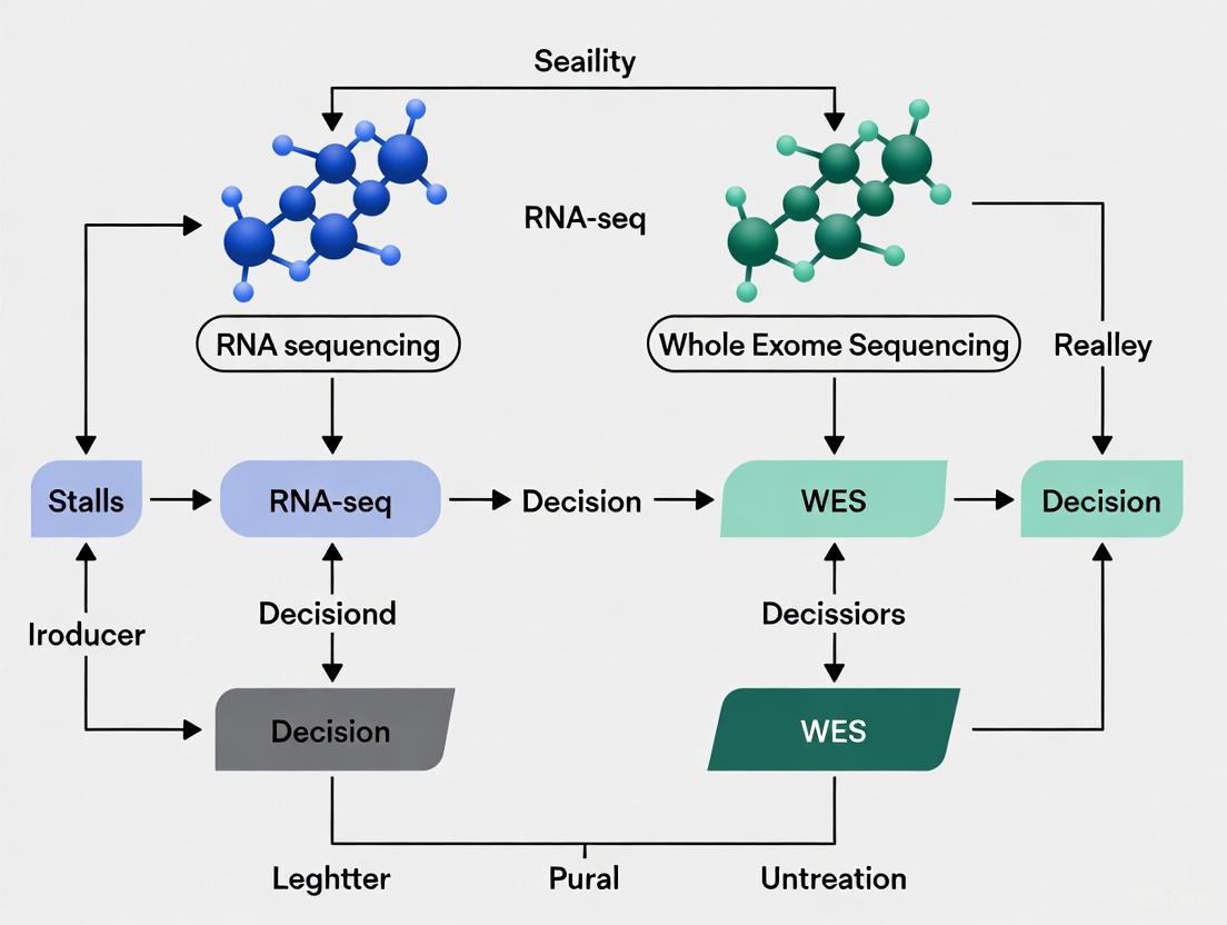
The complementary nature of these workflows is visually summarized in the diagram below.
The Scientist's Toolkit: Essential Research Reagents
Successful implementation of WES and RNA-seq relies on a suite of validated laboratory reagents and bioinformatic tools.
Table 3: Key Reagent Solutions for WES and RNA-seq Workflows
| Item | Function | Example Products (from search results) |
|---|---|---|
| Exome Capture Kits | Enrich sequencing libraries for exonic regions using hybridization-based probes. | Agilent SureSelect [8] [5], Roche KAPA HyperExome [8] [5], Twist Biosciences Exome [5] |
| Nucleic Acid Extraction Kits | Isolate high-quality DNA and/or RNA from various sample types (e.g., blood, FFPE tissue). | Qiagen AllPrep DNA/RNA Kits [4], Promega Maxwell Kits [4] |
| Library Prep Kits | Prepare fragmented DNA or cDNA for sequencing by adding platform-specific adapters. | Illumina TruSeq stranded mRNA kit (RNA) [4], MGI Universal DNA Library Prep Set (DNA) [8] |
| Alignment & Variant Callers | Bioinformatics software to map sequences to a reference genome and identify genetic variants. | BWA (alignment) [4] [8], GATK (variant calling) [5], Strelka2 (somatic variants) [4] |
| Transcriptome Analysis Tools | Software for quantifying gene expression, detecting aberrant splicing, and finding fusions. | STAR (alignment) [4], Kallisto (expression quantification) [4] |
The evidence clearly shows that WES and RNA-seq are not competing technologies but powerful, complementary partners in genetic research and diagnostics. WES serves as an excellent first-tier test for scanning coding regions, while RNA-seq provides the functional evidence needed to interpret variants and diagnose complex cases. For researchers and drug developers, integrating these multi-omic approaches is key to uplifting diagnostic yields, validating new drug targets, and ultimately advancing personalized medicine.
Whole Exome Sequencing (WES) has established itself as a fundamental methodology in genomic research and clinical diagnostics by targeting the protein-coding regions of the genome. While constituting only 1-2% of the entire human genome, the exome harbors an estimated 85% of known disease-causing variants, making WES a powerful and cost-effective approach for identifying pathogenic mutations [9]. This targeted strategy enables researchers and clinicians to focus on the most clinically actionable portions of the genome, providing significant advantages in data management and interpretation compared to whole-genome sequencing.
The diagnostic precision of WES stems from its ability to comprehensively analyze exons, the short, functionally important DNA sequences that represent regions translated into proteins. WES can detect various genetic alterations including single nucleotide variants (SNVs), small insertions and deletions (indels), and copy number variations (CNVs) within these protein-coding regions [9]. In research settings, WES has become particularly valuable for uncovering the genetic basis of rare diseases, neurodevelopmental disorders, and cancer, where protein-altering mutations frequently drive disease pathogenesis.
As sequencing technologies evolve, rigorous performance comparisons between different WES platforms and methodologies have become essential for optimizing research outcomes. Similarly, understanding how WES complements and contrasts with RNA sequencing (RNA-seq) enables researchers to design more effective studies for diagnostic confirmation. This article provides a comprehensive comparison of WES platform performance and experimental approaches, offering researchers detailed methodological frameworks and analytical insights for maximizing the utility of WES in genomic investigations.
Performance Comparison of Major WES Platforms
Experimental Design for Platform Evaluation
A comprehensive 2025 study conducted a systematic comparison of four commercially available WES platforms on the DNBSEQ-T7 sequencer, addressing a significant gap in performance literature for this sequencing system [10]. The investigation evaluated the TargetCap Core Exome Panel v3.0 from BOKE Bioscience (BOKE), IDT's xGen Exome Hyb Panel v2 from Integrated DNA Technologies (IDT), EXome Core Panel from Nanodigmbio Biotechnology (Nad), and Twist Exome 2.0 from Twist Bioscience (Twist) [10].
The experimental design utilized DNA samples from the well-characterized HapMap-CEPH NA12878 and the PancancerLight 800 gDNA Reference Standard (G800) containing more than 720 variants across 330 key cancer genes [10]. Researchers generated 72 libraries using the MGIEasy UDB Universal Library Prep Set (MGI) reagents, with each sample uniquely dual-indexed during PCR amplification using 72 UDB primers from the MGIEasy UDB Primers Adapter Kit Set A [10]. For hybridization capture, the study employed two approaches: manufacturer-specific protocols and a unified MGI enrichment protocol (MGIEasy Fast Hybridization and Wash Kit) to enable direct comparison across platforms [10]. This robust experimental design allowed for systematic assessment of data quality, capture specificity, coverage uniformity, and variant detection accuracy across platforms.
Comparative Performance Metrics
The evaluation revealed that all four platforms exhibited comparable reproducibility and superior technical stability on the DNBSEQ-T7 platform [10]. The table below summarizes the key performance metrics across the evaluated platforms:
Table 1: Performance Comparison of WES Platforms on DNBSEQ-T7
| Platform | Specificity & Uniformity | Variant Detection Accuracy | Protocol Compatibility |
|---|---|---|---|
| BOKE | High coverage uniformity | High detection accuracy Compatible with unified MGI protocol | |
| IDT | High coverage uniformity | High detection accuracy | Compatible with unified MGI protocol |
| Nad | High coverage uniformity | High detection accuracy | Compatible with unified MGI protocol |
| Twist | High coverage uniformity | High detection accuracy | Compatible with unified MGI protocol |
The study established a robust workflow for probe hybridization capture compatible with all four commercial exome kits and the DNBSEQ-Series sequencers [10]. This unified approach demonstrated uniform and outstanding performance across platforms, enhancing broader compatibility regardless of probe brand and simplifying experimental design decisions for researchers.
WES in Integrated Genomic Approaches: Complementarity with RNA-Seq
Synergistic Applications in Cancer Research
The integration of WES with RNA sequencing has demonstrated significant advantages in oncology research, particularly for comprehensive tumor profiling. A 2025 study validated a combined RNA and DNA exome assay across 2,230 clinical tumor samples, revealing that this integrated approach substantially improved detection of clinically relevant alterations [4]. The combined assay enabled direct correlation of somatic alterations with gene expression, recovery of variants missed by DNA-only testing, and enhanced detection of gene fusions [4].
Notably, the integrated RNA-seq and WES approach uncovered clinically actionable alterations in 98% of cases and revealed complex genomic rearrangements that would likely have remained undetected without RNA data [4]. This demonstrates how WES and RNA-seq serve complementary rather than competing roles in comprehensive genomic characterization. Where WES identifies protein-altering mutations across the entire exome, RNA-seq provides functional validation of expression and identifies transcriptomic alterations that may not be evident from DNA analysis alone.
Comparative Diagnostic Utility in Rare Diseases
In rare disease research, RNA-seq has emerged as a valuable ancillary tool following WES, particularly for clarifying variants of uncertain significance. A 2025 study investigated the diagnostic utility of RNA-seq in 53 unrelated individuals with suspected Mendelian disease after standard DNA testing [11]. The researchers employed a hypothesis-driven RNA-seq approach in four specific clinical scenarios: clarifying the impact of putative splice variants, evaluating canonical splice site variants in patients with atypical phenotypes, defining the impact of intragenic copy number variations, and assessing variants within regulatory elements [11].
This approach confirmed a molecular diagnosis and pathomechanism for 45% of participants with a candidate variant, provided supportive evidence for another 21%, and excluded a candidate DNA variant for an additional 24% [11]. These findings underscore how RNA-seq can resolve ambiguous WES findings, particularly for non-coding and splice-affecting variants whose functional consequences may be difficult to predict from DNA sequence alone.
Table 2: Diagnostic Resolution with Hypothesis-Driven RNA-seq Following WES
| Analysis Category | Resolution Outcome | Percentage of Cases |
|---|---|---|
| Candidate Variant Clarification | Molecular Diagnosis Confirmed | 45% |
| Candidate Variant Clarification | Supportive Evidence Provided | 21% |
| Candidate Variant Clarification | Candidate Variant Excluded | 24% |
| Negative WGS Cases | New Putative Diagnosis | Single case |
Experimental Protocols for WES and Integrated Analysis
Standardized WES Laboratory Workflow
The technical workflow for WES requires meticulous attention to each processing step to ensure high-quality results. The following protocol outlines the key laboratory procedures based on validated methodologies from recent studies:
DNA Fragmentation and Library Preparation
- Genomic DNA samples are physically fragmented into fragments ranging from 100-700 bp using a Covaris E210 ultrasonicator or equivalent system [10].
- DNA fragments undergo size selection to obtain 220-280 bp fragments using magnetic bead-based cleanup systems [10].
- End repair, adapter ligation, and pre-PCR amplification are performed using library preparation kits such as the MGIEasy UDB Universal Library Prep Set [10].
- Each sample is uniquely dual-indexed during PCR amplification to enable multiplexing [10].
Hybridization Capture and Enrichment
- For exome capture, several platforms are available including TargetCap (BOKE), xGen Exome Hyb (IDT), EXome Core (Nad), and Twist Exome 2.0 [10].
- Pre-capture libraries are pooled for multiplex hybridization, with input amounts typically ranging from 250-1000 ng per sample depending on multiplexing level [10].
- Hybridization is performed using standardized protocols (e.g., MGIEasy Fast Hybridization and Wash Kit) with incubation times of 1-24 hours depending on the specific platform requirements [10].
- Post-capture amplification is performed using 12 cycles of PCR [10].
Sequencing and Data Generation
- Enriched libraries are converted to single-stranded DNA circles for DNA Nanoball (DNB) generation on DNBSEQ platforms [10].
- Alternatively, libraries can be sequenced on Illumina platforms (e.g., NovaSeq 6000) [4].
- Sequencing is typically performed using paired-end 150 bp reads to achieve minimum coverage of 100x on targeted regions [10].
Figure 1: Standardized WES Laboratory Workflow. Key steps include DNA fragmentation, library preparation, hybridization-based capture, and high-throughput sequencing.
Bioinformatics Analysis Pipeline
The bioinformatics processing of WES data requires a sophisticated pipeline to ensure accurate variant identification:
Primary Analysis and Quality Control
- Raw sequencing reads are processed using tools like FastQC for quality assessment [4].
- Adapter trimming and quality filtering are performed using tools like fastp [11].
- Alignment to reference genome (hg19 or hg38) using aligners such as BWA (DNA) or STAR (RNA) [4] [11].
- PCR duplicate marking using tools like Picard MarkDuplicates [4].
Variant Calling and Annotation
- Germline variant calling using optimized algorithms like Strelka2 or GATK HaplotypeCaller [4].
- Somatic variant calling with tools such as Strelka2, Mutect2, or VarDict [4] [12].
- Variant filtration using parameters including depth (≥10 reads in tumor, ≥20 in normal), VAF (≥0.05 in tumor), and quality scores [4].
- Annotation using population databases (gnomAD, dbSNP) and pathogenicity predictors (SIFT, PolyPhen2) [11].
Integrated Analysis with RNA-seq
- RNA-seq data alignment to transcriptome using Kallisto for expression quantification [4].
- Fusion detection using tools like Arriba [13].
- Splice junction analysis using STAR and custom scripts to identify aberrant splicing [11].
- Expression outlier analysis using Z-scores based on reference cohorts like GTEx [11].
Essential Research Reagents and Platforms
Successful WES implementation requires carefully selected reagents and platforms. The following table details key solutions used in validated experimental protocols:
Table 3: Essential Research Reagents for WES Studies
| Category | Specific Product | Manufacturer | Research Application |
|---|---|---|---|
| Exome Capture Kits | TargetCap Core Exome Panel v3.0 | BOKE Bioscience | Hybridization capture of exonic regions |
| xGen Exome Hyb Panel v2 | Integrated DNA Technologies | Hybridization capture of exonic regions | |
| EXome Core Panel | Nanodigmbio Biotechnology | Hybridization capture of exonic regions | |
| Twist Exome 2.0 | Twist Bioscience | Hybridization capture of exonic regions | |
| Library Preparation | MGIEasy UDB Universal Library Prep Set | MGI | Library construction for DNBSEQ platforms |
| TruSeq stranded mRNA kit | Illumina | RNA library preparation | |
| SureSelect XTHS2 DNA/RNA | Agilent Technologies | Library preparation for FFPE samples | |
| Target Enrichment | MGIEasy Fast Hybridization and Wash Kit | MGI | Unified hybridization protocol |
| Nucleic Acid Extraction | AllPrep DNA/RNA Mini Kit | Qiagen | Simultaneous DNA/RNA isolation from fresh frozen tissue |
| AllPrep DNA/RNA FFPE Kit | Qiagen | Nucleic acid isolation from FFPE samples |
WES maintains its fundamental position in genomic research by providing comprehensive interrogation of the protein-coding genome. Performance comparisons demonstrate that multiple platforms achieve high specificity, uniformity, and detection accuracy, particularly when implemented with standardized protocols [10]. The integration of WES with RNA-seq creates a powerful synergistic approach, enhancing diagnostic resolution in oncology and rare disease research [4] [11].
For researchers designing diagnostic confirmation studies, the combined WES and RNA-seq approach offers distinct advantages, resolving variants of uncertain significance and identifying expressed mutations with greater clinical relevance. As validation frameworks continue to mature [4] [14], this integrated genomic strategy promises to further accelerate precision medicine across diverse research applications.
In the pursuit of diagnostic confirmation for rare diseases and cancer, researchers traditionally rely on DNA-based methods like whole exome sequencing (WES) to identify pathogenic mutations. However, WES alone leaves a significant diagnostic gap, with studies reporting diagnostic yields of 25-50% [2]. This limitation has catalyzed the integration of RNA sequencing (RNA-seq) as a complementary functional approach that captures dynamic gene expression and splicing alterations invisible to DNA-based methods alone. RNA-seq provides a functional lens through which to interpret the genomic landscape, moving beyond static DNA blueprints to reveal active transcriptional programs, aberrant splicing events, and allele-specific expression patterns [2] [11]. This comparison guide objectively evaluates the performance characteristics, diagnostic contributions, and implementation considerations of RNA-seq alongside WES, providing researchers with evidence-based frameworks for deploying these technologies in diagnostic confirmation research.
Technological Comparison: WES and RNA-seq Fundamentals
Core Methodologies and Analytical Outputs
Whole Exome Sequencing (WES) targets the protein-coding regions of the genome (approximately 1-2% of the total genome), providing comprehensive coverage of exonic variants. WES identifies single nucleotide variants (SNVs), small insertions and deletions (INDELs), and copy number variations (CNVs) with clinical diagnostic applications spanning monogenic disorders, cancer genomics, and complex disease research [4] [15]. WES workflows typically involve genomic DNA extraction, exome capture using hybridization-based probes, library preparation, and next-generation sequencing, followed by bioinformatic analysis for variant identification and annotation [15].
RNA Sequencing (RNA-seq) captures and sequences the transcriptome, providing quantitative information about gene expression levels, alternative splicing events, fusion transcripts, and allele-specific expression. RNA-seq enables functional validation of genetic variants by demonstrating their impact on transcriptional output [2] [11]. Methodologically, RNA-seq involves RNA extraction, library preparation (often with poly-A enrichment or ribosomal RNA depletion), sequencing, and specialized bioinformatics pipelines for transcript alignment, quantification, and differential expression analysis [16] [11].
Table 1: Core Technological Capabilities of WES and RNA-seq
| Feature | WES | RNA-seq |
|---|---|---|
| Genomic Coverage | Protein-coding exons (1-2% of genome) | Entire transcriptome (coding and non-coding) |
| Primary Variants Detected | SNVs, INDELs, CNVs | Gene fusions, alternative splicing, expression outliers |
| Functional Information | Static DNA sequence variation | Dynamic gene expression and regulatory consequences |
| Tissue Specificity | Uniform across tissues (germline) | Highly tissue-specific expression patterns |
| Key Analytical Metrics | Coverage depth, variant allele frequency | Transcripts per million (TPM), percent spliced in (PSI) |
| Diagnutive Strength | Coding variant identification | Functional impact assessment of variants |
Complementary Diagnostic Contributions
The integration of WES and RNA-seq creates a powerful diagnostic synergy, with each technology contributing distinct insights to the diagnostic process. WES serves as an excellent first-tier test for identifying potential pathogenic variants in coding regions, while RNA-seq provides functional evidence to confirm or refute their biological impact [11].
In clinical practice, RNA-seq has demonstrated particular utility in specific scenarios following WES: clarifying the impact of putative intronic or exonic splice variants outside canonical splice sites; evaluating canonical splice site variants in patients with atypical phenotypes; defining the impact of intragenic copy number variations on gene expression; and assessing variants within regulatory elements and genic untranslated regions [11]. Hypothesis-driven RNA-seq analyses in these contexts confirmed a molecular diagnosis and pathomechanism for 45% of participants with a candidate variant, provided supportive evidence for a DNA finding for another 21%, and excluded a candidate DNA variant for an additional 24% [11].
Table 2: Diagnostic Performance of WES and RNA-seq in Clinical Studies
| Study Context | WES Diagnostic Yield | RNA-seq Additional Yield | Combined Diagnostic Yield | Reference |
|---|---|---|---|---|
| Neonatal ICU (n=34) | 41% (14/34 patients) | 6% (2/34 patients) | 47% (16/34 patients) | [17] |
| Rare Disease (n=63) | Not specified | 35% (17 additional diagnoses) | Not specified | [2] |
| Undiagnosed Diseases (n=45) | Negative by prior DNA testing | 24% (11/45 patients) | 24% (11/45 patients) | [7] |
| Multi-scenario Rare Disease (n=53) | Candidate variants identified | 45% molecular diagnosis for eligible variants | Significant improvement over WES alone | [11] |
Experimental Data and Performance Validation
Diagnostic Yield Enhancements
Prospective studies demonstrate RNA-seq's capacity to increase diagnostic yields in various clinical contexts. In neonatal intensive care units (NICUs), where rapid diagnosis is critical, implementing rapid WES (rWES) achieved a 41% diagnostic rate with RNA-seq increasing the diagnostic yield by an additional 6%, resulting in a total diagnostic rate of 47% among critically ill newborns [17]. This enhancement translated to tangible clinical benefits, including reduced unnecessary procedures by 15% and shortened hospital stays by 25% [17].
In rare disease diagnostics, Cummings et al. performed RNA-seq on affected muscle tissues from 50 individuals with suspected primary muscle disorders without molecular diagnoses after standard testing. Their approach led to a molecular diagnosis for 17 patients (35% diagnostic yield) who had previously tested negative on genomic testing [2]. The researchers highlighted RNA-seq's particular value in classifying variants of uncertain significance (VUS), identifying second alleles in recessive disorders when WES only returned one pathogenic variant, and detecting deep intronic variants beyond WES resolution [2].
Analytical Validation Frameworks
For clinical implementation, rigorous validation of combined RNA-seq and WES assays is essential. BostonGene's validation of their integrated Tumor Portrait assay established a comprehensive framework involving three critical steps: (1) analytical validation using custom reference samples containing 3,042 SNVs and 47,466 CNVs; (2) orthogonal testing in patient samples; and (3) assessment of clinical utility in real-world cases [4] [14]. When applied to 2,230 clinical tumor samples, this integrated approach enabled direct correlation of somatic alterations with gene expression, recovery of variants missed by DNA-only testing, and improved detection of gene fusions [4]. The assay uncovered clinically actionable alterations in 98% of cases and revealed complex genomic rearrangements that would likely have remained undetected without RNA data [14].
Diagram 1: Integrated WES and RNA-seq Diagnostic Workflow. The parallel analysis of DNA and RNA from patient samples enables comprehensive variant detection and functional validation.
RNA-seq Methodologies for Splicing and Expression Analysis
Splicing Analysis Using Large-Scale Datasets
The analysis of RNA splicing presents particular challenges in large, heterogeneous datasets. MAJIQ v2 represents a suite of algorithms specifically designed to address these challenges by detecting, quantifying, and visualizing splicing variations from complex datasets [18]. This package introduces several key innovations: nonparametric statistical tests for differential splicing (MAJIQ HET), an incremental splicegraph builder, improved intron retention quantification, and the VOILA Modulizer algorithm that parses local splicing variations (LSVs) into classified modules [18].
Splicing quantification in MAJIQ v2 is performed in units of LSVs, which correspond to splits in gene splicegraphs coming into or out of a reference exon. Each LSV edge (splice junction or intron retention) is quantified by its percent spliced in (PSI, Ψ ∈ [0,1]) or changes in relative inclusion between conditions (dPSI, ΔΨ ∈ [-1,1]) [18]. The Bayesian model accounts for read distribution and read stacks, outputting posterior distributions over inclusion levels and confidence metrics. This approach captures not only classical splicing event types but also complex variations involving more than two alternative junctions and unannotated splice variants [18].
Functional Interpretation of RNA-seq Data
The functional interpretation of RNA-seq results typically follows a structured pipeline: (1) raw reads quality check, (2) alignment of reads to a reference genome, (3) aligned reads summarization according to annotation files, (4) differential expression analysis, and (5) gene set analysis and/or functional enrichment analysis [16]. For differential expression analysis, count-based methods like those implemented in DESeq2 are preferred over traditional statistical tests, as RNA-seq data are discrete counts with specific distribution characteristics [16] [17].
Functional enrichment analysis provides biological insight into differentially expressed gene lists through three main approaches: over-representation analysis, functional class scoring, and pathway topology methods [19]. Over-representation analysis tools like clusterProfiler use the hypergeometric distribution to determine whether Gene Ontology categories or pathways are statistically over-represented in significant gene lists compared to background expectations [19]. The Gene Ontology project provides consistent descriptions of gene products across three independent ontologies: biological process, molecular function, and cellular component [19].
Diagram 2: RNA-seq Data Analysis Pipeline. From raw sequencing data to biological interpretation, highlighting key analytical steps and tools for expression and splicing analysis.
The Scientist's Toolkit: Essential Research Reagents and Solutions
Table 3: Essential Research Reagents and Computational Tools for Integrated WES/RNA-seq Studies
| Category | Specific Tools/Reagents | Function | Application Notes |
|---|---|---|---|
| Nucleic Acid Extraction | Qiagen AllPrep DNA/RNA Mini Kit, PAXGene Blood RNA Tubes | Simultaneous DNA/RNA preservation and extraction | Maintains nucleic acid integrity for dual-omics approaches [4] [11] |
| Library Preparation | TruSeq Stranded mRNA Kit, SureSelect XTHS2 Exome Capture | Library construction for RNA-seq and target enrichment for WES | Ensures compatibility with Illumina sequencing platforms [4] [17] |
| Sequencing Platforms | Illumina NovaSeq 6000, NextSeq 500 | High-throughput sequencing | Provides required depth for rare variant detection [4] [17] |
| Alignment Tools | STAR (RNA-seq), BWA (WES) | Read mapping to reference genomes | Handles splice junctions (STAR) and genomic variants (BWA) [4] [11] |
| Variant Callers | Strelka2, Manta (WES) | Somatic and germline variant detection | Optimized for exome sequencing data [4] |
| Splicing Analysis | MAJIQ v2, FRASER2 | Detection of aberrant splicing events | Identifies local splicing variations and intron retention [18] [17] |
| Expression Analysis | DESeq2, OUTRIDER | Differential expression and outlier detection | Statistical analysis of count-based RNA-seq data [17] |
| Functional Enrichment | clusterProfiler | Gene Ontology and pathway analysis | Biological interpretation of significant gene lists [19] |
The integration of RNA-seq with WES represents a paradigm shift in diagnostic confirmation research, moving beyond static genomic information to incorporate dynamic functional evidence. While WES provides comprehensive coverage of protein-coding variants, RNA-seq adds the crucial functional dimension through its ability to detect aberrant splicing, allele-specific expression, and quantitative expression changes [2] [11]. The evidence consistently demonstrates that this combined approach increases diagnostic yields by 6-35% across diverse clinical contexts including rare Mendelian disorders, cancer, and critically ill neonatal populations [2] [17].
For researchers implementing these technologies, several strategic considerations emerge. First, tissue selection for RNA-seq is critical, with optimal results obtained from disease-relevant tissues when possible [2]. Second, hypothesis-driven RNA-seq analysis following WES demonstrates higher diagnostic utility than blinded approaches, particularly for specific scenarios like variant reinterpretation and splice variant characterization [11]. Finally, robust analytical validation frameworks are essential for clinical implementation, incorporating reference standards, orthogonal validation, and real-world clinical correlation [4] [14].
As genomic medicine evolves, the functional lens provided by RNA-seq will increasingly complement DNA-based sequencing, offering not only diagnostic answers but also insights into disease mechanisms that may inform therapeutic strategies. For research applications requiring comprehensive molecular characterization, the combined WES and RNA-seq approach provides a powerful framework for unraveling complex genetic conditions.
For researchers in oncology and rare disease diagnostics, the integration of RNA sequencing (RNA-seq) with DNA-based methods like Whole Exome Sequencing (WES) is transforming genomic analysis. While WES reliably identifies variants in coding regions, it cannot assess whether these variants are functionally expressed. RNA-seq bridges this critical "DNA-to-protein divide," providing functional validation and uncovering alterations invisible to DNA-only approaches. This guide objectively compares the performance of integrated RNA/DNA sequencing against WES alone, supported by recent experimental data and validation studies.
The table below summarizes the core performance differences based on recent large-scale studies:
| Feature | WES (DNA-Only) | Integrated RNA-seq + WES |
|---|---|---|
| Variant Detection Basis | Identifies potential variants in coding regions. [4] | Confirms expression of DNA variants; detects novel, expressed alterations. [3] |
| Actionable Alteration Rate | Lower than integrated approaches. | 98% of cases in a 2,230-tumor cohort. [4] |
| Fusion Gene Detection | Limited or indirect capability. | Improved detection via direct transcriptomic analysis. [4] |
| Identification of Complex Rearrangements | May remain undetected. | Revealed by correlating DNA with RNA data. [4] |
| Functional Relevance | Reports presence, not expression or functional effect. | Filters out non-expressed variants; provides allele-specific expression data. [4] [3] |
| Best Use-Case | Comprehensive cataloging of coding variants. | Functional validation, discovering expressed fusions/splicing variants, guiding personalized treatment. [4] [7] |
Quantitative Performance Benchmarks
Detection Sensitivity and Specificity
The value of RNA-seq is context-dependent, heavily influenced by gene expression levels and the tissue of origin. A foundational study comparing WES and RNA-seq in the same individual found that while RNA-seq captured 81% of exonic variants in well-expressed genes from the relevant tissue, this sensitivity dropped to only 40% when considering all genes indiscriminately. [20] This underscores the necessity of selecting an appropriate tissue for RNA analysis.
A 2025 clinical validation of a combined RNA and DNA exome assay across a large tumor cohort demonstrated its power to uncover clinically actionable alterations in 98% of cases. [4] This integrated approach directly improves diagnostic yield by recovering variants missed by DNA-only testing and improving the detection of gene fusions.
Orthogonal Validation of Variant Pathogenicity
RNA-seq provides a critical functional filter for DNA-based findings. A 2025 study on rare diseases found that RNA-seq was able to reclassify half of the eligible variants identified by genome or exome sequencing, providing the functional evidence needed for more accurate diagnosis. [7] Similarly, in oncology, research shows that integrating RNA-seq data helps confirm that a DNA mutation is actually transcribed, thereby strengthening its claim to clinical relevance. [3] Some variants detected by DNA-seq are not expressed, suggesting they may reside in silent regions of the genome and have lower clinical impact. [3]
Experimental Protocols and Methodologies
Integrated RNA-seq and WES Assay Validation
A 2025 study established a comprehensive, three-step validation framework for a combined assay (Tumor Portrait), which serves as a robust model for clinical implementation. [4]
- Step 1: Analytical Validation. Researchers generated exome-wide somatic reference standards, including 3,042 SNVs and 47,466 CNVs, using cell lines sequenced at varying tumor purities. This provided a ground truth for benchmarking the assay's accuracy. [4]
- Step 2: Orthogonal Testing. Variants identified in patient samples using the integrated assay were confirmed using alternative, established testing methods to verify results. [4]
- Step 3: Clinical Utility Assessment. The validated assay was applied to 2,230 clinical tumor samples to demonstrate its real-world performance in improving detection rates and informing treatment strategies. [4]
Laboratory Procedures: Nucleic acids are co-extracted from tumor samples (FF or FFPE). Libraries are prepared using exome capture kits (e.g., Agilent SureSelect). Sequencing is performed on an Illumina NovaSeq 6000 platform, with stringent QC applied at every stage. [4]
Bioinformatics Analysis: WES data is aligned to hg38 using BWA. RNA-seq data is aligned with STAR. Somatic SNVs and INDELs are called using Strelka2, while variants from RNA-seq data are called using Pisces. [4]
RNA-Seq Specific Somatic Mutation Pipeline
A specialized pipeline for identifying somatic mutations from RNA-seq data in Glioblastoma Multiforme (GBM) was developed to complement WES findings. [21]
- Alignment: The protocol uses a STAR aligner 2-pass procedure to accurately map RNA-seq reads, which is crucial for identifying splice variants and gene fusions. [21]
- Variant Calling: The aligned data is processed through GATK's MuTect2 in tumor-vs-normal mode to call somatic variants. [21]
- Functional and Database Filtering: Identified variants are annotated and filtered against databases like COSMIC (somatic mutations) and dbSNP (germline variants) to prioritize those with potential cancer-driving roles. The functional impact is further predicted using tools like SIFT and FATHMM. [21]
Visualization of Integrated Analysis Workflow
The following diagram illustrates the logical workflow and synergistic relationship between WES and RNA-seq data in a typical integrated analysis pipeline.
The Scientist's Toolkit: Essential Research Reagents & Solutions
The table below details key laboratory and bioinformatics resources essential for implementing the integrated assays described in the cited studies.
| Category | Item | Function & Application |
|---|---|---|
| Wet-Lab Reagents | AllPrep DNA/RNA Mini Kit (Qiagen) | Co-extraction of DNA and RNA from fresh frozen (FF) solid tumors. [4] |
| AllPrep DNA/RNA FFPE Kit (Qiagen) | Co-extraction of DNA and RNA from formalin-fixed paraffin-embedded (FFPE) tissues. [4] | |
| SureSelect XTHS2 DNA/RNA Kit (Agilent) | Library preparation for exome sequencing from both DNA and RNA inputs. [4] | |
| Bioinformatics Tools | STAR Aligner | Fast, splice-aware alignment of RNA-seq reads; often used with a 2-pass method for novel junction discovery. [4] [21] |
| Strelka2 & Manta | Caller for somatic SNVs and small INDELs from WES data. [4] | |
| MuTect2 (GATK) | Widely-used tool for sensitive somatic variant calling; applicable to both WES and RNA-seq data. [21] | |
| Pisces | Variant caller optimized for processing RNA-seq data. [4] | |
| Reference Materials | Cell Line-Derived Reference Standards | Contains predefined SNVs/CNVs for analytical validation and benchmarking of assay performance. [4] |
| High-Confidence Variant Databases (e.g., COSMIC, dbSNP) | Used to annotate and filter variants to prioritize somatic, clinically relevant mutations. [21] |
In clinical diagnostics, DNA-based tests have been the cornerstone of genetic analysis. Whole Exome Sequencing (WES) focuses on sequencing the protein-coding regions of the genome, which constitute approximately 1-2% of the entire genome but harbor the majority of known disease-causing variants [22]. RNA Sequencing (RNA-seq) complements this by capturing the expressed transcriptome, providing functional evidence of how genetic variants actually affect cellular processes [22] [23]. The integration of these technologies is transforming clinical diagnostics by overcoming the limitations of either approach alone, particularly in the interpretation of variants of uncertain significance (VUS) and the detection of aberrant splicing events [22] [24]. This guide provides an objective comparison of their performance, supported by experimental data and detailed methodologies.
Technical Comparison: WES vs. RNA-seq
The table below summarizes the core technical attributes and diagnostic applications of WES and RNA-seq, highlighting their complementary strengths.
Table 1: Technical and Diagnostic Comparison of WES and RNA-seq
| Aspect | Whole Exome Sequencing (WES) | RNA Sequencing (RNA-seq) |
|---|---|---|
| Primary Focus | Genomic DNA from exonic regions (1-2% of genome) [22] | Expressed RNA transcripts (whole transcriptome) [22] |
| Key Applications | Identifying SNVs, INDELs, and small CNVs [4] | Detecting gene fusions, aberrant expression, and allele-specific expression [4] [22] |
| Variant Detection | High-accuracy detection of coding variants [4] | Functional validation of splicing defects and VUS [22] [24] |
| Splicing Analysis | Limited to in silico prediction of splice variants [22] | Direct experimental evidence of splicing aberrations [22] [23] |
| Coverage Gaps | Does not cover 100% of exome; limited non-coding region analysis [25] | Dependent on gene expression levels; may miss lowly expressed genes [3] |
| Diagnostic Yield | ~28-55% for Mendelian disorders [22] | Provides ~15% diagnostic uplift over WES alone [22] |
| Tissue Specificity | Static profile, consistent across cell types | Dynamic profile, highly dependent on tissue type and condition [23] |
Experimental Data and Diagnostic Performance
Diagnostic Yield in Real-World Cohorts
Recent large-scale studies demonstrate the quantitative benefit of integrating WES and RNA-seq. A combined RNA and DNA exome assay applied to 2,230 clinical tumor samples enabled the direct correlation of somatic alterations with gene expression, recovered variants missed by DNA-only testing, and improved the detection of gene fusions, uncovering clinically actionable alterations in 98% of cases [4].
In rare disease diagnostics, a 2025 cohort study implemented a hypothesis-driven RNA-seq approach for patients with specific clinical scenarios following WES. This strategy confirmed a molecular diagnosis and pathomechanism for 45% of participants with a candidate variant, provided supportive evidence for a further 21%, and excluded a candidate DNA variant in 24% of cases [24]. This underscores RNA-seq's high utility as an ancillary test for interpreting specific types of DNA findings.
Complementary Strengths in Variant Interpretation
The synergy between WES and RNA-seq is particularly evident in their ability to resolve different types of molecular defects, as quantified in the table below.
Table 2: Resolution of Aberrant RNA Phenotypes by RNA-seq in Clinical Diagnostics
| Aberrant RNA Phenotype | Function of RNA-seq Analysis | Reported Diagnostic Contribution | Common Experimental Follow-up to WES |
|---|---|---|---|
| Aberrant Splicing [22] | Detects exon skipping, intron retention, and splice site alterations caused by non-canonical variants. | Accounts for ~10% of diagnoses in WES-negative cases [22]. | Analysis of variants of uncertain significance (VUS) in intronic or exonic regions [24]. |
| Aberrant Expression [22] | Identifies gene expression outliers (over- or under-expression) resulting from regulatory variants. | Identifies ~10% of diagnoses in WES-inconclusive cases [22]. | Investigation of promoter or regulatory region variants missed by WES [22]. |
| Mono-allelic Expression (MAE) [22] | Detects the preferential expression of one allele due to epigenetic silencing or NMD. | Explains ~2% of unsolved WES/WGS cases [22]. | Confirmation of allele-specific expression for variants in imprinted genes or with skewed X-inactivation [23]. |
Experimental Protocols for Integrated Analysis
Protocol 1: Combined WES and RNA-seq from a Single Tumor Sample
A validated methodology for integrated profiling from a single specimen involves the following steps [4]:
- Nucleic Acid Isolation: For FFPE tumors, use the AllPrep DNA/RNA FFPE Kit (Qiagen). Quantify and quality-check DNA and RNA extracts using Qubit 2.0, NanoDrop OneC, and TapeStation 4200.
- Library Preparation:
- WES Library: Use 10-200 ng of DNA with the SureSelect XTHS2 DNA kit and the SureSelect Human All Exon V7 exome probe (Agilent Technologies).
- RNA-seq Library: Use 10-200 ng of RNA with the SureSelect XTHS2 RNA kit and the SureSelect Human All Exon V7 + UTR exome probe (Agilent Technologies).
- Sequencing: Perform on an Illumina NovaSeq 6000 platform, monitoring for Q30 > 90% and PF > 80%.
- Bioinformatics Analysis:
- Alignment: Map WES data to hg38 with BWA aligner and RNA-seq data with STAR aligner.
- Variant Calling: Use Strelka2 for somatic SNVs and INDELs in WES data. For RNA-seq variant calling, employ Pisces.
- Fusion Detection: Use specialized algorithms on RNA-seq data to identify gene fusions.
Protocol 2: Hypothesis-Driven RNA-seq for Rare Diseases
For rare diseases where WES has identified a candidate variant, a targeted RNA-seq protocol can be applied [24]:
- Tissue Selection: Utilize Genotype-Tissue Expression Portal to select a clinically accessible tissue where the gene of interest has a median TPM ≥ 5. Prioritize fibroblasts, blood, or muscle biopsy.
- RNA Extraction and QC: Extract total RNA using kits such as PAXGene Blood RNA Kit or RNeasy Mini Kit. Determine RNA quality using TapeStation RNA ScreenTape.
- Library Prep and Sequencing: Spike total RNA with SIRV Set 3 (Lexogen). Prepare libraries using automated NEBNext Poly(A) mRNA Magnetic Isolation Module and NEBNext Ultra II Directional RNA Library Prep kit. Sequence on NovaSeq6000 with paired-end 150 bp runs.
- Bioinformatics Analysis:
- Trim reads with fastp and align to GRCh38 using STAR in two-pass mode.
- Perform splice junction detection using the SJ.out.tab file from STAR. Junctions with ≥5 uniquely mapped reads are analyzed.
- Calculate a junction score and Z-score using a GTEx control cohort. Aberrant junctions are classified as novel, missing, or outlier based on an absolute Z-score ≥ 3.
Essential Research Reagent Solutions
The table below lists key reagents and kits used in the featured experimental protocols, providing a practical resource for laboratory setup.
Table 3: Key Research Reagent Solutions for Integrated WES and RNA-seq
| Product / Kit Name | Function in Workflow | Specific Application Note |
|---|---|---|
| AllPrep DNA/RNA FFPE Kit (Qiagen) [4] | Concurrent isolation of DNA and RNA from a single FFPE tumor sample. | Preserves nucleic acid integrity from challenging, archived samples. |
| SureSelect XTHS2 (Agilent) [4] | Target enrichment for whole exome (DNA) and exome-plus-UTR (RNA). | Enables focused sequencing on clinically relevant genomic regions. |
| NEBNext Ultra II Directional RNA Library Prep (NEB) [24] | Construction of strand-specific RNA-seq libraries. | Critical for accurate transcriptome assembly and fusion detection. |
| SIRV Set 3 (Lexogen) [24] | Spike-in RNA controls for sequencing workflow. | Moners technical performance and normalization across batches. |
| PAXGene Blood RNA Tubes (BD) [24] | Blood collection for RNA stabilization. | Prevents RNA degradation in whole blood samples during transport. |
Market Adoption and Future Directions
The clinical adoption of WES and RNA-seq is accelerating, reflected in market growth and technological integration. The global genomics data analysis market is projected to grow at a CAGR of 15.45%, reaching USD 33.51 billion by 2035 [26]. The WES market specifically is expected to rise from US$ 2.17 billion in 2025 to US$ 6.88 billion by 2032, a CAGR of 17.9% [25]. This growth is fueled by population genomics initiatives (e.g., NIH's All of Us Program), expanding insurance coverage for WES, and growing collaborations between genomics companies and healthcare providers [25]. North America currently leads this market, while the Asia-Pacific region is emerging as a high-growth area due to rapid genomic infrastructure development and initiatives like India's IndiGen program [25].
The future of clinical diagnostics lies in the deeper integration of multi-omic data. Targeted RNA-seq panels are being developed to provide deeper coverage of genes with somatic mutations of interest, improving detection accuracy for rare alleles [3]. Furthermore, the application of AI and machine learning is revolutionizing data interpretation by enabling faster analysis, accurate variant detection, and predictive modeling, thereby enhancing precision medicine outcomes [26]. As these technologies mature and workflows become more standardized, the combined use of WES and RNA-seq is poised to become the benchmark for comprehensive clinical genomic diagnosis.
From Theory to Practice: Implementing Integrated WES and RNA-seq Workflows
Next-generation sequencing (NGS) has revolutionized molecular diagnostics in both rare diseases and oncology. Whole Exome Sequencing (WES) has become a standard approach, focusing on the protein-coding regions that harbor an estimated 85% of known disease-causing variants [27]. However, a significant diagnostic gap remains, with WES alone achieving diagnostic yields typically between 25-50% in rare disease cases [2] and missing key alterations in oncology [28]. This limitation stems primarily from WES's inherent constraint in detecting functional consequences of variants, particularly those affecting RNA splicing, expression, and regulation.
RNA sequencing (RNA-seq) has emerged as a powerful complementary technology that bridges this functional gap. By providing direct evidence of transcript-level disruptions, RNA-seq can identify aberrant splicing events, allele-specific expression imbalances, and gene fusions that DNA-based methods alone cannot resolve [29]. Recent studies demonstrate that integrating RNA-seq with WES increases diagnostic yields by 10-35% across diverse clinical contexts [7] [29]. This guide provides a comprehensive comparison of integrated RNA-seq/WES approaches against alternative testing strategies, supported by experimental data and methodological protocols for diagnostic confirmation research.
Technology Comparison: WES, RNA-seq, and Their Integration
Individual Technology Profiles
Whole Exome Sequencing (WES) targets the approximately 2% of the genome that codes for proteins, providing comprehensive coverage of exonic regions. This method efficiently identifies single nucleotide variants (SNVs), small insertions and deletions (indels), and some copy number variations (CNVs) [27]. However, WES cannot assess non-coding regulatory regions, has limited capability to detect structural variants (SVs) and gene fusions, and provides no functional data on how identified variants affect RNA expression or processing [27].
RNA Sequencing (RNA-seq) profiles the transcriptome by capturing and sequencing RNA molecules. This approach detects gene fusions, alternative splicing events, aberrant gene expression, and allele-specific expression [2] [29]. Unlike WES, RNA-seq provides functional evidence for variant impact but does not reliably detect non-expressed genomic variants or variants in regulatory regions that may affect gene expression [30].
Whole Transcriptome Sequencing (WTS), a comprehensive form of RNA-seq, analyzes the entire complement of RNA transcripts without relying on pre-defined annotations. WTS offers greater resolution for splice variants and can identify novel transcripts and regulatory non-coding RNAs, though it requires higher sequencing depth for accurate gene expression quantification [30].
Comparative Performance Metrics
Table 1: Diagnostic Performance of Genomic Testing Strategies
| Testing Approach | Typical Diagnostic Yield | Variant Types Detected | Key Limitations |
|---|---|---|---|
| WES Alone | 25-50% [2] | SNVs, small indels, some CNVs | Cannot detect non-coding variants, provides no functional data |
| Targeted Panel Alone | ~56% (PID study) [31] | Pre-defined SNVs, indels in targeted genes | Limited to pre-selected genes, quickly becomes outdated |
| WGS Alone | Higher than WES [27] | SNVs, indels, CNVs, SVs, non-coding variants | Higher cost, data interpretation challenges for non-coding variants |
| RNA-seq/WTS Alone | 35% (muscle disorders) [2] | Fusions, splicing defects, expression outliers | Limited to expressed genes, misses regulatory variants |
| Integrated WES + RNA-seq | 10-35% increase over DNA-only methods [7] [29] | Combines WES variants with functional RNA evidence | Requires specialized validation, higher computational burden |
Advantages of Integrated RNA-seq and WES Approach
Enhanced Diagnostic Resolution
Integrating RNA-seq with WES from a single sample significantly improves diagnostic resolution across multiple dimensions. In rare disease diagnostics, this combined approach provides functional evidence that enables reclassification of variants of uncertain significance (VUS). A study of 30 previously unsolved rare disease cases demonstrated that RNA-seq contributed to diagnostic resolution in 27% of cases (10 definitively, 1 likely) by detecting aberrant splicing events including exon skipping, cryptic splice-site activation, and intron retention [29].
In oncology, combined RNA-seq and WES testing identified clinically actionable alterations in 98% of 2,230 clinical tumor samples, recovering variants missed by DNA-only testing and improving fusion detection [4]. This integrated approach uncovered complex genomic rearrangements that would likely have remained undetected without RNA data, demonstrating its superior clinical utility.
Economic and Workflow Considerations
Despite initial perceptions of higher costs, integrated RNA-seq/WES testing can provide economic advantages compared to sequential or tiered testing approaches. A 2025 economic modeling study in non-small cell lung cancer (NSCLC) demonstrated that compared to sequential single-gene testing, comprehensive profiling using whole-exome and whole-transcriptome sequencing (WES/WTS) reduced costs by $14,602 per patient while providing minimal survival benefits [28].
In primary immunodeficiency (PID) testing, cost analysis based on current commercial pricing reveals that a WES-only strategy would save $300-$950 per patient compared to a tiered approach beginning with targeted panels, depending on diagnostic yield [31]. These findings challenge the traditional perception that targeted panels are more cost-effective, particularly when considering the potential for reduced diagnostic odysseys.
Experimental Validation and Protocols
Analytical Validation Framework
Robust validation of integrated RNA-seq/WES assays requires a comprehensive approach. A 2025 study established a three-step validation framework: (1) analytical validation using custom reference samples containing 3,042 SNVs and 47,466 CNVs; (2) orthogonal testing in patient samples; and (3) assessment of clinical utility in real-world cases [4]. This rigorous methodology ensures both technical accuracy and clinical relevance.
For somatic variant detection in oncology, the Association of Molecular Pathology (AMP) recommends determining positive percentage agreement and positive predictive value for each variant type, establishing requirements for minimal depth of coverage, and using an adequate number of samples to establish test performance characteristics [32]. This error-based approach identifies potential sources of errors throughout the analytical process and addresses them through test design and quality controls.
Laboratory Methodologies
Nucleic Acid Extraction: Successful integration begins with high-quality nucleic acid extraction. For fresh frozen solid tumors, the AllPrep DNA/RNA Mini Kit enables simultaneous isolation of both DNA and RNA from a single sample [4]. For formalin-fixed paraffin-embedded (FFPE) samples, the AllPrep DNA/RNA FFPE Kit is recommended, with DNA and RNA quantity and quality measured using Qubit, NanoDrop, and TapeStation systems [4].
Library Preparation: For WES, the SureSelect Human All Exon V7 exome probe provides comprehensive exome coverage [4]. For RNA-seq, library construction can utilize either the TruSeq stranded mRNA kit for fresh frozen tissue or the SureSelect XTHS2 RNA kit for FFPE samples [4]. The SureSelect Human All Exon V7 + UTR exome probe enables targeted RNA capture, enhancing detection of relevant transcripts.
Sequencing and Analysis: Sequencing is typically performed on Illumina NovaSeq 6000 systems with Q30 scores >90% [4]. Bioinformatics pipelines align WES data to the human genome (hg38) using BWA aligner, while RNA-seq data is aligned using STAR aligner [4]. Somatic variant calling employs optimized Strelka and Manta algorithms, with specialized filtration parameters to ensure variant accuracy [4].
Table 2: Key Research Reagent Solutions for Integrated Assays
| Reagent/Kit | Manufacturer | Primary Function | Application Notes |
|---|---|---|---|
| AllPrep DNA/RNA Mini Kit | Qiagen | Simultaneous DNA/RNA extraction from single sample | Maintains nucleic acid integrity; ideal for fresh frozen specimens |
| AllPrep DNA/RNA FFPE Kit | Qiagen | Co-extraction from FFPE tissue | Optimized for challenging, cross-linked samples |
| SureSelect Human All Exon V7 | Agilent Technologies | Exome capture for WES | Comprehensive exonic region coverage |
| SureSelect XTHS2 RNA Kit | Agilent Technologies | Library prep for RNA-seq | Suitable for degraded FFPE-derived RNA |
| TruSeq stranded mRNA kit | Illumina | RNA library preparation | Ideal for high-quality RNA from fresh frozen tissue |
Workflow Visualization
Integrated RNA-seq and WES Workflow from a Single Sample
Diagnostic Confirmation Applications
Variant Interpretation and Classification
RNA-seq provides functional evidence that significantly enhances variant interpretation, particularly for splice-altering variants. In rare disease diagnostics, RNA-seq has been shown to reclassify half of eligible variants identified through exome or genome sequencing, providing critical evidence for pathogenicity [7]. The molecular mechanisms resolved through RNA-seq include exon skipping (46% of variants), intron retention (15%), cryptic splice-site activation (8%), and multiple splicing effects (15%) [29].
For cancer diagnostics, integrating RNA-seq with WES enables direct correlation of somatic alterations with gene expression patterns, recovery of variants missed by DNA-only testing, and improved detection of gene fusions [4]. This approach also reveals allele-specific expression of oncogenic drivers, providing functional validation of putative pathogenic variants.
Tissue-Specific Considerations
The choice of tissue for RNA-seq significantly impacts diagnostic success. In rare disease diagnostics, resolution varies by tissue source: fibroblast-derived RNA resolved 27% of cases, blood-derived RNA resolved 55%, and both tissues contributed in 18% of cases [29]. Disease-relevant tissues often provide superior diagnostic information, as demonstrated in muscle disorders where sequencing affected muscle tissues identified pathogenic variants not detectable in blood [2].
Complementary Value of RNA-seq and WES Integration
Integrated RNA-seq and WES testing from a single sample represents a significant advancement in genomic diagnostics, overcoming limitations of either method alone. The combined approach provides functional validation of DNA variants through direct transcriptome assessment, leading to improved diagnostic yields across rare diseases and oncology. While implementation requires specialized validation frameworks and bioinformatic capabilities, the clinical utility and potential cost savings support its adoption as a primary testing approach in complex diagnostic cases. As validation guidelines continue to evolve and experience grows, integrated RNA-seq/WES testing is poised to become a standard of care in precision medicine.
Next-generation sequencing (NGS) has revolutionized genomic research and clinical diagnostics, with RNA Sequencing (RNA-seq) and Whole Exome Sequencing (WES) emerging as pivotal technologies for diagnostic confirmation. The selection between these approaches involves critical trade-offs in diagnostic yield, technical performance, and clinical utility [4] [22]. RNA-seq provides functional evidence for variant interpretation by capturing dynamic transcriptome information, including aberrant splicing, allele-specific expression, and gene expression outliers [22]. In contrast, WES comprehensively targets the protein-coding regions of the genome, identifying single nucleotide variants (SNVs), insertions/deletions (INDELs), and copy number variations (CNVs) across more than 20,000 genes [4]. Effective implementation of either technology depends on a meticulously optimized workflow encompassing nucleic acid isolation, library preparation, and sequencing, with specific protocols tailored to sample type and research objectives [33] [34]. This guide objectively compares experimental methodologies and performance data for these critical workflow components to inform researchers and drug development professionals.
Nucleic Acid Isolation and Extraction Methods
The initial step of nucleic acid isolation is fundamental to sequencing success, with method selection dictated by sample type, quality, and intended downstream applications. Efficient extraction is crucial for obtaining high-quality DNA or RNA free from contaminants that inhibit library preparation.
DNA Extraction Techniques
DNA extraction methods vary significantly in their mechanism, throughput, and suitability for different sample types.
- Solid-Phase Extraction: This column-based method uses a silica membrane to bind DNA under specific pH and salt conditions. Contaminants are removed through washing, and pure DNA is eluted in a low-salt buffer. It is widely used in commercial kits for its rapid and efficient purification [35].
- Magnetic Bead-Based Purification: A modification of solid-phase extraction, this method uses silica-coated magnetic beads that bind DNA. A magnet aggregates the particles, allowing contaminants to be washed away. This technique is time- and cost-effective as it eliminates repeated centrifugation and vacuum filtration steps, making it suitable for automation and high-throughput workflows [35] [33].
- Guanidinium Thiocyanate-Phenol-Chloroform Extraction: This solution-based technique separates DNA into an aqueous layer through a series of organic extractions. The DNA is then collected via centrifugation and precipitated. While effective, it involves handling hazardous chemicals like phenol and chloroform [35].
- Automated Extraction Systems: Robotic systems streamline the extraction process, increasing scalability, reliability, and reproducibility while reducing the need for highly trained personnel. Although representing a higher initial investment, automation enhances consistency for large-scale sequencing projects [35].
Table 1: Comparison of DNA Extraction Methods
| Method | Principle | Throughput | Advantages | Limitations |
|---|---|---|---|---|
| Solid-Phase Extraction [35] | Silica-membrane binding | Medium | Rapid, efficient, many commercial kits | Multiple centrifugation steps |
| Magnetic Bead-Based [35] [33] | Silica-coated magnetic beads | High | Amenable to automation, fewer centrifugation steps, cost-effective | Requires specialized magnetic equipment |
| Phenol-Chloroform [35] | Organic phase separation | Low | High purity, does not require specialized columns | Uses toxic chemicals, labor-intensive |
| Automated Systems [35] | Robotic handling of other methods | Very High | High reproducibility, scalable, reduced manual labor | High initial cost |
RNA Extraction Considerations
RNA isolation requires stringent conditions to prevent degradation by ubiquitous RNases. The quality of RNA, especially from challenging sample types like Formalin-Fixed Paraffin-Embedded (FFPE) tissues, is a critical determinant for successful RNA-seq. The AllPrep DNA/RNA Kit (Qiagen) allows for the simultaneous isolation of both DNA and RNA from a single sample, which is invaluable for integrated analysis [4]. For FFPE samples specifically, the AllPrep DNA/RNA FFPE Kit (Qiagen) is designed to overcome issues related to cross-linking and fragmentation [4].
RNA quality is typically assessed using metrics such as the RNA Integrity Number (RIN) or the DV200 value (the percentage of RNA fragments larger than 200 nucleotides). While FFPE samples often have low DV200 values, samples with a DV200 ≥ 30% are generally considered usable for RNA-seq protocols [34].
Library Preparation Methods and Kits
Library preparation converts purified nucleic acids into sequencing-ready libraries and is a major source of technical variability. The choice of kit significantly impacts gene detection, quantification, and the ability to work with degraded or low-input samples.
DNA Library Preparation for WES
WES library preparation involves fragmenting DNA, ligating adapters, and enriching exonic regions via hybridization capture.
- Illumina DNA Prep (DN): This kit has demonstrated high-quality results with low GC bias, making it suitable for sequencing bacteria with varying genomic characteristics [36].
- Roche KAPA HyperPlus (KP) & NEBNext Ultra II FS (NN): These kits also perform well, producing high-quality genome assemblies with minimal GC bias, as evidenced by sequencing performance across different bacterial species [36].
- Nextera XT (XT): In comparative studies, this kit exhibited significant GC bias and lower sequencing quality for samples with low GC content, making it less robust for diverse applications [36].
- Santa Cruz Reaction (SCR): This DIY library build method is highly effective for retrieving degraded DNA from museum specimens and can be implemented at high throughput for low cost, offering a valuable alternative to commercial kits for challenging samples [33].
RNA Library Preparation for RNA-seq
RNA-seq library prep kits must effectively deplete ribosomal RNA (rRNA) and preserve strand orientation to accurately quantify gene expression.
- Illumina Stranded Total RNA Prep Ligation with Ribo-Zero Plus (Kit B): This kit demonstrates superior performance in rRNA depletion (0.1% rRNA content vs. 17.45% in a competitor) and achieves better alignment rates with lower duplication rates (~10.73% vs. ~28.48%). It requires a standard input of 100-200 ng of RNA [34] [4].
- TaKaRa SMARTer Stranded Total RNA-Seq Kit v2 (Kit A): The key advantage of this kit is its ability to work with very low input RNA (as low as 1 ng, a 20-fold reduction), achieving comparable gene expression quantification to Kit B, albeit with increased required sequencing depth. It shows higher rRNA content and duplication rates [34].
- TruSeq Stranded Total RNA Kit: This kit provides strand information and can detect both coding and non-coding RNA, making it a comprehensive choice for total transcriptome analysis [37].
Table 2: Comparison of RNA-seq Library Preparation Kits
| Kit Name | Input Requirement | Key Strengths | Key Weaknesses |
|---|---|---|---|
| Illumina Stranded Total RNA Prep [34] | 100-200 ng | Excellent rRNA depletion, high library yield, superior alignment rates | Requires more input RNA |
| TaKaRa SMARTer Stranded Total RNA-Seq [34] | 1-10 ng | Works with extremely low input RNA, comparable expression quantification | Higher rRNA content, higher duplication rate |
| TruSeq Stranded Total RNA Kit [37] | 10-200 ng | Detects coding and non-coding RNA, provides strand information | Standard input requirements |
Sequencing Platforms and Data Analysis
Following library preparation, pooled libraries are sequenced on high-throughput platforms, and the resulting data undergoes a rigorous bioinformatic analysis to generate biologically meaningful results.
Sequencing Platforms
The Illumina NovaSeq 6000 and HiSeq 2500/2000 systems are workhorses for both WES and RNA-seq, capable of generating the high coverage required for these applications [4] [37]. For targeted panels or smaller-scale projects, benchtop sequencers like the MGI DNBSEQ-G50RS and Illumina MiSeq offer efficient and cost-effective solutions [38]. Key quality control metrics monitored during sequencing include the percentage of bases with a quality score (Q30) above 90% and a cluster passing filter (PF) greater than 80% [4].
Bioinformatics Analysis
The analytical workflow differs for WES and RNA-seq data.
- WES Analysis: After demultiplexing, reads are aligned to a reference genome (e.g., hg38) using aligners like BWA [4]. Variant calling for SNVs and INDELs is performed using tools such as Strelka2 and Manta [4]. For targeted panels, proprietary software like Sophia DDM can use machine learning for rapid variant analysis and visualization [38].
- RNA-seq Analysis: RNA-seq reads are typically aligned with the STAR aligner or quantified directly against a transcriptome using Kallisto [4]. Downstream analysis focuses on identifying differentially expressed genes (using tools like DESeq2), detecting aberrant splicing events, and identifying allele-specific expression [22]. Pathway enrichment analysis with databases like KEGG is used to interpret the biological significance of gene expression changes [34].
Comparative Performance in Diagnostic Confirmation
The core thesis of evaluating RNA-seq versus WES is clarified by their direct comparison in clinical diagnostic settings, particularly in solving Mendelian disorders and characterizing cancer.
Diagnostic Yield and Technical Performance
Table 3: Diagnostic Yield Comparison: WES vs. RNA-seq
| Metric | Whole Exome Sequencing (WES) | RNA Sequencing (RNA-seq) | Combined Approach |
|---|---|---|---|
| Theoretical Diagnostic Yield [22] | 28% - 55% | - | - |
| Diagnostic Uplift from RNA-seq [22] | - | ~15% over WES/WGS | - |
| Key Detected Aberrations | SNVs, INDELs, CNVs [4] | Aberrant splicing, mono-allelic expression, aberrant expression [22] | All of the above plus gene fusions [4] |
| Actionable Findings in Cancer [4] | - | - | 98% of cases (in a cohort of 2230) |
Integrated Assays and Panel Alternatives
Combining WES with RNA-seq in a single assay provides a powerful tool for comprehensive genomic profiling. One such integrated assay demonstrated a 98% rate of uncovering clinically actionable alterations in a cohort of 2230 tumor samples. It improved the detection of gene fusions and allowed for the recovery of variants missed by DNA-only testing [4].
As an alternative to genome-scale testing, targeted NGS panels offer a focused and cost-effective approach. One study developed a 61-gene oncopanel that demonstrated 99.99% repeatability and 99.98% reproducibility. A significant advantage was the reduction of turnaround time from 3 weeks (with outsourced testing) to just 4 days, which is critical for timely clinical decision-making [38].
The Scientist's Toolkit: Essential Research Reagents and Materials
Successful execution of NGS workflows relies on a suite of trusted reagents and kits. The following table details key solutions used in the experiments cited throughout this guide.
Table 4: Essential Research Reagent Solutions for NGS Workflows
| Item Name | Function in Workflow | Specific Application Example |
|---|---|---|
| AllPrep DNA/RNA FFPE Kit (Qiagen) [4] | Concurrent isolation of DNA and RNA from FFPE tissue. | Nucleic acid extraction from archived clinical specimens for integrated WES and RNA-seq [4]. |
| Monarch PCR & DNA Cleanup Kit (NEB) [33] | Purification and size selection of DNA fragments. | DNA extraction and cleanup from insect museum specimens for degraded DNA protocols [33]. |
| NEBNext Ultra II FS DNA Library Prep Kit (NEB) [36] | Preparation of Illumina-compatible sequencing libraries from fragmented DNA. | Library construction for bacterial whole-genome sequencing; demonstrated low GC bias [36]. |
| TruSeq Stranded Total RNA Kit (Illumina) [37] | Preparation of strand-specific RNA-seq libraries. | Library construction for transcriptome analysis, enabling detection of coding and non-coding RNA [37]. |
| SureSelect Human All Exon V7 (Agilent) [4] | Hybridization-based capture of exonic regions. | Target enrichment for Whole Exome Sequencing in a combined RNA-DNA clinical assay [4]. |
| AmpliTaq Gold Mastermix (Thermo Fisher) [33] | PCR amplification with uracil tolerance. | Indexing PCR amplification for libraries built from museum specimens containing deaminated bases [33]. |
| SPRI/QuantBio SparQ Beads [33] | Magnetic bead-based clean-up and size selection of DNA libraries. | Post-ligation and post-amplification purification steps in various library prep protocols [33]. |
Experimental Workflow and Pathway Diagrams
The following diagrams summarize the logical relationships and key decision points in the NGS workflows discussed.
Core NGS Wet-Lab Workflow
Method Selection Decision Pathway
Detailed Experimental Protocols
To ensure reproducibility, here are the detailed methodologies for key experiments cited.
- Sample Lysis: Detach one or two legs from insect specimens and incubate overnight at 56°C in 90μL of lysis Buffer C (200mM Tris pH8, 25mM EDTA pH8, 0.05% Tween-20, 0.4 mg/ml Proteinase K).
- Silica-Based Extraction (Rohland Method):
- Add a 10:1 ratio of Binding Buffer D to lysate.
- Add silica beads to capture DNA.
- Wash beads twice with an ethanol-based buffer.
- Dry beads for approximately 5 minutes.
- Elute DNA in a low-salt buffer or ultra-pure water.
- Quality Control: Quantify DNA using a Qubit Fluorometer and assess fragment size distribution using an Agilent Tapestation system.
- Nucleic Acid Isolation: Extract DNA and RNA from fresh frozen (FF) or FFPE tumor samples using the AllPrep DNA/RNA Kit or AllPrep DNA/RNA FFPE Kit (Qiagen). Quantity using Qubit and NanoDrop, and assess integrity via TapeStation.
- WES Library Prep:
- Fragment 10-200 ng of genomic DNA.
- Perform end-repair, A-tailing, and adapter ligation using the SureSelect XTHS2 DNA library prep kit (Agilent).
- Enrich exonic regions by hybridizing with the SureSelect Human All Exon V7 biotinylated probe set (Agilent).
- Capture and amplify the library.
- RNA-seq Library Prep:
- For FF samples, use the TruSeq stranded mRNA kit (Illumina). For FFPE, use the SureSelect XTHS2 RNA kit (Agilent).
- Perform rRNA depletion (for total RNA protocols) or poly-A selection (for mRNA protocols).
- Carry out cDNA synthesis, fragmentation, adapter ligation, and PCR amplification.
- Sequencing: Pool libraries and sequence on an Illumina NovaSeq 6000 platform, targeting a minimum of 90% of bases with Q30 > 30.
- Sample Preparation: Isolate RNA from six melanoma FFPE samples. Assess RNA quality and DV200 values.
- Library Preparation:
- Kit A (TaKaRa): Follow the SMARTer Stranded Total RNA-Seq Kit v2 protocol with low input RNA (as low as 1 ng).
- Kit B (Illumina): Follow the Illumina Stranded Total RNA Prep Ligation with Ribo-Zero Plus protocol with standard input RNA (100 ng).
- Sequencing and Analysis:
- Sequence all libraries on an Illumina platform.
- Assess metrics: total reads, alignment rate, rRNA content, duplication rate, and exonic mapping rate.
- Perform differential expression analysis (e.g., using DESeq2) and pathway enrichment analysis (e.g., with KEGG) to compare the concordance of results between the two kits.
The advancement of precision oncology and genetic disease research hinges on the accurate detection of molecular alterations. Next-generation sequencing (NGS) technologies, particularly RNA Sequencing (RNA-seq) and Whole Exome Sequencing (WES), have become cornerstone methodologies in diagnostic confirmation research. While WES provides a comprehensive view of coding variants across approximately 20,000 genes, RNA-seq delivers a dynamic snapshot of gene expression and transcriptomic alterations [4] [39]. Historically, these technologies have been deployed independently, but emerging evidence demonstrates that their integration significantly enhances diagnostic yield beyond what either method can achieve alone [4] [28] [40]. This comparative guide evaluates the performance, protocols, and clinical utility of bioinformatics pipelines for alignment, variant calling, and expression quantification within the context of combined RNA-seq and WES approaches, providing researchers and drug development professionals with actionable insights for implementing these technologies.
Comparative Analysis of Assay Performance
Detection Capabilities Across Genomic Alteration Types
Different sequencing technologies and analytical pipelines exhibit distinct strengths and weaknesses in detecting various types of genomic alterations. The table below summarizes the detection capabilities of DNA-only (WES) versus integrated RNA-DNA sequencing approaches.
Table 1: Detection Capabilities of WES vs. Integrated RNA-DNA Sequencing
| Alteration Type | WES (DNA-Only) | Integrated RNA & WES | Key Performance Findings |
|---|---|---|---|
| Single Nucleotide Variants (SNVs) | Strong detection | Enhanced detection | RNA-seq recovers somatic variants missed by DNA-only testing and confirms expression of DNA-identified variants [4] [3]. |
| Insertions/Deletions (INDELs) | Strong detection | Enhanced detection | Combined approach improves accuracy, with RNA-seq validating functional relevance of small INDELs [4]. |
| Copy Number Variations (CNVs) | Strong detection | Comparable detection | WES surpasses targeted panels in identifying arm-level CNVs; RNA provides orthogonal confirmation [4] [41]. |
| Gene Fusions | Limited detection | Superior detection | RNA-seq dramatically improves fusion detection, identifying clinically actionable fusions missed by DNA [4] [28]. |
| Gene Expression | Not detected | Comprehensive detection | Gene expression signatures predict immunotherapy outcomes and enable tumor microenvironment analysis [4] [42]. |
| Alternative Splicing | Not detected | Comprehensive detection | RNA-seq identifies aberrant splicing events, a known disease mechanism [3] [40]. |
Diagnostic Yield and Clinical Utility
The ultimate value of a genomic assay is measured by its ability to provide clinically actionable findings. Recent large-scale studies have quantified the diagnostic advantages of integrated sequencing.
Table 2: Diagnostic Yield and Clinical Impact of Sequencing Approaches
| Metric | WES Alone | Integrated RNA & WES | Study Context |
|---|---|---|---|
| Diagnostic Yield | 44.1% (Phase I) | 58.1% (After Phase II RNA-seq) | 236 patients with developmental and epileptic encephalopathy [40]. |
| Cases with Actionable Alterations | Not specified | 98% of cases | 2230 clinical tumor samples [4]. |
| Increase in Fusion Detection | Baseline | 2.3% to 13.0% more patients identified | Non-small cell lung cancer, depending on fusion prevalence [28]. |
| Economic Impact | Higher cost per diagnosis | Cost reduction of $400-$1,724 per patient | Lower total costs due to improved therapy matching in NSCLC [28]. |
Experimental Protocols for Validation and Comparison
Analytical Validation Using Reference Standards
Robust validation is critical for clinical implementation. The integrated WES/RNA-seq assay described in Communications Medicine employed a rigorous three-step validation framework [4]:
- Reference Sample Generation: Custom reference materials were developed, containing 3,042 known SNVs and 47,466 CNVs. These standards were sequenced across multiple runs using cell lines at varying tumor purity levels to establish performance metrics across different experimental conditions [4].
- Orthogonal Confirmation: Findings from the combined assay were verified in patient-derived samples using established, independent testing methods to confirm technical accuracy [4].
- Clinical Utility Assessment: The validated assay was applied to a large cohort of 2,230 clinical tumor samples to demonstrate real-world performance and diagnostic impact [4].
This multi-step protocol ensures that the bioinformatics pipeline is analytically valid, clinically relevant, and capable of detecting a comprehensive range of genomic alterations with high sensitivity and specificity.
Benchmarking of Bioinformatics Pipelines for RNA-seq
A comprehensive study published in Scientific Reports systematically evaluated 192 distinct RNA-seq analysis pipelines to assess their performance in gene expression quantification and differential expression analysis [43]. The experimental protocol serves as a model for rigorous pipeline comparison:
- Sample Preparation: The study utilized two human multiple myeloma cell lines (KMS12-BM and JJN-3) under different drug treatment conditions and controls, generating 18 total samples sequenced on an Illumina HiSeq 2500 platform with paired-end 101bp reads [43].
- Pipeline Construction: Researchers constructed pipelines from all possible combinations of:
- 3 Trimming algorithms (Trimmomatic, Cutadapt, BBDuk)
- 5 Alignment tools (including STAR, HISAT2, and others)
- 6 Counting methods (including featureCounts, HTSeq, and others)
- 3 Pseudoaligners (including Kallisto and Salmon)
- 8 Normalization approaches [43]
- Validation Method: Performance was benchmarked using:
- qRT-PCR on 32 candidate genes
- A set of 107 housekeeping genes constitutively expressed across tissues
- Non-parametric statistics to measure precision and accuracy of gene expression signals [43]
This systematic approach provides researchers with a validated framework for selecting optimal bioinformatics tools based on their specific experimental needs.
Integrated Bioinformatics Workflow for Combined RNA-seq and WES Analysis
The synergistic power of combined RNA and DNA sequencing is realized through an integrated bioinformatics workflow that processes both data types in parallel, followed by integrative analysis. The following diagram illustrates this comprehensive pipeline.
Diagram: Integrated bioinformatics workflow for combined RNA-seq and WES analysis, demonstrating parallel processing of DNA and RNA data streams with integrative analysis to generate a comprehensive clinical report.
The Scientist's Toolkit: Essential Research Reagents and Computational Solutions
Successful implementation of integrated RNA-seq and WES pipelines requires both wet-lab reagents and sophisticated computational tools. The following table details key components of the research toolkit.
Table 3: Essential Research Reagents and Computational Tools for Integrated Sequencing
| Category | Tool/Reagent | Specific Function | Application Notes |
|---|---|---|---|
| Wet-Lab Reagents | AllPrep DNA/RNA Kit (Qiagen) | Simultaneous extraction of DNA and RNA from same sample | Preserves sample integrity and enables direct correlation of genotypes with expression [4]. |
| SureSelect XTHS2 (Agilent) | Exome capture for both DNA and RNA | Target enrichment for comprehensive variant detection and expression analysis [4]. | |
| TruSeq Stranded mRNA Kit (Illumina) | RNA library preparation | Maintains strand specificity for accurate transcriptome mapping [4] [43]. | |
| Alignment Tools | BWA | DNA read alignment | Standard for WES data; implements Burrows-Wheeler Transform for efficient mapping [4] [39]. |
| STAR | RNA read alignment | Specifically designed for spliced alignment across exon junctions [4] [44]. | |
| Variant Callers | Strelka2 | Somatic SNV/INDEL calling | Optimized for paired tumor-normal WES data with high sensitivity [4]. |
| Pisces | RNA variant calling | Detects expressed mutations from RNA-seq data [4]. | |
| GATK HaplotypeCaller | Germline variant calling | Industry standard for germline SNVs and INDELs [39] [41]. | |
| Expression Tools | Kallisto | Transcript quantification | Pseudoalignment for fast, accurate estimation of transcript abundances [4] [43]. |
| Integrated Pipelines | RnaXtract | Bulk RNA-seq analysis pipeline | All-in-one solution for gene expression, variants, and cell-type composition [42]. |
| RUM (RNA-Seq Unified Mapper) | Comprehensive RNA alignment | Combines genome and transcriptome alignment for improved accuracy [44]. |
The integration of RNA-seq with WES represents a paradigm shift in genomic analysis for diagnostic confirmation research. While WES alone provides extensive coverage of coding variants, the addition of RNA-seq substantially increases diagnostic yield by detecting expressed variants, gene fusions, and splicing alterations that frequently elude DNA-only approaches [4] [40]. This combined methodology enables direct correlation of somatic alterations with gene expression impacts, recovery of variants missed by DNA-only testing, and provides a more comprehensive view of the molecular drivers of disease [4] [3].
For researchers and drug development professionals, the implementation of integrated pipelines requires careful consideration of analytical validation frameworks, benchmarking of bioinformatics tools, and selection of appropriate reagents. The experimental data and protocols presented herein demonstrate that despite increased computational complexity, the combined approach offers superior clinical utility and can be cost-effective through improved therapy matching and reduced diagnostic odysseys [28] [40]. As precision medicine continues to evolve, the convergence of multi-omic data streams through robust bioinformatics pipelines will be essential for unlocking the full potential of genomic medicine.
Patients with rare neurodevelopmental disorders (NDDs) and congenital conditions often face a long and uncertain "diagnostic odyssey" in search of a definitive etiology. Next-generation sequencing (NGS) technologies have revolutionized this diagnostic landscape, with whole-exome sequencing (WES) and RNA sequencing (RNA-seq) emerging as powerful tools for identifying underlying genetic causes. WES examines the protein-coding regions of the genome, which harbor approximately 85% of known disease-causing variants, while RNA-seq analyzes the transcriptome to reveal functional consequences of genetic variation on gene expression and splicing. Understanding the relative strengths, limitations, and appropriate applications of these technologies is crucial for clinicians and researchers seeking to optimize diagnostic pathways for patients with rare diseases. This review systematically compares the diagnostic performance of WES and RNA-seq, providing evidence-based insights into their clinical applications for diagnosing NDDs and congenital conditions.
Diagnostic Performance: Yield Comparison Across Technologies
Established Diagnostic Yields of Whole-Exome Sequencing
Whole-exome sequencing has demonstrated significant diagnostic utility across diverse rare disease populations. In a comprehensive study of 3,040 consecutive clinical cases, WES achieved an overall diagnostic yield of 28.8%, with yield varying substantially by clinical indication [45]. The highest diagnostic rates were observed for patients with disorders involving hearing (55%), vision (47%), and skeletal muscle system (40%) [45]. Analysis of family trios (proband plus both parents) significantly improved diagnostic yield (31.0%) compared to proband-only testing (23.6%), highlighting the value of familial segregation analysis [45].
For neurodevelopmental disorders specifically, WES maintains robust diagnostic performance. A study of 87 families with NDDs reported a diagnostic yield of 36% (31/87 families) using WES, with de novo mutations representing the most common genetic alteration (48% of diagnosed cases) [46]. Similarly, in a cohort of 54 pediatric patients with rare NDDs, Trio-WES (both parents and child) identified diagnostic variants in 24 patients (44.4%), demonstrating its effectiveness as a first-line test [47].
Complementary Value of RNA Sequencing
RNA sequencing serves as a powerful complement to DNA-based methods by functionally validating variants and identifying pathogenic mechanisms invisible to WES alone. In a study of patients with suspected primary muscle disorders who remained undiagnosed after standard genetic testing, RNA-seq on affected muscle tissues achieved a remarkable diagnostic yield of 35%, delivering molecular diagnoses for 17 previously undiagnosed patients [2]. The technology proved particularly valuable for identifying pathogenic deep intronic variants in collagen VI-related dystrophy that had escaped detection by both WES and whole-genome sequencing (WGS) [2].
The diagnostic uplift provided by RNA-seq is especially significant for cases where WES identifies variants of uncertain significance (VUS) or yields negative results. A 2025 study evaluating blood RNA-seq in rare disease diagnostics reported that RNA-seq provided a 60% (6/10) diagnostic uplift for cases with candidate splicing VUS [48]. Even in cases without prior candidate variants, RNA-seq achieved a 2.7% (3/111) diagnostic uplift, demonstrating its value across different diagnostic scenarios [48].
Comparative Performance in Head-to-Head Studies
Direct comparisons between WES and RNA-seq reveal their complementary strengths. A retrospective study of patients with pediatric-onset neurological phenotypes and negative or inconclusive prior WES found that WGS with RNA-seq resulted in a definite diagnosis in an additional 25% of cases, with 60% of these solved cases arising from variants missed by WES [49]. This demonstrates RNA-seq's ability to resolve clinically ambiguous cases after exhaustive DNA-based testing.
Table 1: Diagnostic Yield Comparison Across Genetic Testing Approaches
| Testing Method | Cohort Description | Sample Size | Diagnostic Yield | Key Findings | Citation |
|---|---|---|---|---|---|
| WES (various indications) | Mixed rare diseases | 3,040 cases | 28.8% | Higher yield for trio (31.0%) vs. proband-only (23.6%) | [45] |
| WES (NDDs) | Families with neurodevelopmental disorders | 87 families | 36% | De novo mutations most common (48% of diagnoses) | [46] |
| Trio-WES + CNVseq | Pediatric NDDs | 54 patients | 44.4% | Combination significantly higher than WES alone | [47] |
| RNA-seq (muscle tissue) | Suspected muscle disorders, previously undiagnosed | 50 patients | 35% | Identified deep intronic variants missed by WES/WGS | [2] |
| WGS + RNA-seq | Pediatric neurology, negative/inconclusive WES | 20 families | 25% | Majority (60%) from variants missed by WES | [49] |
| Blood RNA-seq | ES/GS unsolved with splicing VUS | 10 cases | 60% uplift | Effective VUS reclassification | [48] |
Technological Capabilities and Variant Detection
Variant Detection Profiles
The differential diagnostic yields between WES and RNA-seq reflect their distinct technological capabilities for detecting various variant types. WES primarily identifies single nucleotide variants (SNVs), small insertions/deletions (indels), and copy number variants (CNVs) within coding regions and canonical splice sites. RNA-seq, in contrast, detects functional consequences of variants—including aberrant splicing, allele-specific expression, and abnormal expression levels—regardless of their genomic location.
Table 2: Variant Detection Capabilities of WES vs. RNA-seq
| Variant Type | WES Detection | RNA-seq Detection | Clinical Utility |
|---|---|---|---|
| Coding SNVs/Indels | Excellent | Moderate (depends on expression level) | Primary WES strength; RNA can validate functional impact |
| Canonical splice site variants | Good | Excellent | Both detect well; RNA provides functional confirmation |
| Deep intronic variants | Poor (unless targeted) | Excellent | Key RNA-seq advantage; identifies cryptic splice events |
| Gene fusions | Limited | Excellent | RNA-seq superior for detection and confirmation |
| Copy number variants | Good | Good (via expression changes) | Complementary approaches |
| Aberrant splicing | Indirect (prediction) | Direct observation | RNA-seq provides functional evidence |
| Allele-specific expression | No | Yes | Unique RNA-seq capability |
| Non-coding RNAs | No | Yes | Emerging diagnostic application |
| Expression outliers | No | Yes | Direct quantification of expression defects |
Advantages of RNA-seq for Specific Diagnostic Challenges
RNA-seq provides particular value for resolving specific diagnostic challenges that frequently limit WES effectiveness. It enables functional validation of splice-altering variants, which account for approximately 9% of pathogenic variants in rare disease genes but are often classified as VUS by DNA-based methods alone [2] [48]. By directly demonstrating aberrant splicing patterns, RNA-seq facilitates VUS reclassification and pathogenicity confirmation.
Additionally, RNA-seq detects allele-specific expression (ASE) patterns indicative of various regulatory mechanisms, including genetic imprinting, nonsense-mediated decay (NMD), and epigenetic regulation [2]. In recessive disorders, RNA-seq can identify the second pathogenic allele when WES returns only one heterozygous variant due to coverage limitations or unconventional mutation mechanisms [2].
For deep intronic variants, RNA-seq has proven uniquely powerful, as demonstrated by the discovery of a recurrent de novo deep intronic pathogenic variant in COL6A1 in four patients with collagen VI-related dystrophy who were completely negative after extensive diagnostic workup including WGS and clinical WES [2]. This variant created a novel splice site resulting in pseudo-exon inclusion that would have remained undetectable by conventional DNA-based methods.
Methodological Approaches and Workflows
Whole-Exome Sequencing Methodology
Standard WES protocols begin with genomic DNA extraction from patient samples (typically blood or saliva), followed by library preparation using hybridization-based capture of exonic regions. The basic workflow includes:
WES laboratory procedures typically utilize 100-200 ng of genomic DNA fragmented to 200-500bp fragments, followed by adapter ligation and hybridization with exome capture kits such as the SureSelect Human All Exon (Agilent) or xGen Exome Research Panel (IDT) [4] [47]. After capture enrichment, libraries are sequenced on platforms such as Illumina NovaSeq or HiSeq systems with a typical target coverage of 100-150x to ensure comprehensive variant detection [50] [47].
Bioinformatic analysis involves alignment to a reference genome (GRCh37/hg19 or GRCh38/hg38) using tools like BWA, followed by variant calling with GATK for SNVs/indels and specialized tools like Canvas or Manta for CNV detection [4] [50]. Variant prioritization incorporates population frequency filtering (typically MAF < 0.5%), prediction of functional impact, and phenotype-gene matching using resources like OMIM and Human Phenotype Ontology (HPO) terms [46] [47].
RNA Sequencing Methodology
RNA-seq protocols begin with RNA extraction from relevant tissues, with critical importance placed on sample quality and integrity. The standard workflow includes:
For RNA-seq, sample quality is paramount, with RNA integrity number (RIN) > 7.0 typically required for reliable results [4]. Library preparation approaches include oligo-dT enrichment for mRNA sequencing or rRNA depletion for broader transcriptome coverage. The TruSeq Stranded mRNA kit (Illumina) is commonly used, followed by sequencing on Illumina platforms to a depth of 20-100 million reads depending on the application [4] [48].
Bioinformatic analysis utilizes specialized tools such as STAR aligner for read mapping and pipelines like DROP for detecting aberrant expression (AE) and splicing (AS) outliers [48]. Expression quantification with tools like Kallisto enables identification of significant expression outliers compared to control datasets, while splicing analysis with FRASER identifies abnormal splicing patterns [4] [48]. Functional validation of findings often employs RT-PCR and Sanger sequencing to confirm aberrant splicing events [2].
Essential Research Reagents and Solutions
Table 3: Key Research Reagents and Solutions for WES and RNA-seq
| Category | Specific Product | Manufacturer | Application | Function |
|---|---|---|---|---|
| Nucleic Acid Extraction | AllPrep DNA/RNA Mini Kit | Qiagen | Simultaneous DNA/RNA extraction | Preserves molecular integrity for dual analysis |
| PAXgene Blood RNA Tube | BD Biosciences | Blood RNA stabilization | Preserves RNA profile for transcriptome studies | |
| Library Preparation | SureSelect XTHS2 | Agilent Technologies | Exome capture | Comprehensive target enrichment |
| TruSeq Stranded mRNA Kit | Illumina | RNA-seq library prep | Maintains strand orientation information | |
| Target Enrichment | SureSelect Human All Exon V7 | Agilent Technologies | WES target capture | Covers coding exons with high specificity |
| xGen Exome Research Panel v1.0 | Integrated DNA Technologies | WES target capture | Alternative comprehensive exome coverage | |
| Sequencing | NovaSeq 6000 | Illumina | High-throughput sequencing | Enables large-scale study designs |
| HiSeq 4000/5000 | Illumina | Cost-effective sequencing | Suitable for smaller cohort studies |
Tissue Considerations and Practical Implementation
Tissue Selection for RNA Sequencing
A critical consideration for diagnostic RNA-seq is the selection of appropriate tissue sources, as gene expression and splicing patterns are highly tissue-specific. While blood offers a minimally invasive sampling option, disease-relevant tissues (such as muscle, skin fibroblasts, or other affected organs) often provide superior diagnostic information for certain conditions [2] [51].
In the study of primary muscle disorders, RNA-seq on affected muscle tissues was essential for diagnosis, as muscle disease genes were not well-expressed in more easily-accessible tissues [2]. The researchers utilized diseased muscle samples obtained from biopsies as part of standard clinical protocol, comparing them to skeletal muscle RNA-seq samples from the GTEx project as reference [2].
For neurodevelopmental disorders, fibroblasts derived from skin biopsies have proven valuable, as they can capture expression and splicing patterns relevant to neurological function that may not be apparent in blood [51]. However, recent advances have demonstrated that blood RNA-seq still provides substantial diagnostic value, particularly for splicing variant assessment, with one study reporting a 60% diagnostic uplift for cases with splicing VUS [48].
Integration into Diagnostic Pathways
Current evidence supports a sequential or integrated approach to genetic testing rather than considering WES and RNA-seq as mutually exclusive options. A proposed diagnostic pathway for rare NDDs incorporates:
- First-line testing with WES or WGS, which identifies causative variants in 28-36% of cases [46] [50]
- RNA-seq as a reflex test for unsolved cases, particularly those with:
- Suspicious splicing VUS requiring functional validation
- Strong clinical evidence of genetic etiology without identified variants
- Suspected regulatory defects not captured by WES
- Tissue selection guided by clinical presentation and accessibility
This integrated approach maximizes diagnostic yield while considering resource utilization and patient burden. The 2025 study on blood RNA-seq recommended an RNA-complementary approach as the preferred strategy for clinical utility, where RNA-seq follows ES/GS to refine VUS interpretation and identify cryptic splicing defects [48].
The diagnostic evaluation of neurodevelopmental disorders and congenital conditions has been transformed by next-generation sequencing technologies. WES provides a robust first-tier test with diagnostic yields of 28-36% across diverse rare disease populations, effectively detecting coding SNVs, indels, and CNVs. RNA-seq serves as a powerful complementary tool that increases diagnostic yield by 8-35% in selected cohorts, with particular strength in functional validation of splicing variants, detection of deep intronic mutations, and resolution of VUS.
Future diagnostic pipelines will likely leverage the synergistic potential of combined DNA and RNA analysis, potentially as integrated WES/RNA-seq assays that provide comprehensive variant detection and functional characterization in a single workflow [4]. As long-read sequencing technologies mature and multi-omics approaches advance, the diagnostic odyssey for patients with rare diseases will continue to shorten, bringing closer the promise of precision medicine for all.
This guide provides an objective comparison of RNA sequencing (RNA-seq) and Whole Exome Sequencing (WES) for identifying key oncogenic features, framing the evaluation within the broader thesis of optimizing genomic tools for diagnostic confirmation in cancer research.
Performance Benchmarking: RNA-seq vs. WES
The table below summarizes the capabilities of RNA-seq and WES across different oncogenic features, based on current experimental data.
| Oncogenic Feature | RNA-seq Utility & Performance | WES Utility & Limitations | Supporting Experimental Data |
|---|---|---|---|
| Gene Fusions | High utility. Detects known and novel expressed oncogenic fusions. Identified 2.8% prevalence of clinically relevant fusions (e.g., FGFR3, EGFR) in a cohort of 13,655 HNC tumors [52]. | Limited utility. Primarily designed for exonic regions; cannot reliably detect structural variations or intergenic breakpoints [52] [53]. | Combined dataset of 13,655 HNC tumors; fusion calling with STAR-Fusion and Arriba tools [52]. |
| Splicing Alterations | High utility. Provides functional evidence for splice-altering variants (exon skipping, intron retention). Contributed to diagnostic resolution in 27% of rare genetic disorder cases; exon skipping was the most common mechanism (46%) [54]. | Limited utility. Cannot detect transcript-level consequences of non-coding or intronic variants. Often leaves splice-altering variants classified as VUS [54]. | Retrospective review of 30 cases from Utah Penelope Program and Undiagnosed Diseases Network [54]. |
| Tumor Mutation Burden (TMB) | Feasible with sufficient depth. RNA-seq from FFPE samples with high coverage (mean 68 MGMRs) achieved a ~1.0 AUC for high/low TMB classification and a 0.95 Spearman correlation with WES-derived TMB [55]. | Standard method. The established approach for TMB assessment, though it can be limited by panel size and tumor purity [55]. | Analysis of 73 experimental WES/RNA-seq pairs from FFPE samples [55]. |
| Somatic SNV Detection | Complementary utility. Can identify expressed somatic mutations missing from WES. In GBM, RNA-seq-only data uncovered novel somatic mutations in known pathways and showed better representation of COSMIC database mutations [21]. High-precision calling from scRNA-seq is possible with specialized tools (e.g., RESA, avg. precision: 0.77) [56]. | Primary utility. Robust detection of coding somatic SNVs. However, may miss mutations in poorly covered exons or due to tumor heterogeneity [21] [56]. | Benchmarking using GBM tumor data from TCGA and a novel pipeline (STAR aligner + MuTect2) [21]. Evaluation on 19 scRNA-seq datasets with matched WES [56]. |
| Tissue of Origin (TOO) | High diagnostic utility. Whole transcriptome data is a gold standard for TOO prediction. In CUP, WGTS (WGS + RNA-seq) informed TOO diagnosis in 71% of otherwise undiagnosed cases [53]. | Moderate utility. Can inform TOO via mutational signatures and driver mutation patterns, but is inferior to transcriptome data [53]. | WGTS applied to 73 CUP tumors; comparison to 386-523 gene panel testing [53]. |
Detailed Experimental Protocols and Workflows
Protocol for Fusion Gene Detection in Head and Neck Cancer
This protocol, which identified a 2.8% prevalence of oncogenic fusions, can be adapted for other solid tumors [52].
- RNA-seq Library Prep: Stranded, ribosomal RNA-depleted or poly-A selected libraries are prepared from tumor RNA.
- Sequencing: Conducted to a sufficient depth (e.g., 50-100 million paired-end reads).
- Bioinformatic Analysis:
- Fusion Calling: Process raw RNA-seq data through two specialized fusion detection tools, such as STAR-Fusion and Arriba, to balance sensitivity and specificity [52].
- Filtering and Annotation: Filter results for high-confidence fusions and annotate using databases like OncoKB to identify those classified as "Oncogenic" or "Likely Oncogenic" [52].
- Validation: Confirm positive calls using an orthogonal method (e.g., RT-PCR).
The following diagram illustrates the core workflow for identifying clinically actionable gene fusions.
Protocol for Somatic SNV Calling from scRNA-seq Data
The RESA framework enables high-precision detection of expressed somatic mutations from single-cell data, crucial for studying intratumor heterogeneity [56].
- Data Acquisition: Full-length scRNA-seq data (e.g., SMART-seq2) is required for adequate coverage across the gene body.
- The RESA Workflow [56]:
- Initial Variant Calling: Align reads using STAR 2-pass mode and call variants with GATK. Perform a parallel, independent alignment and variant calling pipeline (e.g., Minimap2 + Strelka) to reduce bias.
- Annotation and Filtering:
- Retain only exonic SNVs.
- Remove variants found in RNA editing databases (e.g., REDIportal) and population germline variant databases (e.g., gnomAD).
- Apply quality filters for read depth, variant quality, and strand bias.
- Recurrence Analysis: Implement a cross-cell recurrence filter to distinguish true somatic mutations (which often recur in a clonal population) from random technical artifacts.
- Model Refinement (RESA-jLR): Use a joint logistic regression classifier to model quality and sequence-related features, further expanding the pool of high-confidence somatic SNVs.
The multi-step RESA workflow is designed to maximize precision in a noisy data environment.
The Scientist's Toolkit: Research Reagent Solutions
The table below details key reagents and materials used in the featured experiments.
| Item Name | Function/Application | Example Use Case in Featured Research |
|---|---|---|
| Formalin-Fixed Paraffin-Embedded (FFPE) Tissue | Stable, archival source of tumor nucleic acids; the preferred biomaterial in clinical settings due to availability [55]. | Used for parallel WES and RNA-seq to validate RNA-seq-based TMB estimation [55]. |
| Ribosomal RNA Depletion Kits | Remove abundant ribosomal RNA during library prep to enrich for coding and non-coding RNA transcripts. | Used in the Utah Penelope Program cohort for RNA-seq on whole blood [54]. |
| Poly-A Selection Kits | Enrich for polyadenylated mRNA molecules during library preparation. | Used for the Undiagnosed Diseases Network (UDN) cohort RNA-seq libraries [54]. |
| STAR Aligner | A fast and accurate splice-aware aligner for RNA-seq data. | Used for read alignment in somatic mutation calling from bulk and single-cell RNA-seq data [54] [21] [56]. |
| Qiagen RNeasy FFPE Kit | RNA extraction from FFPE tissue sections, optimized to handle cross-linked and fragmented RNA. | Used to isolate RNA from FFPE slices for TMB estimation studies [55]. |
Navigating Challenges and Enhancing Analysis in Multi-Omic Diagnostics
Next-generation sequencing technologies, particularly whole-exome sequencing (WES) and whole-genome sequencing (WGS), have revolutionized the identification of genetic variants associated with human disorders. However, despite their power, these DNA-based methods leave a significant portion of cases undiagnosed. Most studies report a diagnostic yield of 25–50% for WES, meaning many patients remain without a molecular diagnosis after extensive testing [2]. A primary factor limiting diagnostic success is the high prevalence of variants of uncertain significance (VUS) – genetic changes whose clinical impact remains unknown [22]. The limited ability to interpret noncoding variants and those affecting RNA processing creates a critical bottleneck in genetic diagnosis and precision medicine.
The transcriptome serves as a dynamic intermediary between the static DNA blueprint and functional proteins, capturing the cell's active genetic processes at a specific time and place [22]. RNA sequencing (RNA-seq) technologies leverage this by directly probing the functional consequences of genetic variants, providing a powerful tool to resolve VUS cases. This guide objectively compares the performance of RNA-seq against WES for diagnostic confirmation, providing researchers and drug development professionals with experimental data and methodologies for implementing this approach in their work.
The VUS Problem: Scope and Challenges in Clinical Genomics
Classification Challenges and Limitations of DNA-Only Analysis
The American College of Medical Genetics and Genomics (ACMG) established a 28-criterion guideline for variant classification, incorporating population data, functional evidence, computational predictions, and segregation data [22]. Despite this structured framework, several factors contribute to the VUS challenge:
- Noncoding Variant Interpretation: While approximately 85% of disease-causing variants are located in coding regions, our understanding of noncoding variants remains limited [6]. These variants often lack sufficient evidence for confident classification.
- Splicing Variant Complexity: An estimated 10% of pathogenic variants affect RNA splicing [22]. While variants in canonical splice sites (±1, 2) are relatively straightforward to interpret, the effect of deep intronic, exonic, and synonymous variants on splicing is difficult to predict from DNA sequence alone.
- In Silico Prediction Limitations: Computational tools like GeneSplicer, SPANR, and VEP perform reasonably well for canonical splice sites but often fail to accurately predict the effects of synonymous, missense, and deep intronic variants on splicing [22].
Diagnostic Yield Gaps Between WES and WGS
While WGS provides more comprehensive genomic coverage than WES, its diagnostic improvement is modest. The diagnostic yield of WGS exceeds that of WES by only about 5% [22]. This minor difference reflects fundamental challenges in clinical interpretation of noncoding variants detected by WGS. Coding variants still constitute more than 90% of the pathogenic/likely pathogenic variants in clinical databases, leaving WGS with a significant interpretation burden for the additional noncoding variants it detects [22].
RNA-seq as a Solution: Mechanisms and Methodologies
How RNA-seq Complements Genomic Sequencing
RNA sequencing directly probes three fundamental aberrant RNA phenotypes that provide functional evidence for variant classification [22]:
- Aberrant Expression: Significantly decreased or increased transcript levels compared to normal physiological ranges, potentially indicating promoter variants or nonsense-mediated decay (NMD).
- Aberrant Splicing: Disruption of normal splicing patterns including exon skipping, exon extension, and intron retention caused by variants in splice regions.
- Monoallelic Expression (MAE): Preferential expression of one allele over the other, potentially indicating epigenetic silencing, promoter variants, or NMD.
Table 1: Aberrant RNA Phenotypes Detectable by RNA-seq
| RNA Phenotype | Underlying Mechanisms | Functional Consequences |
|---|---|---|
| Aberrant Expression | Promoter variants, NMD, epigenetic silencing | Significantly reduced or elevated transcript levels |
| Aberrant Splicing | Splice-site disruption, exonic splicing regulators | Exon skipping, intron retention, pseudoexon inclusion |
| Monoallelic Expression | Imprinting, X-inactivation, NMD, promoter variants | Skewed allele-specific expression patterns |
Key Experimental Considerations for RNA-seq
Tissue Selection and Relevance
The choice of tissue for RNA analysis is critical, as gene expression is highly tissue-specific. Cummings et al. emphasized a two-fold rationale for sequencing disease-relevant tissues: the ability to evaluate tissue-dependent expression and splicing profiles, and overcoming the issue of disease genes not being well-expressed in easily accessible tissues [2]. In their study of muscle disorders, they sequenced diseased muscle samples obtained from biopsies and compared them to 184 skeletal muscle RNA-seq samples from the GTEx project as a reference panel.
Reference Datasets and Quality Control
Establishing appropriate reference datasets is essential for distinguishing pathological RNA phenotypes from normal variation. The Cummings study employed a robust methodology using identical parameters and pipelines to compare patient samples against quality-matched reference samples from the GTEx consortium [2]. This approach enabled them to define parameters for subsequent analysis of undiagnosed cases based on findings from positive controls.
Comparative Performance: RNA-seq vs. WES/WGS
Diagnostic Yield Improvements
Multiple studies have demonstrated that RNA-seq significantly improves diagnostic yields over DNA-only approaches:
Table 2: Diagnostic Yield Improvements with RNA-seq
| Study | Patient Population | WES/WGS Yield | RNA-seq Additional Yield | Overall Improvement |
|---|---|---|---|---|
| Cummings et al. [2] | Suspected muscle disorders (n=50) | Not diagnostic | 35% (17/50 patients) | 35% |
| Baylor Genetics [7] | Consecutive clinical cases (n=3594) | Eligible cases for RNA-seq | 50% variant reclassification | Not specified |
| Yépez et al. [22] | WES/WGS unsolved cases | Not diagnostic | ~10% (aberrant expression) | ~10% |
| Multiple studies [22] | Various rare diseases | Not specified | Average 15% diagnostic uplift | 15% mean improvement |
A 2025 study from Baylor Genetics demonstrated that RNA-seq provided functional evidence to reclassify half of eligible VUS cases identified by genome and exome sequencing, offering critical clarity for clinical interpretation [7]. Interestingly, their research also revealed that over a third of RNA-seq eligible cases had noncoding variants found by genome sequencing that would likely be missed if exome sequencing had been ordered [7].
Variant Detection Sensitivity and Specificity
The sensitivity of RNA-seq for variant detection depends heavily on gene expression levels. A systematic comparison of high-coverage WGS and RNA-seq in the same individual found that although only 40% of exonic variants identified by WGS were captured using RNA-seq, this number rose to 81% when concentrating on genes known to be well-expressed in the source tissue [20]. This highlights the critical importance of tissue selection for RNA-seq analysis.
False positive rates can be problematic in RNA-seq data, especially at higher coverage levels [20]. However, specificity improves significantly when analysis is restricted to genes without paralogs and with adequate expression levels. When SNVs were restricted to exons in genes without annotated paralogs, specificity rose from 0.47 to 0.54, and further improved to 0.72 for PBMC-expressed genes without paralogs [20].
Experimental Protocols and Workflows
Integrated DNA and RNA Sequencing Workflow
The following diagram illustrates a comprehensive integrated workflow for combining WES and RNA-seq data to resolve VUS cases:
Integrated DNA and RNA Sequencing Workflow for VUS Resolution
Detailed Methodological Protocols
Tissue-Specific RNA-seq Protocol (Muscle Disorders)
The protocol employed by Cummings et al. provides a robust framework for disease-specific RNA-seq [2]:
- Sample Collection: Obtain diseased tissue samples (e.g., muscle biopsies) following standard clinical protocols for undiagnosed patients.
- RNA Extraction: Isolate high-quality RNA using standardized kits (e.g., AllPrep DNA/RNA Mini Kit for fresh frozen tissue).
- Library Preparation: Construct sequencing libraries using stranded mRNA kits (e.g., TruSeq stranded mRNA kit for fresh frozen tissue).
- Sequencing: Perform high-throughput sequencing on platforms such as Illumina NovaSeq 6000.
- Data Processing: Align reads to the reference genome using STAR aligner and quantify gene expression with tools like Kallisto.
- Quality Control: Implement stringent QC measures including RIN scores assessment, DNA contamination control, and sample identity verification.
Aberrant Splicing Detection Methodology
The detection of aberrant splicing events requires specialized analytical approaches:
- Junction Read Analysis: Identify novel splice junctions and quantify known junctions using tools like LeafCutter or rMATS.
- Outlier Detection: Compare splicing patterns in patient samples against reference datasets to identify statistically significant outliers.
- Experimental Validation: Confirm putative splicing defects using orthogonal methods such as RT-PCR.
- Pathogenicity Assessment: Evaluate the impact of aberrant splicing on protein function and conservation of affected domains.
In the Cummings study, this approach identified various splicing defects including exon skipping in TTN and RYR1, pseudo-exon inclusion in DMD, and deep intronic variants creating novel splice sites in COL6A1 [2].
Key Research Reagent Solutions for RNA-seq Studies
Table 3: Essential Research Reagents and Platforms for RNA-seq Analysis
| Reagent/Platform | Type | Primary Function | Example Applications |
|---|---|---|---|
| TruSeq stranded mRNA kit (Illumina) | Library Prep | mRNA sequencing library construction | Strand-specific RNA-seq library preparation [4] |
| SureSelect XTHS2 (Agilent) | Library Prep | Library construction from FFPE tissue | RNA-seq from challenging clinical samples [4] |
| SureSelect Human All Exon (Agilent) | Exome Capture | Exome-wide capture for RNA | Targeted RNA-seq with exome coverage [4] |
| Qubit Flex (Thermo Fisher) | Quality Control | Nucleic acid quantification | Accurate measurement of DNA/RNA concentration [8] |
| Bioanalyzer System (Agilent) | Quality Control | Fragment size analysis | Assessment of RNA integrity (RIN scores) [8] |
| DNBSEQ-G400 (MGI Tech) | Sequencing Platform | High-throughput sequencing | PE100 sequencing for transcriptome analysis [8] |
| NovaSeq 6000 (Illumina) | Sequencing Platform | Clinical-grade sequencing | CLIA-certified RNA-seq applications [4] |
Implementation in Clinical and Research Settings
Validation Frameworks for Clinical Adoption
The clinical implementation of RNA-seq requires rigorous validation frameworks. BostonGene developed a three-step validation process for their integrated RNA-seq and WES assay [4] [14]:
- Technical Benchmarking: Using custom reference samples containing 3,042 SNVs and 47,466 CNVs across multiple sequencing runs.
- Orthogonal Testing: Comparison with established clinical methods using patient samples.
- Real-World Validation: Assessment of clinical utility in 2,230 clinical tumor samples.
This approach enabled CLIA, CAP, and NYSDOH approvals for their combined assay, demonstrating that standardized validation of integrated RNA-DNA sequencing is feasible for clinical application [4].
Tissue-Specific Considerations and Challenges
The effectiveness of RNA-seq for VUS resolution depends heavily on tissue-specific factors:
- Tissue Accessibility: Clinically accessible tissues (blood, fibroblasts) may not express relevant genes for neurological or muscular disorders.
- Sample Quality: RNA from formalin-fixed paraffin-embedded (FFPE) tissue is often degraded, requiring specialized protocols.
- Developmental Timing: Some genes are only expressed during specific developmental stages, limiting analysis in adult patients.
- Reference Datasets: Establishment of tissue-matched normative reference databases is essential for identifying true outliers.
RNA-seq represents a powerful complementary approach to WES and WGS for resolving the VUS challenge in genetic diagnosis. By providing direct functional evidence of variant impact at the transcript level, RNA-seq increases diagnostic yields by an average of 15% beyond DNA-only approaches [22]. The technology is particularly valuable for identifying splicing defects, allele-specific expression, and aberrant expression outliers that escape detection or interpretation by genomic sequencing alone.
For researchers and drug development professionals, implementing RNA-seq requires careful consideration of tissue relevance, experimental design, and analytical frameworks. As validation standards improve and costs decrease, integrated DNA-RNA sequencing approaches are poised to become standard practice in genetic diagnosis, ultimately ending more diagnostic odysseys and delivering precise molecular answers to patients and families.
The continued refinement of RNA-seq technologies, reference datasets, and analytical methods will further enhance its utility for variant interpretation. As demonstrated by recent large-scale studies [4] [7], the systematic integration of transcriptomic data into genomic analysis pipelines represents the future of comprehensive genetic testing.
Optimizing Bioinformatic Tools for Splicing Defect and Fusion Detection
In the pursuit of precision oncology and rare disease diagnosis, next-generation sequencing (NGS) has become an indispensable tool. However, a key challenge remains: choosing the optimal sequencing approach and bioinformatic tools to reliably detect functionally relevant genomic alterations. While whole exome sequencing (WES) effectively identifies protein-coding variants, it provides limited information on transcriptional consequences. RNA sequencing (RNA-seq) closes this gap by revealing expressed mutations, fusion genes, and splicing defects—the very alterations that drive disease pathogenesis [3] [57].
The integration of RNA-seq with WES represents a powerful approach that substantially improves detection of clinically relevant alterations in cancer and genetic diseases [4]. This combined strategy enables direct correlation of somatic alterations with gene expression, recovery of variants missed by DNA-only testing, and improved detection of gene fusions and splicing abnormalities [4] [14]. However, the full potential of this integrated approach can only be realized through careful selection and optimization of bioinformatic tools specifically designed for splicing defect and fusion detection.
This guide provides a comprehensive comparison of bioinformatic tools for detecting splicing defects and gene fusions, presenting experimental data and methodologies to inform researchers, scientists, and drug development professionals in their diagnostic confirmation research.
Comparative Performance of Fusion Detection Tools
Benchmarking Fusion Prediction Accuracy
Fusion genes are critical drivers in many cancers, making their accurate detection essential for diagnosis and treatment selection. A comprehensive benchmark study evaluated 23 fusion detection methods using both simulated and real RNA-seq data from 60 cancer cell lines [58]. The results revealed substantial variation in performance across tools.
Table 1: Performance Comparison of Leading Fusion Detection Tools
| Tool | Strategy | Sensitivity (%) | Precision (%) | Execution Time | Best Use Case |
|---|---|---|---|---|---|
| STAR-Fusion | Read-mapping | 89.2 | 95.7 | Fast | Routine clinical detection |
| Arriba | Read-mapping | 90.5 | 94.3 | Fast | High-confidence fusion calls |
| STAR-SEQR | Read-mapping | 88.7 | 93.9 | Fast | Clinical applications |
| FusionCatcher | Read-mapping | 85.1 | 91.2 | Moderate | Comprehensive discovery |
| JAFFA-Hybrid | Hybrid | 82.6 | 89.4 | Slow | Fusion isoform reconstruction |
| TrinityFusion | De novo assembly | 68.3 | 96.1 | Very slow | Novel fusion discovery |
The benchmark study demonstrated that read-mapping approaches generally outperformed de novo assembly-based methods in both speed and sensitivity [58]. STAR-Fusion, Arriba, and STAR-SEQR emerged as the most accurate and fastest methods for fusion detection on cancer transcriptomes. These tools leverage chimeric and discordant read alignments to predict fusions with high confidence while effectively filtering false positives.
Notably, de novo assembly-based methods like TrinityFusion, while slower and less sensitive, proved valuable for reconstructing fusion isoforms and identifying tumor viruses—important considerations for certain research applications [58]. The lower sensitivity of assembly-based methods was particularly evident for lowly expressed fusions, though performance improved substantially with longer read lengths.
Impact of Read Length and Expression Levels
Fusion detection sensitivity is significantly affected by fusion expression level and sequencing read length [58]. Most tools show markedly better performance with longer reads (101 bp versus 50 bp), particularly for detecting low-abundance fusions. This read length effect is most pronounced for de novo assembly-based methods, which benefit substantially from the increased continuity provided by longer reads.
Experimental Protocol: Fusion Detection Benchmarking The benchmark analysis followed this rigorous methodology [58]:
- Data Generation: Created ten simulated RNA-seq datasets (five with 50 bp reads, five with 101 bp reads), each containing 500 simulated fusion transcripts across a broad expression range.
- Tool Execution: Ran each of the 23 methods with their recommended aligners and parameters.
- Accuracy Assessment: Calculated precision and recall against known ground truth, scoring true and false positives based on minimum fusion evidence support.
- Real-Data Validation: Applied tools to RNA-seq from 60 cancer cell lines with partially known fusion truth sets.
- Performance Metrics: Generated precision-recall curves and calculated area under the curve (AUC) as overall accuracy measures.
This experimental design allowed comprehensive assessment of each method's sensitivity to read length, expression levels, and computational requirements—critical considerations for clinical and research applications.
Advanced Detection of Splicing Defects
FRASER: A Specialized Tool for Aberrant Splicing Detection
While fusion genes represent one class of transcriptional alterations, aberrant splicing events constitute another major mechanism of disease pathogenesis. FRASER (Find RAre Splicing Events in RNA-seq) is an algorithm specifically designed to detect aberrant splicing from RNA-seq data, addressing limitations of previous methods [59].
Unlike earlier approaches, FRASER captures not only alternative splicing but also intron retention events, which typically doubles the number of detected aberrant events [59]. The method employs a count-based statistical test while automatically controlling for widespread latent confounders such as batch effects, sample preparation differences, and biological covariations.
Key Advantages of FRASER:
- Comprehensive Event Detection: Identifies exon skipping, alternative donor/acceptor usage, and intron retention events
- Confounder Control: Automatically models and corrects for technical and biological covariations using a denoising autoencoder approach
- Statistical Rigor: Implements beta-binomial distribution for outlier calling, providing false discovery rate control
- Annotation Independence: Works de novo without complete dependence on existing splice site annotations
Experimental Validation of Splicing Detection Tools
FRASER was extensively validated against existing methods using the GTEx dataset comprising 7,842 RNA-seq samples from 48 tissues [59]. The benchmark demonstrated FRASER's substantial improvements over previous methods in detecting simulated splicing outliers.
Table 2: Performance Comparison of Splicing Detection Methods
| Method | Splicing Events Detected | FDR Control | Confounder Correction | Intron Retention Detection |
|---|---|---|---|---|
| FRASER | All types | Yes (beta-binomial) | Automatic (autoencoder) | Yes |
| LeafCutterMD | Alternative splicing only | Yes (multivariate) | Limited | No |
| SPOT | Alternative splicing only | Yes | Principal components | No |
| Z-score cutoff | All types | No (arbitrary cutoff) | PCA regression | Variable |
Experimental Protocol: Splicing Defect Detection The FRASER methodology involves these key steps [59]:
- Splice Site Mapping: Create a de novo splice site map by calling introns supported by sufficient split reads, independent of genome annotation.
- Metric Calculation: Compute four splicing metrics (ψ5, ψ3, θ5, θ3) that quantify alternative acceptor usage, alternative donor usage, donor splicing efficiency, and acceptor splicing efficiency.
- Confounder Control: Fit a low-dimensional latent space for each tissue separately using PCA on logit-transformed splicing metrics.
- Outlier Detection: Model observations using the latent space and identify statistically significant outliers using the beta-binomial distribution.
- Validation: Compare against known pathogenic splicing events and artificial outliers.
This approach enabled FRASER to identify a pathogenic intron retention in MCOLN1 causing mucolipidosis that was missed by other methods [59].
Integrated RNA-seq and WES Validation Framework
Clinical Validation of Combined Sequencing Approaches
The true power of multi-optic integration emerges when RNA-seq and WES are systematically combined and validated. A recent large-scale study developed and validated an assay integrating RNA-seq and WES across 2,230 clinical tumor samples [4]. This research provides practical validation guidelines for implementing integrated RNA and DNA sequencing in clinical oncology.
The validation framework established in this study involved three critical steps [4] [14]:
- Analytical Validation: Using custom reference samples containing 3,042 small mutations (SNVs/INDELs) and 47,466 copy number variations (CNVs) across multiple sequencing runs of cell lines at varying purities.
- Orthogonal Testing: Comparing results with established clinical methods in patient samples to verify concordance.
- Clinical Utility Assessment: Demonstrating real-world impact through application to 2,230 clinical tumor samples.
This integrated approach enabled direct correlation of somatic alterations with gene expression, recovery of variants missed by DNA-only testing, and improved detection of gene fusions [4]. The study found that up to 50% of relevant protein-coding mutations detected by RNA-seq were below the WES detection threshold, highlighting the complementary nature of both approaches.
Experimental Workflow for Integrated Analysis
The following diagram illustrates the comprehensive workflow for integrated RNA-seq and WES analysis, from sample preparation through clinical reporting:
Integrated RNA-seq and WES Analysis Workflow
This workflow demonstrates the comprehensive nature of validated integrated analysis, from sample processing through clinical reporting. The approach employs specific, optimized tools at each analytical step while maintaining rigorous quality control throughout the process.
The Scientist's Toolkit: Essential Research Reagents and Materials
Successful implementation of splicing defect and fusion detection requires not only bioinformatic tools but also carefully selected laboratory reagents and materials. The following table details essential components for robust integrated RNA-seq and WES analyses:
Table 3: Essential Research Reagent Solutions for Integrated Sequencing
| Category | Specific Product/Kit | Function | Key Considerations |
|---|---|---|---|
| Nucleic Acid Extraction | AllPrep DNA/RNA Mini Kit (Qiagen) | Simultaneous DNA/RNA extraction from single sample | Preserves molecular relationships between DNA and RNA |
| FFPE Extraction | AllPrep DNA/RNA FFPE Kit (Qiagen) | Nucleic acid extraction from archival samples | Optimized for cross-linked, fragmented material |
| WES Library Prep | SureSelect XTHS2 DNA Kit (Agilent) | Whole exome library preparation | Target: SureSelect Human All Exon V7 |
| RNA-seq Library Prep | TruSeq stranded mRNA kit (Illumina) or SureSelect XTHS2 RNA kit (Agilent) | RNA sequencing library preparation | Strandedness crucial for accurate fusion detection |
| Exome Capture | SureSelect Human All Exon V7 + UTR (Agilent) | Comprehensive exonic region capture | Includes UTR regions for enhanced splicing analysis |
| Sequencing Platform | NovaSeq 6000 (Illumina) | High-throughput sequencing | Enables deep coverage for variant detection |
| Quality Control | Qubit 2.0, NanoDrop OneC, TapeStation 4200 | Nucleic acid quantification and quality assessment | Critical for library preparation success |
These reagents and instruments form the foundation of reliable integrated sequencing workflows. The selection of matched DNA and RNA extraction methods is particularly important for maintaining sample integrity and enabling direct comparison between genomic variants and their transcriptional consequences [4].
The comprehensive comparison of bioinformatic tools presented in this guide demonstrates that strategic tool selection significantly impacts detection accuracy for splicing defects and gene fusions. RNA-seq provides essential functional validation of DNA-level findings, bridging the gap between "DNA as potential" and actualized transcriptional consequences [3].
For fusion detection, read-mapping tools like STAR-Fusion and Arriba offer the best combination of speed and accuracy for most clinical applications [58]. For splicing defect identification, FRASER provides superior sensitivity, especially for intron retention events, while properly controlling for technical confounders [59]. The integration of both RNA-seq and WES delivers a more complete picture of a patient's cancer biology, with demonstrated clinical utility—98% of tumors in a 2,230-sample cohort showed at least one actionable mutation when this approach was employed [4] [14].
Successful implementation requires careful attention to the entire workflow—from sample collection through bioinformatic analysis—using the recommended reagents, tools, and validation frameworks outlined in this guide. As sequencing technologies continue to evolve, these bioinformatic approaches will play an increasingly critical role in translating genomic data into clinically actionable insights for precision medicine.
In the evolving landscape of genomic diagnostics, a fundamental challenge persists: whole exome sequencing (WES) identifies potential disease-causing variants but leaves a substantial diagnostic gap, with reported yields of only 25-50% [2]. This limitation has prompted the integration of RNA sequencing (RNA-seq) as a complementary functional tool that can illuminate the molecular consequences of genetic variants. At the heart of this integrated approach lies a critical factor that directly determines diagnostic success: appropriate tissue selection for transcriptome analysis.
The principle of tissue specificity is not merely theoretical—it has demonstrable clinical impact. As Cummings et al. demonstrated in their work on primary muscle disorders, sequencing disease-relevant tissues was essential for evaluating tissue-dependent expression and splicing profiles, ultimately achieving a 35% diagnostic yield in previously undiagnosed cases [2]. This review systematically examines the interplay between tissue specificity and RNA-seq efficacy, providing evidence-based guidance for researchers navigating the transition from genomic discovery to functional confirmation in diagnostic workflows.
Technical Comparison: WES versus RNA-seq in Diagnostic Confirmation
Complementary Diagnostic Strengths
Table 1: Comparative Analysis of WES and RNA-seq in Diagnostic Applications
| Parameter | Whole Exome Sequencing (WES) | RNA Sequencing (RNA-seq) |
|---|---|---|
| Primary Focus | Identifies variants in coding regions [22] | Analyzes transcriptome expression and structure [22] |
| Diagnostic Yield | 25-50% for Mendelian disorders [2] | Adds ~15% diagnostic uplift post-WES/WGS [22] [24] |
| Variant Types Detected | SNVs, INDELs, small CNVs [4] | Aberrant splicing, allele-specific expression, expression outliers, gene fusions [2] [4] [22] |
| Tissue Consideration | Minimal (germline DNA largely consistent across tissues) | Critical (expression highly tissue-specific) [2] [24] |
| Key Applications | Initial variant discovery, coding variant identification | Functional validation of VUS, solving WES-negative cases, detecting aberrant splicing [2] [60] |
| Limitations | Limited non-coding coverage, poor structural variant detection [6] | Dynamic expression, tissue accessibility challenges, RNA stability issues [24] [61] |
Quantitative Performance Metrics Across Studies
Table 2: Documented Diagnostic Performance of RNA-seq Following Inconclusive WES
| Study Context | Cohort Size | Diagnostic Yield | Key Findings Related to Tissue Selection |
|---|---|---|---|
| General Rare Disease Cohort [24] | 53 probands | 45% diagnosis via hypothesis-driven RNA-seq | Clinically accessible tissues (blood, fibroblasts) sufficient when gene expressed; targeted tissue selection based on GTEx data |
| Primary Muscle Disorders [2] | 50 undiagnosed patients | 35% (17 patients) | Disease-relevant muscle tissue essential; easily accessible tissues insufficient for muscle gene expression |
| ATP6AP1-CDG Cases [60] | 3 patients | 100% after inconclusive WES | Fibroblasts enabled detection of aberrant splicing from deep intronic variants |
| Mendelian Disorders [22] | 8 studies (meta-analysis) | Mean 15% diagnostic uplift | Success depended on investigating tissues where the candidate gene is expressed |
The Scientific Basis for Tissue-Specific RNA-seq
Three Aberrant RNA Phenotypes in Disease Diagnosis
RNA-seq identifies disease-causing alterations through three primary aberrant RNA phenotypes, each with distinct tissue considerations:
Aberrant Expression: Significant deviation from normal expression ranges, potentially indicating promoter variants or nonsense-mediated decay [22]. For example, Yépez et al. identified underexpression of UFM1 in a patient with a homozygous promoter deletion after WES was inconclusive [22].
Allele-Specific Expression (ASE): Preferential expression of one allele, potentially revealing epigenetic silencing or variants affecting transcription [22]. This phenomenon can cause heterozygous variants in recessive disorders to behave like homozygous variants at the transcript level [22].
Aberrant Splicing: Disruption of normal splicing patterns due to variants in canonical splice sites, deep intronic regions, or exonic regulatory elements [2] [22]. This accounts for at least 10% of pathogenic variants [22], with RNA-seq proving particularly valuable for characterizing variants of uncertain significance (VUS) [2].
Tissue-Specific Expression Impacts Diagnostic Sensitivity
The diagnostic sensitivity of RNA-seq is directly proportional to the expression levels of candidate genes in the sampled tissue. As demonstrated in a 2025 study, selecting tissues where the gene of interest is robustly expressed (≥5 TPM based on GTEx data) was a critical factor in achieving a 45% diagnostic rate in hypothesis-driven RNA-seq [24]. This approach stands in stark contrast to using easily accessible but potentially irrelevant tissues, which may not express the disease-relevant genes, as was the case with muscle disorders where easily-accessible tissues failed to express crucial muscle disease genes [2].
Experimental Design Framework for Tissue Selection
Decision Workflow for Tissue Selection in Diagnostic RNA-seq
The following diagram outlines a systematic approach for selecting appropriate tissues for RNA-seq in diagnostic confirmation research:
Practical Implementation of Tissue Selection
Implementing an effective tissue selection strategy requires both bioinformatic and clinical considerations:
Leverage Public Expression Databases: The Genotype-Tissue Expression (GTEx) Portal provides essential reference data for determining which tissues adequately express your candidate genes [24]. A practical threshold of ≥5 TPM (transcripts per million) can guide tissue selection decisions.
Prioritize Clinically Accessible Tissues: When multiple tissues show adequate expression, prioritize those that are clinically accessible. Blood, fibroblasts, and lymphoblastoid cell lines (LCLs) represent the most feasible options for human studies [24].
Consider Disease-Relevant Tissues: For disorders with tissue-specific pathogenesis, directly affected tissues may be necessary. In muscle disorders, for example, only muscle tissue expressed the relevant genes at sufficient levels for detection [2].
Experimental Protocols for Diagnostic RNA-seq
Comprehensive RNA-seq Workflow for Diagnostic Confirmation
The following diagram illustrates the complete RNA-seq workflow from sample collection to diagnostic interpretation:
Detailed Methodological Considerations
Sample Collection and Quality Control
Tissue Collection: Collect blood in PAXgene Blood RNA tubes or process immediately for cell culture establishment [24]. For fibroblast lines, establish from skin biopsies using clinical fibroblast service protocols [24].
RNA Extraction and QC: Extract total RNA using Qiagen RNeasy Mini Kit (for fibroblasts/LCLs) or PAXGene Blood RNA Kit (for blood) [24]. Assess RNA quality using TapeStation RNA ScreenTape, requiring RIN >7 for optimal results [24] [61]. Include spike-in controls like SIRV Set 3 (diluted 1:1000) for process validation [24].
Library Preparation and Sequencing
Library Construction: Use automated NEBNext Poly(A) mRNA Magnetic Isolation Module and NEBNext Ultra II Directional RNA Library Prep kit [24]. Stranded protocols are essential for determining transcript orientation and analyzing non-coding RNAs [61].
Sequencing Parameters: Sequence on Illumina NovaSeq6000 with paired-end 150bp runs [24]. Target 20-30 million reads per sample for standard bulk RNA-seq, though 3'-mRNA-seq methods may require only 3-5 million reads [62].
Bioinformatics Analysis
Alignment and Quantification: Trim reads with fastp (v0.24.0) and align to GRCh38 using STAR (v2.7.0f) in two-pass mode [24]. Perform gene and isoform quantification with RSEM (v1.3.3) [24].
Aberrance Detection: Perform splice junction detection using SJ.out.tab files from STAR, considering junctions with ≥5 uniquely mapped reads [24]. Calculate Z-scores using GTEx control cohorts for the relevant tissue, with absolute Z-score ≥3 indicating aberrant junctions [24]. For expression outliers, use Z-score >2 compared to GTEx controls [24].
Essential Research Reagents and Platforms
Table 3: Key Research Reagent Solutions for Diagnostic RNA-seq
| Reagent/Platform | Primary Function | Application Context |
|---|---|---|
| PAXgene Blood RNA Tubes [24] | RNA stabilization during blood collection | Preserves RNA integrity in clinical blood samples |
| Qiagen RNeasy Mini Kit [24] | Total RNA extraction from cells/fibroblasts | Standardized RNA purification for consistent yields |
| NEBNext Ultra II Directional RNA Library Prep [24] | Stranded RNA-seq library construction | Maintains transcript strand information |
| SIRV Set 3 Spike-in Controls [24] | Process validation and normalization | Technical controls for library prep and sequencing |
| SureSelect XTHS2 Exome Capture [4] | Target enrichment for WES | Integrated RNA-DNA exome sequencing |
| TruSeq Stranded mRNA Kit [4] | mRNA sequencing library prep | Focused on poly-A transcript capture |
The evidence consistently demonstrates that appropriate tissue selection is a decisive factor in maximizing the diagnostic utility of RNA-seq following inconclusive WES. The 15-35% diagnostic uplift achieved through RNA-seq is contingent upon analyzing tissues where candidate genes are adequately expressed [2] [22] [24]. While clinically accessible tissues like blood and fibroblasts often suffice, disease-relevant tissues remain essential for certain disorders [2].
As genomic medicine evolves, the strategic integration of WES and RNA-seq—with careful attention to tissue selection—will continue to bridge the diagnostic gap for rare diseases. This approach not only increases diagnostic yields but also reveals novel disease mechanisms, ultimately advancing both patient care and our fundamental understanding of genetic disorders.
This guide provides an objective comparison of Whole Exome Sequencing (WES) and RNA Sequencing (RNA-seq) for diagnostic confirmation research, focusing on their respective capabilities and limitations in managing alignment errors and identifying RNA editing sites. The evaluation is grounded in experimental data and recent studies.
Technical artifacts in next-generation sequencing, such as alignment errors and misinterpreted RNA editing events, present significant challenges in diagnostic confirmation. Alignment errors occur when sequenced reads are incorrectly mapped to the reference genome, potentially leading to false variant calls. These errors are compounded in RNA-seq due to the presence of intronic sequences, splicing variants, and the complexity of the transcriptome. Furthermore, bona fide RNA editing sites—post-transcriptional modifications that alter the RNA sequence—can be misclassified as genomic variants or technical noise if not properly identified. The choice between WES and RNA-seq significantly impacts the ability to distinguish these biological signals from technical artifacts, ultimately affecting diagnostic yield and reliability.
Comparative Diagnostic Performance: WES vs. RNA-seq
Multiple studies have quantified the diagnostic improvement gained by integrating RNA-seq with standard genomic testing. The table below summarizes key performance metrics from recent research.
Table 1: Diagnostic Yield of WES and RNA-seq in Rare Disease Studies
| Study Focus / Population | WES Diagnostic Yield | Additional Yield from RNA-seq | Combined Diagnostic Yield | Key Findings |
|---|---|---|---|---|
| General Mendelian Disorders [2] | 25–50% | ~10% (via reanalysis) | ~35–60% | RNA-seq identified deep intronic and structural variants missed by WES. |
| Suspected Muscle Disorders [2] | Not Diagnosed | 35% (17/50 patients) | 35% | RNA-seq on muscle tissue clarified variants and identified new pathogenic mechanisms. |
| Rare Disease Cohort (Ancillary Testing) [24] | Candidate variant identified | 45% (confirmed diagnosis) | N/A | Hypothesis-driven RNA-seq confirmed molecular diagnosis in specific clinical scenarios. |
| Rare Disease Cohort (WGS-Negative) [24] | 0% (WGS used) | ~5% (1 new finding) | ~5% | Limited utility as a first-line test after negative WGS. |
The data demonstrates that RNA-seq serves as a powerful ancillary test, particularly for cases where WES or WGS has identified a candidate variant of uncertain significance. Its highest utility is in clarifying the impact of splice-affecting variants, deep intronic mutations, and copy number variations on gene expression [24]. However, its yield as a first-line test after a negative WGS appears to be more limited.
Experimental Protocols for Benchmarking Sequencing Technologies
To objectively compare the performance of WES and RNA-seq, researchers employ standardized experimental and computational workflows. The following protocols are cited in key studies.
Protocol for Identifying RNA Editing Sites with RNA-seq
The identification of RNA editing sites presents a significant challenge, as these true biological signals must be distinguished from technical artifacts like alignment errors and sequencing errors. The following workflow, derived from validated studies, outlines a robust methodology for this purpose.
Detailed Methodology:
- Sample Preparation and Sequencing: Isolate high-quality total RNA (RIN >7 is recommended) [63]. For comprehensive transcriptome analysis, use ribosomal RNA (rRNA) depletion instead of poly-A selection to capture non-coding RNAs [64] [65]. Strand-specific library preparation is crucial for determining the origin of transcripts and reducing alignment ambiguity [63].
- Read Preprocessing and Alignment: Perform quality control on raw reads using tools like FASTQC. Trim low-quality bases and adapters with tools like FASTP [66]. Align the high-quality reads to the reference genome using splice-aware aligners such as STAR or HISAT2 [66] [24]. The choice of aligner significantly impacts the rate of misalignment, especially around splice junctions.
- Variant Calling and Filtration: Initial variant calling can be performed using tools like GATK or specialized RNA-editing detection tools like REDItools [66] [4]. This step requires rigorous filtration to remove false positives. Key filtration steps include:
- Removing known genomic polymorphisms using databases like dbSNP.
- Filtering out variants supported by low-quality reads or low allelic frequency.
- Comparing with matched DNA sequencing data (if available) to exclude somatic DNA mutations [67].
- Annotation and Validation: Annotate remaining candidate variants using databases of known RNA editing sites, such as REDIportal (which contains over 4.5 million events in humans) [66] [67]. Validation can be performed experimentally (e.g., with RT-PCR) or computationally by assessing the proportion of A-to-G mismatches (a hallmark of A-to-I editing), which indicates high prediction accuracy [68] [69].
Protocol for the CADRES Pipeline for Differential RNA Editing
The Calibrated Differential RNA Editing Scanner (CADRES) is a sophisticated pipeline designed to precisely identify differential C>U RNA editing sites, effectively filtering out interference from sequencing artifacts and DNA mutations [67].
Table 2: Key Steps in the CADRES Pipeline
| Phase | Step | Description | Tools/Purpose |
|---|---|---|---|
| RDD Phase | Read Mapping & Alignment | Prepare high-quality alignment files from WGS/WES and RNA-seq. | Picard tools for data quality and alignment integrity. |
| (RNA-DNA Difference) | Boost Recalibration | Joint DNA-RNA mutation calling to create a library of de novo RNA editing sites. | GATK4 MuTect2; creates a "known site" reference for BQSR. |
| Base Quality Score Recalibration (BQSR) | Recalibrates base quality scores in RNA-seq data, protecting de novo RNA editing sites from being filtered out. | Prevents downgrading of genuine RNA variants. | |
| RRD Phase | Final Mutation Calling | Re-performs mutation calling on recalibrated data with rigorous filters. | Isolates high-confidence, bona fide RNA editing sites. |
| (RNA-RNA Difference) | Differential Analysis | Identifies sites with statistically significant differences in editing depth between two biological conditions. | Generalised Linear Mixed Model (GLMM) in the rMATS framework. |
| Output | Classification of Differential Variants on RNA (DVRs). | Sites crucial for studying biological variations. |
The strength of CADRES lies in its two-phase approach. The RDD phase systematically excludes variants that originate from the genome, while the RRD phase pinpoints which of the genuine RNA edits are dynamically regulated, making it particularly useful for understanding disease mechanisms [67].
Performance Comparison of Bioinformatics Tools
The accuracy of RNA editing site identification is highly dependent on the computational tools used. The table below benchmarks several established methods based on published data.
Table 3: Benchmarking of RNA Editing Detection Tools
| Tool / Method | Data Type | Key Principle | Reported Performance | Notable Strengths |
|---|---|---|---|---|
| DeepRed [68] | Short-read RNA-seq | Deep learning model using primitive RNA sequences without prior-knowledge filters. | 97.9% AUC on test set; 97.9% PPV on experimental data. | High accuracy; applicable to species with poor genome annotations. |
| L-GIREMI [69] | Long-read RNA-seq (PacBio/ONT) | Uses mutual information and linkage patterns in long reads to identify editing sites. | 98.1% of predicted sites were A-to-G type, indicating high accuracy. | Enables analysis of co-editing on single RNA molecules. |
| CADRES [67] | Paired DNA & RNA-seq | Combines DNA/RNA variant calling with statistical analysis of editing depth. | Improved specificity in identifying C>U edits over other methods. | Effectively filters out A3B-mediated DNA mutations. |
| REDItools [66] | Short-read RNA-seq | Common pipeline for initial screening of RNA editing candidates. | Performance depends heavily on pre-processing and alignment steps. | Flexible and widely used for large-scale profiling. |
Successful implementation of the aforementioned protocols requires a suite of reliable reagents and computational resources.
Table 4: Essential Reagents and Resources for RNA-seq Studies
| Category | Item | Specific Example / Tool | Function in Workflow |
|---|---|---|---|
| Wet-Lab Reagents | RNA Stabilization Reagent | PAXgene Blood RNA Tubes [24] | Preserves RNA integrity at sample collection. |
| RNA Extraction Kit | Qiagen RNeasy Mini Kit [24] | Isolves high-quality total RNA from tissues/cells. | |
| rRNA Depletion Kit | RiboMinus (Thermo Fisher) [64] | Depletes abundant ribosomal RNA to enrich for mRNA and ncRNA. | |
| Stranded Library Prep Kit | NEBNext Ultra II Directional RNA Library Prep [24] | Creates strand-specific sequencing libraries. | |
| Bioinformatics Tools | Splice-Aware Aligner | STAR [66] [24], HISAT2 [66] | Aligns RNA-seq reads across splice junctions. |
| RNA Editing Detector | REDItools [66], L-GIREMI [69], DeepRed [68] | Identifies and quantifies RNA editing sites from aligned reads. | |
| Variant Caller | GATK [67] [4], Pisces [4] | Calls nucleotide variants from sequencing data. | |
| Reference Databases | RNA Editing Database | REDIportal [66] [67] | Repository of known RNA editing sites for validation. |
| Genomic Polymorphism DB | dbSNP [68] [69] | Filters out common genomic SNPs from RNA variants. | |
| Tissue Expression Atlas | GTEx Portal [2] [24] | Informs tissue selection for RNA-seq based on gene expression. |
The integration of RNA-seq with WES significantly enhances diagnostic capabilities by managing technical artifacts and uncovering a layer of genomic regulation invisible to DNA-based methods alone. While WES remains a powerful first-line tool, RNA-seq provides decisive diagnostic confirmation in specific scenarios, particularly for interpreting splice variants and deep intronic mutations. The choice of experimental protocol and bioinformatics tools, such as CADRES for differential editing or DeepRed/L-GIREMI for direct detection, is critical for accurate results. As standardized validation frameworks for combined assays emerge [4], the integrated RNA-DNA sequencing approach is poised to become a mainstay in clinical diagnostics and personalized medicine, ultimately improving patient care and treatment strategies.
In the field of genetic diagnostics, next-generation sequencing techniques, primarily whole-exome sequencing (WES), have revolutionized the identification of causal variants in Mendelian disorders and cancer. However, the diagnostic yield of WES analysis rarely exceeds 50%, leaving a significant proportion of patients without a conclusive genetic diagnosis [70] [23]. A key challenge is the functional interpretation of detected variants. While whole-genome sequencing (WGS) provides more comprehensive genomic coverage, its diagnostic yield over WES improves by only about 5%, underscoring the limitation of relying solely on DNA-level information [22]. The high fraction of variants of uncertain significance (VUS) and the difficulty in interpreting non-coding variants have urged scientists to implement RNA sequencing (RNA-seq) in the diagnostic approach as a high-throughput assay to complement genomic data with functional evidence [22].
RNA-seq directly probes the transcriptome, providing a functional readout of the genome. It can identify aberrant gene expression, mono-allelic expression, and aberrant splicing events caused by genetic variants [22] [23]. By integrating somatic mutation data from WES or WGS with gene expression profiles from RNA-seq, researchers can directly correlate the presence of genomic alterations with their functional consequences on transcription, thereby improving diagnostic yield and enabling novel discoveries in disease mechanisms. This guide compares the performance and applications of integrated RNA and DNA sequencing approaches against DNA-only methods, providing a framework for their implementation in research and clinical diagnostics.
Performance Comparison: Diagnostic Yield of Sequencing Approaches
The primary advantage of integrating RNA-seq with DNA-sequencing is the significant increase in diagnostic yield. The following table summarizes the performance improvements reported across multiple studies.
Table 1: Diagnostic Yield of Sequencing Approaches
| Sequencing Approach | Reported Diagnostic Yield | Key Advantages | Study Context |
|---|---|---|---|
| WES Alone | 28% - 55% [23] | Interprets protein-coding regions reliably [23] | Mendelian disorders [23] |
| WGS Alone | ~5% increase over WES [22] | Detects non-exonic and structural variants missed by WES [49] | Mendelian disorders [22] |
| WGS + RNA-seq | 25% of WES-inconclusive cases solved [49] | 60% of solved cases involved variants missed by WES [49] | Pediatric-onset neurological disorders [49] |
| RNA-seq after WES/WGS | 15% mean diagnostic uplift (range: 8-36%) [22] [70] | Identifies aberrant expression, splicing, and mono-allelic expression [22] [70] | Diverse rare disorders [70] |
| Combined RNA/DNA Exome Assay | 98% of cases with clinically actionable alterations [4] | Improved fusion detection & recovery of variants missed by DNA-only testing [4] | Pan-cancer cohort (2,230 samples) [4] |
The integration of RNA-seq is particularly effective for identifying splicing defects. It is estimated that at least 10% of pathogenic variants impact RNA splicing, and RNA-seq can directly probe these aberrations, overcoming the limitations of in silico prediction tools [22] [23]. In cancer, combined RNA and DNA sequencing enhances the detection of gene fusions and complex genomic rearrangements, contributing to a finding of clinically actionable alterations in 98% of cases in a large tumor cohort [4].
Experimental Protocols for Integrated Analysis
Implementing a robust integrated DNA and RNA sequencing workflow requires standardized laboratory and computational procedures. The following section details the protocols validated in large-scale studies.
Laboratory Workflow and Nucleic Acid Handling
The foundational step for a successful integrated analysis is the simultaneous extraction and high-quality preparation of both DNA and RNA from the same patient sample.
Table 2: Key Research Reagent Solutions for Integrated Sequencing
| Reagent / Kit | Function | Application Note |
|---|---|---|
| AllPrep DNA/RNA Kit (Qiagen) | Concurrent isolation of genomic DNA and total RNA from a single sample. | Preserves paired nucleic acids from precious biospecimens; suitable for FFPE and fresh frozen tissue [4]. |
| TruSeq Stranded mRNA Kit (Illumina) | Library preparation for RNA-seq from fresh frozen tissue. | Selects for polyadenylated RNA; strand-specificity allows accurate transcript assembly [4] [70]. |
| SureSelect XTHS2 Kit (Agilent) | Library preparation for WES and RNA-seq from FFPE tissue. | Optimized for degraded nucleic acids common in clinical archives; enables exome capture [4]. |
| SureSelect Human All Exon V7 (Agilent) | Exome capture probe set for WES. | Provides comprehensive coverage of coding regions for variant discovery [4] [70]. |
| NovaSeq 6000 System (Illumina) | High-throughput sequencing platform. | Generates the required depth for both WES (>100x) and RNA-seq (>22 Gbp) [49] [4]. |
For DNA sequencing, the process involves shearing genomic DNA, end-repair, adapter ligation, exome capture, and sequencing. For RNA sequencing, the process starts with RNA fragmentation, followed by reverse transcription to cDNA, adapter ligation, and sequencing. When working with formalin-fixed paraffin-embedded (FFPE) samples, using kits specifically designed for cross-linked and degraded nucleic acids is critical for success [4].
Bioinformatics and Computational Analysis Pipeline
The analytical workflow for integrated sequencing involves multiple steps to process the raw data and correlate somatic alterations with expression profiles. The following diagram illustrates a generalized computational workflow.
Diagram 1: Computational Workflow for Integrated DNA & RNA Analysis
Key steps in the bioinformatics pipeline include:
- Quality Control (QC): Raw sequencing reads (FASTQ files) are checked for quality using tools like FastQC. Adapters and low-quality bases are trimmed with tools like Trimmomatic or CutAdapt [23].
- Alignment: DNA-seq reads are aligned to a reference genome (e.g., hg38) using aligners like BWA. RNA-seq requires splice-aware aligners like STAR, which can map reads across exon-exon junctions [4] [23].
- Variant Calling and Expression Quantification:
- DNA: Somatic single nucleotide variants (SNVs) and insertions/deletions (INDELs) are called from aligned reads using tools like Strelka2 with a paired tumor/normal design [4].
- RNA: Gene expression is quantified (e.g., with Kallisto), and aberrant events are detected using specialized frameworks like the DROP pipeline, which integrates modules for aberrant expression, splicing, and mono-allelic expression [4] [70].
- Data Integration: This is the crucial step for correlation. Statistical models like xseq are used to systematically quantify the impact of somatic mutations on gene expression profiles. xseq uses a hierarchical Bayes approach to infer whether a mutation influences the expression of connected genes in a network, providing probabilities for cis- and trans-effects [71].
Analytical Validation and Clinical Utility
For an integrated assay to be clinically actionable, it must undergo rigorous validation. A combined RNA and DNA exome assay was validated in three steps using a cohort of 2,230 clinical tumor samples [4]:
- Analytical Validation: Custom reference samples containing 3,042 SNVs and 47,466 copy number variations (CNVs) were sequenced across multiple runs to establish accuracy, precision, and sensitivity.
- Orthogonal Testing: Results from the integrated assay were confirmed in patient samples using established, independent methods.
- Clinical Utility Assessment: The assay was applied to real-world cases to demonstrate its impact on diagnostic resolution.
This validation confirmed that the integrated approach could recover variants missed by DNA-only testing and uncover complex genomic rearrangements [4]. The following diagram illustrates the key analytical concepts that RNA-seq brings to variant interpretation.
Diagram 2: Resolving Variants of Uncertain Significance (VUS) with RNA-seq
Case Studies Demonstrating Clinical Impact
- Solving Neurological Disorders: In a study of 22 patients with pediatric-onset neurological phenotypes and negative/inconclusive prior WES, WGS with RNA-seq provided a definite diagnosis for an additional 25% of cases. Strikingly, 60% of these solved cases were due to variants that were missed by the initial WES [49]. This includes structural variants and intronic single nucleotide variants inaccessible to WES.
- Mitochondrial Disease Diagnostics: In a cohort of 303 individuals with suspected mitochondrial disorders, RNA-seq on skin fibroblasts established a genetic diagnosis for 16% of the 205 WES-inconclusive cases. The detection of aberrant expression, such as a 50% reduction in a transcript, together with mono-allelic expression, allowed for the diagnosis of dominant disorders caused by haploinsufficiency [70].
Successful implementation of an integrated sequencing strategy requires a combination of laboratory reagents, bioinformatics tools, and computational resources.
Table 3: Essential Research Reagent Solutions and Tools
| Category | Item | Brief Function Description |
|---|---|---|
| Wet-Lab Reagents | AllPrep DNA/RNA Kit (Qiagen) | Simultaneous purification of DNA and RNA from a single sample. |
| TruSeq Stranded mRNA / DNA Prep Kits (Illumina) | Library preparation for RNA-seq and WES, respectively. | |
| Agilent SureSelect Exome Capture | Enrichment for exonic regions during WES library prep. | |
| Bioinformatics Tools | STAR | Spliced alignment of RNA-seq reads to a reference genome. |
| Strelka2, GATK | Calling somatic and germline variants from DNA-seq data. | |
| DROP Pipeline | An integrated pipeline for detecting aberrant expression, splicing, and mono-allelic expression from RNA-seq data [70]. | |
| xseq | A hierarchical Bayes model to quantify the impact of somatic mutations on gene expression profiles [71]. | |
| Reference Data | GENCODE | Reference transcriptome for gene annotation and quantification. |
| MSigDB Hallmark Pathways | Curated gene sets for pathway-level analysis and interpretation [72]. | |
| gnomAD | Population frequency database for variant filtering and annotation. |
Evidence and Efficacy: Validating and Comparing Diagnostic Performance
Next-generation sequencing (NGS) has revolutionized molecular diagnostics, with Whole Exome Sequencing (WES) and RNA Sequencing (RNA-seq) emerging as pivotal technologies. While WES targets the protein-coding exons to identify genetic variants, RNA-seq analyzes the transcriptome to reveal functional consequences. A comprehensive validation framework encompassing analytical, orthogonal, and clinical validation is essential to establish these tests for diagnostic confirmation research and clinical application [4] [14]. This guide objectively compares the performance of RNA-seq and WES within this structured validation framework, providing researchers and drug development professionals with the experimental data and protocols necessary for rigorous evaluation.
Performance Comparison: RNA-seq vs. WES
The diagnostic utility of RNA-seq and WES varies significantly based on the clinical context and the type of genomic alteration. The following tables summarize key performance metrics from recent studies.
Table 1: Diagnostic Yield in Rare Diseases
| Scenario | Technology | Diagnostic Yield | Key Findings | Source |
|---|---|---|---|---|
| Suspected Mendelian Disorders | WES | 25-50% | Established first-line diagnostic yield; leaves many cases unsolved. | [2] |
| Undiagnosed after WES | WES Reanalysis | ~10% | Incremental gain from periodic data re-review. | [2] |
| Primary Muscle Disorders | RNA-seq (on tissue) | 35% (17/50 patients) | Provided molecular diagnosis for cases unrevealing by prior WES/WGS. | [2] |
| Specific Clinical Scenarios* | Hypothesis-driven RNA-seq | 45% (15/33 probands) | Confirmed molecular diagnosis and pathomechanism. | [24] |
*Scenarios include clarifying non-canonical splice variants, assessing canonical splice sites with atypical phenotypes, defining impact of intragenic CNVs, and evaluating variants in regulatory/UTR regions [24].
Table 2: Detection Capabilities for Different Variant Types
| Variant Type | WES Performance | RNA-seq Performance | Key Context |
|---|---|---|---|
| Single Nucleotide Variants (SNVs) & INDELs | High accuracy in exonic regions [73]. | Can rescue WES-missed variants; up to 50% of protein-coding mutations found by RNA-seq were below WES detection threshold [14]. | WES is robust for exonic SNVs/INDELs. RNA-seq complements by detecting low-expression alleles. |
| Copy Number Variations (CNVs) | Identifies relatively small (<10 Kb) CNVs [2]. | Can define impact of intragenic CNVs on gene expression [24]. | Both can detect CNVs; RNA-seq adds functional layer. |
| Gene Fusions | Limited detection. | Improves detection of gene fusions [4]. | RNA-seq is superior for fusion detection. |
| Splicing Defects (canonical splice sites) | Can detect variants but cannot confirm functional impact. | Can confirm disruption of normal splicing, enabling pathogenic classification [2]. | RNA-seq is critical for functional validation. |
| Splicing Defects (deep intronic) | Limited detection and interpretation. | Reliably identifies deep intronic variants creating pseudo-exons [2]. | RNA-seq reveals a category of variants often missed by WES. |
| Aberrant Gene Expression | Not detected. | Identifies mono-allelic expression, and up- or down-regulated expression [2] [23]. | RNA-seq provides a unique diagnostic dimension. |
Validation Methodologies and Experimental Protocols
A robust validation framework for integrated assays requires multiple lines of evidence, as exemplified by recent large-scale studies [4] [14].
The Three Pillars of Assay Validation
1. Analytical Validation: Establishes the fundamental accuracy, precision, and sensitivity of the test under controlled conditions.
- Objective: To demonstrate the test can reliably detect what it claims to detect.
- Methods: Use of well-characterized reference standards and cell lines. For example, one study employed exome-wide somatic reference standards with 3,042 SNVs and 47,466 CNVs across multiple sequencing runs at varying tumor purities [4].
- Key Metrics: Sensitivity, specificity, reproducibility, and accuracy for each variant type (SNVs, INDELs, CNVs, fusions, expression).
2. Orthogonal Validation: Confirms results using a different methodological principle.
- Objective: To provide independent verification of the test's findings.
- Methods: Comparing NGS results with established standard-of-care techniques. For instance:
3. Clinical Validation: Assesses the test's performance and utility in a real-world patient population.
- Objective: To determine the test's ability to provide clinically actionable information that improves patient outcomes.
- Methods: Applying the assay to a large, well-defined clinical cohort. One such study analyzed 2,230 clinical tumor samples to assess the assay's impact on identifying actionable alterations and its integration into clinical workflows [4]. The diagnostic yield in specific patient subgroups is a key outcome measure [2] [24].
Detailed Experimental Protocol for an Integrated RNA-seq and WES Workflow
The following workflow is adapted from validated clinical assays [4] [24].
Integrated Assay Validation Workflow
I. Sample Acquisition and Nucleic Acid Extraction
- Sample Types: Fresh frozen (FF) or formalin-fixed paraffin-embedded (FFPE) tumor tissue with matched normal sample (e.g., blood, saliva) [4].
- Extraction: Co-isolation of DNA and RNA from a single tumor sample using kits like the AllPrep DNA/RNA Mini Kit (Qiagen) to ensure analyte compatibility [4].
- Quality Control (QC): DNA/RNA quantity and quality are measured using Qubit, NanoDrop, and TapeStation. RNA Integrity Number (RIN) > 7 is often required for RNA-seq [4] [24].
II. Library Preparation and Sequencing
- WES Library Prep: 10-200 ng of DNA is used with exome capture kits (e.g., Agilent SureSelect Human All Exon V7). Target enrichment is performed via hybridization [4] [1].
- RNA-seq Library Prep: 10-200 ng of RNA is used. Poly-A selection is performed to enrich for mRNA (e.g., Illumina TruSeq stranded mRNA kit). For degraded FFPE RNA, capture-based protocols (e.g., SureSelect XTHS2 RNA) can be used [4].
- Sequencing: Libraries are sequenced on an Illumina NovaSeq 6000 platform to achieve sufficient depth (e.g., >100x for WES) [4].
III. Bioinformatics Analysis
- Alignment and QC: WES data is aligned to GRCh38 (hg38) using BWA. RNA-seq data is aligned with a splice-aware aligner like STAR. Stringent QC metrics are applied (e.g., Q30 > 90%, mapping rate > 80%) [4] [24] [23].
- Variant Calling:
- WES: Somatic SNVs/INDELs are called using Strelka2 with paired tumor-normal analysis. CNVs and structural variants are called using tools like Manta [4].
- RNA-seq: Gene expression is quantified with Kallisto or RSEM. Fusion detection is performed using STAR or dedicated tools. Aberrant splicing analysis involves calculating splice junction Z-scores against a normal reference panel (e.g., GTEx) [4] [24].
- Integrated Analysis: Variants from WES and transcriptional aberrations from RNA-seq are combined to generate a unified report, highlighting actionable findings [4] [14].
Essential Research Reagent Solutions
The table below details key reagents and tools critical for implementing and validating integrated RNA-seq and WES assays.
Table 3: Essential Research Reagents and Tools
| Item | Function | Example Products & Tools |
|---|---|---|
| Nucleic Acid Co-isolation Kit | Simultaneous extraction of DNA and RNA from a single sample, preserving the relationship between genome and transcriptome. | AllPrep DNA/RNA Mini Kit (Qiagen) [4] |
| Exome Capture Kit | Enriches for protein-coding regions from the genomic DNA library for WES. | SureSelect Human All Exon (Agilent) [4] |
| RNA Library Prep Kit | Prepares mRNA sequencing libraries; poly-A selection for fresh tissue, capture-based for FFPE. | TruSeq stranded mRNA kit (Illumina), SureSelect XTHS2 RNA (Agilent) [4] |
| Splice-Aware Aligner | Aligns RNA-seq reads across exon-exon junctions, crucial for detecting splicing variants and fusions. | STAR [4] [24] |
| Variant Caller (WES) | Identifies somatic single nucleotide variants and small insertions/deletions from tumor-normal pairs. | Strelka2, Manta [4] |
| Gene Quantification Tool | Calculates gene and isoform expression levels from RNA-seq data. | Kallisto, RSEM [4] [24] |
| Reference Standards | Provides ground truth for analytical validation, containing known SNVs, INDELs, and CNVs. | Custom references from cell lines (e.g., with 3042 SNVs, 47,466 CNVs) [4] |
| Normal Transcriptome Reference | A panel of normal RNA-seq samples to statistically define aberrant splicing and expression. | Genotype-Tissue Expression (GTEx) project data [2] [24] |
The establishment of comprehensive validation frameworks is paramount for the adoption of RNA-seq and WES in diagnostic confirmation research. While WES remains a powerful first-tier test for identifying exonic variants, RNA-seq provides an indispensable complementary role by interpreting VUS, detecting deep intronic and structural variants, and revealing functional regulatory changes. The synergistic application of both technologies, validated through rigorous analytical, orthogonal, and clinical studies, significantly enhances diagnostic yield and provides a more complete molecular portrait of disease. For researchers and drug developers, this integrated approach, supported by standardized protocols and reagents, is critical for advancing precision medicine and accelerating the development of targeted therapeutics.
This comparison guide provides an objective analysis of diagnostic performance between Whole Exome Sequencing (WES) alone and WES complemented by RNA Sequencing (RNA-seq). Comprehensive data synthesized from recent clinical studies reveals that integrating RNA-seq with WES consistently improves diagnostic yield by 10-36% across diverse patient populations, primarily by resolving variants of uncertain significance (VUS) and detecting aberrant splicing events invisible to DNA-level analysis. This guide presents quantitative comparisons, detailed experimental methodologies, and practical resources to inform research and clinical development in genomic medicine.
Quantitative Diagnostic Yield Comparison
Table 1: Comprehensive Diagnostic Yield Analysis of WES vs. WES + RNA-seq
| Study & Population Description | Cohort Size | WES-Only Diagnostic Yield | WES + RNA-seq Diagnostic Yield | Absolute Yield Increase | Primary Mechanisms Identified |
|---|---|---|---|---|---|
| Neurological Disorders (Pediatric, WES-negative) [49] | 20 families | 0% (Pre-selected negative) | 25% | 25% | Structural variants, intronic variants, splicing defects |
| Rare Mendelian Disorders (Muscle Diseases) [2] | 50 undiagnosed patients | 0% (Pre-selected negative) | 35% (17/50 patients) | 35% | Aberrant splicing, deep intronic variants, allele-specific expression |
| General Rare Disease (Blood RNA-seq) [48] | 121 patients (111 no candidate, 10 VUS) | 0% (Pre-selected negative/VUS) | 2.7% (no candidate) & 60% (VUS refinement) | 2.7-60% (context-dependent) | Splicing defect resolution (VUS), aberrant expression |
| Suspected Mendelian Disorders (Hypothesis-driven RNA-seq) [11] | 33 probands with candidate variants | N/A (Candidate variants known) | 45% molecular diagnosis rate | N/A | Splice variant impact, CNV effect on expression, regulatory element variants |
| Neuromuscular Disorders (Exome-negative) [74] | 25 patients | 0% (Pre-selected negative) | 36% (9/25 patients) | 36% | Exon skipping, intron inclusion, transcriptional repression |
Detailed Experimental Protocols and Methodologies
Standard WES and RNA-seq Integrated Workflow
The following workflow represents a consolidated protocol derived from multiple clinical studies included in this analysis [49] [11] [48]:
Sample Collection & Quality Control
- DNA Source: Blood, saliva, or tissue samples collected in EDTA tubes or similar DNA-stabilizing buffers
- RNA Source: Blood collected in PAXgene Blood RNA tubes (BD Biosciences); tissue biopsies flash-frozen or stored in RNAlater; fibroblast lines established from skin biopsies [11] [48] [74]
- Quality Metrics: DNA/RNA quantity measured via Qubit fluorometry; RNA integrity (RIN) assessed via TapeStation; minimum RIN > 7 required for sequencing [11]
Library Preparation & Sequencing
- WES Library Prep: TruSeq Nano DNA HT Library Prep Kit (Illumina); hybrid capture-based exome enrichment using SureSelect (Agilent) or similar platforms [49] [4]
- RNA-seq Library Prep: TruSeq Stranded Total RNA Kit (Illumina) with ribodepletion; or oligo-dT enrichment for mRNA selection [49] [48]
- Sequencing Parameters: Illumina NovaSeq 6000 platform; 2×150 bp paired-end reads; minimum 30x coverage for WES; target 20-100 million reads per sample for RNA-seq [49] [11]
Bioinformatic Analysis
- Alignment: WES to GRCh37/38 using BWA or DRAGEN; RNA-seq to reference transcriptome using STAR two-pass method [49] [48] [4]
- Variant Calling: GATK best practices for WES SNVs/indels; CNV detection via ExomeDepth or similar; somatic calling with Strelka2 [4]
- Transcriptomic Analysis: Splicing analysis (FRASER, DROP); expression quantification (RSEM, Kallisto); outlier detection against GTEx reference panels [11] [48]
Tissue-Specific Methodological Considerations
Table 2: Tissue Selection Strategies for Diagnostic RNA-seq
| Tissue Type | Appropriate Disease Applications | Advantages | Limitations | Expression Reference |
|---|---|---|---|---|
| Whole Blood | Immune disorders, systemic conditions [48] | Minimally invasive, standardized collection | Limited expression of tissue-specific genes | GTEx blood references |
| Cultured Myotubes (from fibroblast transdifferentiation) | Neuromuscular disorders [74] | Faithfully reflects muscle transcriptome | 4-6 week differentiation protocol required | Muscle-specific expression panels |
| Skin Fibroblasts | Metabolic disorders, broad applications [11] | Accessible, proliferative in culture | May not reflect tissue-specific splicing | Fibroblast expression databases |
| Muscle Biopsy | Primary muscle disorders [2] | Direct disease-relevant tissue | Invasive procedure, requires specialized collection | GTEx muscle references |
Visualized Workflows and Analytical Pathways
Diagram 1: Comparative Study Design for WES vs. WES + RNA-seq
Diagram 2: Molecular Mechanisms Revealed by Integrated Analysis
The Scientist's Toolkit: Essential Research Reagents and Platforms
Table 3: Key Experimental Reagents and Platforms for Integrated Sequencing
| Category | Specific Product/Platform | Application Note | Supporting Citation |
|---|---|---|---|
| RNA Stabilization | PAXgene Blood RNA Tubes (BD Biosciences) | Preserves RNA profile for blood transcriptomics | [11] [48] |
| Nucleic Acid Extraction | Qiagen AllPrep DNA/RNA Kits | Simultaneous DNA/RNA extraction from same sample | [4] |
| WES Library Prep | Illumina TruSeq Nano DNA HT Kit | High-throughput exome library preparation | [49] |
| RNA-seq Library Prep | NEBNext Ultra II Directional RNA Kit | Strand-specific transcriptome libraries | [11] |
| Exome Capture | Agilent SureSelect Human All Exon V7 | Comprehensive exonic region targeting | [4] |
| Sequencing Platform | Illumina NovaSeq 6000 | Production-scale sequencing for cohort studies | [49] [11] |
| Bioinformatic Pipelines | DROP (Detection of RNA Outliers Pipeline) | Aberrant splicing/expression detection | [48] |
| Reference Databases | GTEx (Genotype-Tissue Expression) | Tissue-specific expression reference norms | [2] [11] |
Critical Analysis and Research Implications
The consolidated data demonstrates that WES + RNA-seq significantly outperforms WES alone in diagnosing genetically elusive conditions, particularly for cases involving suspected splicing defects or non-coding regulatory variants. The diagnostic uplift ranges from 10% in general rare disease cohorts to 35-36% in preselected WES-negative cases with specific clinical presentations [2] [49] [74].
The key advantage of integrated approach lies in its ability to functionally validate VUS by demonstrating actual transcript-level consequences including pseudoexon inclusion (e.g., DMD, COL6A1), exon skipping, and allele-specific expression [2] [11]. This functional evidence enables pathogenicity reclassification according to established guidelines [48]. Research applications should prioritize RNA-seq for: (1) resolving splicing VUS with SpliceAI scores >0.2; (2) investigating disorders with high prevalence of non-coding mutations; and (3) studying diseases where relevant tissue is accessible for transcriptomic analysis [11] [48].
Future methodological developments should focus on standardized analytical pipelines, improved reference databases for rare tissues, and computational methods for integrating DNA and RNA evidence for variant classification.
Whole exome sequencing (WES) has revolutionized clinical genetics by enabling the analysis of protein-coding regions where an estimated 85% of disease-causing variants reside [49]. However, a significant diagnostic gap remains, with WES failing to identify pathogenic variants in many cases of suspected genetic disorders. This limitation stems from several fundamental technical constraints: WES does not cover 100% of the exome, lacks sensitivity for detecting structural variants (SVs) and copy number variations (CNVs), and cannot assess functional transcriptional consequences of identified variants [75]. Furthermore, WES primarily determines variant presence without revealing functional consequences on gene expression or splicing [3].
RNA sequencing (RNA-seq) has emerged as a powerful complementary technology that bridges this diagnostic gap by providing functional evidence for variant interpretation. By analyzing the transcriptome, RNA-seq can validate the expression of DNA-level variants, identify aberrant splicing events, detect gene fusions, and reveal allele-specific expression [49] [4]. This multi-omic approach strengthens variant classification and provides mechanistic insights into pathogenicity that would remain obscured with DNA-based methods alone. The integration of RNA-seq with WES represents a paradigm shift in diagnostic genomics, particularly for cases where initial WES results are negative or inconclusive.
Experimental Approaches for Integrated Genomic Analysis
Laboratory Methodologies for Combined RNA and DNA Sequencing
Implementing integrated WES and RNA-seq requires specialized wet-lab protocols to ensure high-quality nucleic acid extraction, library preparation, and sequencing. For comprehensive analysis, matched DNA and RNA are typically extracted from the same patient sample using specialized kits that preserve both molecular types. The AllPrep DNA/RNA Mini Kit has been successfully utilized for simultaneous extraction from fresh frozen solid tumors, while the AllPrep DNA/RNA FFPE Kit is employed for formalin-fixed paraffin-embedded tissues, with quality assessments performed via Qubit fluorometry and TapeStation analysis [4].
For WES library preparation, the SureSelect XTHS2 DNA kit with the SureSelect Human All Exon V7 exome probe provides targeted capture of coding regions. RNA-seq libraries can be prepared using either enrichment-based methods (TruSeq stranded mRNA kit) that target polyadenylated transcripts or depletion-based approaches (SureSelect XTHS2 RNA kit with SureSelect Human All Exon V7 + UTR probe) that remove ribosomal RNA while retaining both coding and non-coding RNA species [4] [30]. The selection between these methods depends on RNA quality and experimental goals; enrichment-based methods are preferred for high-quality RNA focusing on protein-coding genes, while depletion-based approaches enable analysis of degraded samples and detection of non-coding RNAs [30].
Sequencing is typically performed on Illumina platforms (NovaSeq 6000) with a minimum of 30x coverage for WES and targeted yields of 22 gigabase pairs for RNA-seq to ensure sufficient depth for variant calling and expression quantification [49] [4]. This integrated approach enables direct correlation of somatic alterations with gene expression profiles and recovery of variants missed by DNA-only testing.
Bioinformatics Frameworks for Multi-Omic Data Integration
The analytical pipeline for integrated WES and RNA-seq data requires sophisticated bioinformatic tools to maximize variant detection accuracy while controlling false positive rates. For WES data, alignment to the human genome (hg38) is typically performed using BWA aligner, followed by variant calling with Strelka2 for single nucleotide variants (SNVs) and small insertions/deletions (INDELs), while Manta improves structural variant detection [4]. Copy number variations (CNVs) are identified using tools like mosdepth for coverage analysis.
RNA-seq data analysis presents unique challenges due to transcriptional noise and technical artifacts. The STAR aligner maps RNA sequencing reads to the reference genome, while Kallisto quantifies transcript abundance using pseudoalignment [4]. For variant calling from RNA-seq data, specialized tools like Pisces are employed with stringent filtration parameters, including minimum depth thresholds (tumor depth ≥10 reads), variant allele frequency cutoffs (VAF ≥0.05), and complex filters based on quality scores to eliminate false positives resulting from RNA editing sites or misalignment near splice junctions [3] [4].
Functional validation incorporates additional quality control measures, including assessment of strand specificity, DNA contamination control via RSeQC, and sample identity verification through HLA typing and SNV concordance in housekeeping genes [4]. This comprehensive bioinformatics framework enables researchers to distinguish clinically relevant expressed variants from transcriptionally silent or technically artifacts.
Table 1: Essential Research Reagents and Platforms for Integrated WES and RNA-Seq Studies
| Category | Specific Product | Application Note |
|---|---|---|
| Nucleic Acid Extraction | AllPrep DNA/RNA Mini Kit (Qiagen) | Simultaneous DNA/RNA extraction from fresh frozen tissue [4] |
| DNA Library Prep | SureSelect XTHS2 DNA Kit (Agilent) | Exome capture with SureSelect Human All Exon V7 probe [4] |
| RNA Library Prep | TruSeq Stranded mRNA Kit (Illumina) | Enrichment-based method for high-quality RNA [4] |
| RNA Library Prep | SureSelect XTHS2 RNA Kit (Agilent) | Depletion-based method with rRNA removal [4] |
| Sequencing Platform | NovaSeq 6000 (Illumina) | Production-scale sequencing with 2×150bp reads [4] |
| Alignment Tool | BWA (DNA), STAR (RNA) | Genome alignment optimized for respective data types [4] |
| Variant Caller | Strelka2 (DNA), Pisces (RNA) | Somatic variant detection with false positive control [4] |
Case Studies: Revealing Missed Pathogenic Variants Through Multi-Omic Integration
Elucidating Structural Variants in Bardet-Biedl Syndrome
Whole exome sequencing routinely misses complex structural variants due to its reliance on hybridization-based capture and limited ability to resolve repetitive or non-coding regions. A compelling case demonstrating this limitation involved a European patient with a severe Bardet-Biedl syndrome (BBS) phenotype, an autosomal recessive ciliopathy affecting multiple organs [76]. Initial genetic analyses using targeted exome sequencing (TES) and whole exome sequencing (WES) failed to identify biallelic pathogenic variants in known BBS genes despite a compelling clinical presentation.
The diagnostic breakthrough came through whole genome sequencing (WGS), which revealed a previously missed large deletion encompassing the first exons of the BBS5 gene, combined with a second pathogenic variant [76]. The BBS5 protein constitutes one of eight subunits forming the BBSome, a protein complex essential for protein trafficking within cilia. Functional validation on the patient's cells confirmed the variant's pathogenicity through demonstration of ciliary structure and function defects, including abnormalities in the Sonic Hedgehog signaling pathway [76]. This case highlights a critical limitation of WES in detecting structural variations and demonstrates how more comprehensive genomic approaches can resolve diagnostically challenging cases.
Improving Diagnostic Yield in Neurological Disorders
A retrospective study of patients with pediatric-onset neurological phenotypes further quantified the value of integrating WGS with RNA-seq. The cohort included 22 patients from 20 families with negative or inconclusive WES results despite clinical presentations strongly suggestive of underlying genetic conditions [49]. Implementing duo/trio-based WGS with blood-based RNA-seq achieved a definitive molecular diagnosis in an additional 25% of cases, with 60% of these solved cases arising from variants missed by the original WES analysis [49].
Notably, WGS enabled detection of variant types inaccessible to WES, including structural variants, intronic mutations affecting splicing, and complex rearrangements. Meanwhile, RNA-seq provided functional validation of transcriptional consequences, such as the abnormal exon splicing of ACAD9 in a case of spinocerebellar ataxia with optic atrophy, which resulted from a homozygous splice site variant (c.244+3A>G) [49]. For this case, RNA-seq analysis demonstrated aberrant splicing that would not have been detectable through DNA-based methods alone, highlighting how functional genomics complements variant discovery.
Enhancing Clinical Actionability in Precision Oncology
In oncology, integrating RNA-seq with WES has demonstrated significant utility in uncovering clinically actionable alterations missed by DNA-only approaches. A large-scale validation study of 2,230 clinical tumor samples implemented a combined RNA and DNA exome assay, demonstrating that the integrated approach enabled direct correlation of somatic alterations with gene expression, recovered variants missed by DNA-only testing, and improved detection of gene fusions [4]. The combined assay uncovered clinically actionable alterations in 98% of cases, including complex genomic rearrangements that would likely have remained undetected without transcriptomic data [4].
Targeted RNA-seq approaches have proven particularly valuable for verifying expressed variants in cancer specimens. In one study, targeted RNA-seq uniquely identified variants with significant pathological relevance that were missed by DNA-seq, demonstrating its potential to uncover clinically actionable mutations [3]. Conversely, variants detected by DNA-seq but not expressed at the RNA level may have lower clinical relevance for therapeutic targeting, highlighting the importance of functional validation for precision oncology treatment decisions [3].
Diagram 1: Integrated WES and RNA-seq analysis workflow for revealing cryptic variants. This workflow demonstrates how RNA-seq complements WES by detecting variant types missed by DNA-only approaches and providing functional validation.
Comparative Performance Data: Quantifying the Added Value of RNA-Seq
Diagnostic Yield Metrics Across Testing Modalities
Multiple studies have quantitatively demonstrated the enhanced diagnostic sensitivity achieved through integrating RNA-seq with genomic approaches. Baylor Genetics reported that RNA-seq enabled reclassification of half of eligible variants identified through genome and exome sequencing in a cohort of 3,594 consecutive cases, providing critical functional evidence for variant interpretation [7]. Notably, their research found that over a third of RNA-seq eligible cases had noncoding variants detected by genome sequencing that would likely have been missed if exome sequencing alone had been performed [7].
In rare disease diagnostics, transcriptome sequencing (TxRNA-seq) has demonstrated remarkable efficacy in resolving previously undiagnosed cases. Researchers working with the Undiagnosed Diseases Network implemented TxRNA-seq in 45 patients with previously undiagnosed clinical presentations across multiple specialties, achieving positive diagnostic results in 11 cases (24%) through direct transcript-level assessment of pathogenic mechanisms that DNA-based methods had not detected [7]. This significant diagnostic yield highlights the value of functional genomic approaches for complex rare disease cases that remain elusive after standard genetic testing.
Table 2: Diagnostic Yield Comparisons Across Genomic Testing Strategies
| Testing Methodology | Cohort Description | Additional Diagnostic Yield | Key Limitations Addressed |
|---|---|---|---|
| WES alone | Various pediatric neurological disorders [49] | Baseline (0% in study cohort) | Limited structural variant detectionIncomplete exome coverage |
| WGS + RNA-seq | 22 patients with negative WES [49] | 25% (5/20 families) | Non-coding variantsStructural variantsSplicing defects |
| TxRNA-seq | 45 undiagnosed rare disease patients [7] | 24% (11/45 cases) | Functional validationSplicing analysisExpression quantification |
| Integrated WES/RNA-seq | 2230 clinical tumor samples [4] | 98% actionable findings | Fusion detectionVariant expressionAllele-specific expression |
Analytical Performance of Integrated Sequencing Approaches
Rigorous analytical validation studies have established performance benchmarks for combined RNA and DNA sequencing assays. One comprehensive evaluation developed custom reference samples containing 3,042 SNVs and 47,466 CNVs to establish accuracy metrics across multiple sequencing runs at varying tumor purities [4]. The validation framework incorporated three critical steps: (1) analytical validation using reference standards, (2) orthogonal testing in patient samples, and (3) assessment of clinical utility in real-world cases [4].
Targeted RNA-seq approaches have demonstrated particular value in clinical oncology applications. In one study, targeted RNA-seq panels achieved high accuracy for expressed variant detection while maintaining controlled false positive rates, with variants called using thresholds including variant allele frequency (VAF) ≥2%, total read depth (DP) ≥20, and alternative allele depth (ADP) ≥2 [3]. This approach uniquely identified variants with significant pathological relevance that were missed by DNA-seq alone, while also confirming expression of DNA-level variants and filtering out transcriptionally silent mutations that may have lower clinical relevance for therapeutic decisions [3].
The accumulating evidence from diverse clinical contexts consistently demonstrates that RNA-seq significantly enhances the detection and interpretation of pathogenic variants missed by WES alone. By providing functional validation of DNA-level findings, detecting novel transcriptional events, and enabling more accurate variant classification, RNA-seq addresses fundamental limitations inherent to DNA-based testing approaches. The diagnostic yield improvements of 24-25% in previously negative cases represent substantial advances for patients navigating diagnostic odysseys [7] [49].
For researchers and clinicians, these findings support the integration of RNA-seq as a complementary technology in genomic testing workflows, particularly for cases with strong clinical evidence of genetic disease but negative initial WES results. The comprehensive detection of clinically actionable alterations in 98% of tumor samples through combined RNA and DNA sequencing further underscores the utility of multi-omic approaches in precision oncology [4]. As genomic medicine continues to evolve, methodologies that combine diverse molecular perspectives will be essential for unraveling complex genetic diagnoses and delivering on the promise of personalized medicine.
The advancement of next-generation sequencing (NGS) has fundamentally transformed the diagnostic and therapeutic landscape for genetic disorders and cancer. Among the available technologies, Whole Exome Sequencing (WES) has served as a primary tool for investigating the protein-coding regions of the genome. However, its limitations in interpreting variants of uncertain significance (VUS) and detecting non-coding and splicing variants have prompted the adoption of RNA Sequencing (RNA-seq) as a complementary functional assay. This guide provides an objective comparison of the clinical utility of WES versus WES integrated with RNA-seq, quantifying their impact across diagnosis, therapy selection, and patient outcomes to inform researcher and clinical decision-making.
Diagnostic Yield: A Quantitative Comparison
The most direct measure of clinical utility is diagnostic yield—the percentage of cases in which a test successfully identifies a definitive molecular cause. The integration of RNA-seq with WES consistently resolves cases that remain inconclusive after WES alone.
Table 1: Diagnostic Yield of WES vs. WES with RNA-seq
| Patient Cohort / Study | WES Diagnostic Yield | WES + RNA-seq Diagnostic Yield | Absolute Increase | Key Findings |
|---|---|---|---|---|
| Pediatric Neurological Disorders [49] | 0% (Pre-screened negative/inconclusive) | 25% | +25% | 60% of solved cases had variants missed by WES. |
| Suspected Mitochondrial Diseases [70] | Inconclusive by prior WES | 16% | +16% | Aberrant expression and mono-allelic expression were major contributors. |
| Mixed Mendelian Disorders (8-study mean) [22] | Baseline | +15% (mean) | +15% | RNA-seq provides a mean diagnostic uplift of 15% over genomic data. |
| WES-Inconclusive Cases (Multiple) [23] | 0% (Inconclusive) | 10-35% | +10-35% | RNA-seq increases the diagnostic rate by up to 35% in unresolved cases. |
The data demonstrates that RNA-seq delivers a significant and reproducible diagnostic uplift. This is primarily because RNA-seq moves beyond simple variant detection to provide functional validation of the impact of genetic variants on the transcriptome.
Impact on Therapy Selection and Clinical Management in Oncology
In precision oncology, the goal of molecular profiling is to identify actionable alterations that can inform treatment decisions. Combining WES with RNA-seq provides a more comprehensive molecular portrait than either test alone, leading to more informed therapy recommendations.
Table 2: Impact on Therapy Recommendations in Oncology
| Sequencing Approach | Therapy Recommendations per Patient (Median) | Key Actionable Alterations Detected | Clinical Utility |
|---|---|---|---|
| Targeted Gene Panel | 2.5 | SNVs, Indels, limited CNVs and fusions [13] | Foundational but limited scope. |
| WES/WGS + Transcriptome Sequencing (TS) | 3.5 | SNVs, Indels, CNVs, SVs, TMB, MSI, HRD scores, gene expression, fusions [13] | More comprehensive recommendations; ~1/3 of TRs relied on biomarkers not covered by the panel. |
| Integrated WES + RNA-seq (Lymphoma) | N/A | TP53/CDKN2A alterations, cell-of-origin, LymphGen subtype, tumor microenvironment [77] | Identified clinically significant findings (e.g., resistance mutations) and matched patients to clinical trials. |
A direct comparative study of WES/Whole Genome Sequencing (WGS) with Transcriptome Sequencing (TS) versus a 523-gene panel in rare tumors found that approximately half of the therapy recommendations were identical [13]. Critically, however, one-third of the therapy recommendations from WES/WGS+TS were based on biomarkers not covered by the panel, such as complex biomarkers like tumor mutational burden (TMB), mutational signatures, and high-level copy number alterations [13]. This directly translates to clinical benefit, as two out of ten molecularly informed therapy implementations in this cohort were based on biomarkers absent from the panel [13].
Clinical Workflow and Turnaround Time
For a test to be clinically useful, it must not only be comprehensive but also feasible within a clinical timeframe. A pilot study on lymphoma patients demonstrated that a comprehensive WES and RNA-seq assay (BostonGene Tumor Portrait test) achieved a median turnaround time of 8 days from sample to clinical report, with 76% of reports delivered in ≤9 days [77]. This demonstrates that integrated genomic and transcriptomic analysis can be performed rapidly enough to guide clinical decision-making.
Experimental Protocols for Integrated Sequencing
To achieve the diagnostic and therapeutic benefits outlined above, robust and validated experimental protocols are essential. The following methodology is adapted from published clinical studies [49] [4] [70].
Sample Preparation and Nucleic Acid Isolation
- Source Material: The process can begin with various sample types, including fresh frozen (FF) tumor tissue, formalin-fixed paraffin-embedded (FFPE) tissue, peripheral blood, or skin fibroblasts.
- DNA/RNA Co-Extraction: For concurrent WES and RNA-seq, nucleic acids are often co-extracted from a single tumor sample using kits like the AllPrep DNA/RNA Mini Kit (Qiagen) to ensure matched analysis [4] [77].
- Germline Control: A normal sample (e.g., blood, saliva, or PBMCs) is always sequenced in parallel to distinguish somatic (tumor-specific) variants from germline polymorphisms. DNA from this sample is typically isolated with kits such as the QIAamp DNA Blood Mini Kit (Qiagen) [4].
- Quality Control (QC): Rigorous QC is critical. DNA and RNA quantity and quality are assessed using Qubit fluorometry and TapeStation or BioAnalyzer systems. For RNA, an RNA Integrity Number (RIN) is determined; high RIN is required for reliable transcriptomic analysis [70].
Library Preparation and Sequencing
- WES Library Prep: Libraries for WES are prepared from 10-200 ng of extracted DNA. The process involves fragmentation, end-repair, A-tailing, and adapter ligation. Exome capture is performed using commercial kits like the SureSelect Human All Exon V7 (Agilent) or TruSeq Nano DNA HT Library Prep Kit (Illumina) to enrich for exonic regions [49] [4].
- RNA-seq Library Prep: Libraries are prepared from 10-200 ng of total RNA. A critical step is the removal of ribosomal RNA (rRNA) or enrichment for polyadenylated (polyA+) mRNA to focus sequencing on protein-coding transcripts. Common kits include the TruSeq Stranded mRNA Kit or TruSeq Stranded Total RNA Kit (Illumina) [49] [4] [70].
- Sequencing: The prepared libraries from both DNA and RNA are pooled and sequenced on high-throughput platforms, most commonly the Illumina NovaSeq 6000, to generate a minimum of 100-150 billion base pairs of data per sample [49] [4].
Analytical Approaches and Bioinformatics Pipelines
The raw sequencing data undergoes a multi-step bioinformatic process to generate interpretable results.
WES Data Analysis
- Alignment: Processed DNA reads are aligned to a human reference genome (e.g., hg38) using aligners like BWA-MEM [4].
- Variant Calling: Somatic single nucleotide variants (SNVs) and small insertions/deletions (Indels) are identified using callers such as Strelka2 or GATK. Structural variants (SVs) and copy number variations (CNVs) are detected with tools like Manta and cn.MOPS [4] [70].
- Annotation: Identified variants are annotated for population frequency, functional impact, and pathogenicity using tools like the Ensembl Variant Effect Predictor (VEP) and classified according to ACMG/AMP guidelines [22] [70].
RNA-seq Data Analysis
- Alignment and Quantification: Raw RNA reads are aligned using splice-aware aligners (e.g., STAR or HISAT2) that can map reads across exon-exon junctions. Gene expression levels are then quantified (e.g., as FPKM or TPM) using tools like Kallisto [4] [23].
- Aberration Detection: This is the core of the functional analysis.
- Aberrant Expression: Tools like DESeq2 or OUTRIDER are used to identify genes that are significant expression outliers (over- or under-expressed) compared to a reference set of controls [22] [70].
- Aberrant Splicing: Analysis pipelines (e.g., DROP) detect abnormal splicing events, such as exon skipping, intron retention, or the use of cryptic splice sites [70].
- Allelic Imbalance/Mono-allelic Expression (MAE): By counting reads at heterozygous SNP positions, algorithms can determine if one allele is expressed significantly more than the other, suggesting regulatory variants or nonsense-mediated decay (NMD) [22] [70].
The Scientist's Toolkit: Essential Research Reagents and Solutions
Successful implementation of integrated WES and RNA-seq requires a suite of trusted reagents and platforms.
Table 3: Essential Research Reagent Solutions for Integrated Sequencing
| Category | Product/Kit Examples | Primary Function |
|---|---|---|
| Nucleic Acid Extraction | AllPrep DNA/RNA Kit (Qiagen), RNeasy Mini Kit (Qiagen) [4] [70] | Simultaneous isolation of high-quality DNA and RNA from a single sample. |
| WES Library Prep | TruSeq Nano DNA HT Kit (Illumina), SureSelect XTHS2 (Agilent) [49] [4] | Preparation of sequencing-ready libraries from genomic DNA, with enrichment for exonic regions. |
| RNA-seq Library Prep | TruSeq Stranded mRNA Kit (Illumina), SureSelect XTHS2 RNA (Agilent) [4] [70] | Preparation of stranded RNA-seq libraries, typically via poly-A selection or rRNA depletion. |
| Exome Capture | SureSelect Human All Exon V7 (Agilent) [4] [70] | Probe-based hybridization to capture and enrich the ~1-2% of the genome that is protein-coding. |
| Sequencing Platform | Illumina NovaSeq 6000 [49] [4] | High-throughput sequencing to generate the massive data required for WES and transcriptome coverage. |
| QC Instrumentation | Agilent TapeStation/2100 BioAnalyzer, Qubit Fluorometer (Thermo Fisher) [4] [70] | Assessment of nucleic acid and library quality, quantity, and size distribution. |
The quantitative data from recent clinical studies makes a compelling case for the superior clinical utility of integrating RNA-seq with WES. The consistent 15-35% diagnostic uplift in genetically unresolved cases and the ability to generate more numerous and novel therapy recommendations in oncology underscore RNA-seq's role as an indispensable functional assay. While WES remains a powerful first-tier test, its limitations in interpreting VUS and detecting splicing defects are effectively addressed by RNA-seq. For researchers and clinicians aiming to maximize diagnostic yield and therapeutic insight, a combined WES and RNA-seq approach represents the new gold standard in comprehensive genomic profiling.
The integration of genomic data into clinical diagnostics represents a cornerstone of precision oncology. Two powerful approaches, Whole Exome Sequencing (WES) and RNA Sequencing (RNA-seq), offer complementary insights. This guide objectively compares their performance, revealing that while a combined WES/Whole Transcriptome Sequencing (WTS) approach offers the highest diagnostic yield and can be cost-saving, the choice between them depends on specific research goals, budget, and workflow constraints. Data demonstrates that an integrated RNA and DNA sequencing strategy enhances the detection of clinically actionable alterations beyond what either method can achieve alone [4] [12].
Quantitative Performance Comparison
The table below summarizes key performance metrics and characteristics of WES, RNA-seq, and their combined use.
Table 1: Comparative Analysis of Genomic Profiling Approaches
| Feature | Whole Exome Sequencing (WES) | RNA Sequencing (RNA-seq) | Combined WES & RNA-seq (WES/WTS) |
|---|---|---|---|
| Primary Target | Protein-coding exons (~1-2% of genome) [49] | Entire transcriptome (all RNA transcripts) [30] | Exome and transcriptome |
| Key Detectable Alterations | SNVs, INDELs, CNVs, TMB, MSI [4] | Gene fusions, alternative splicing, gene expression, viral transcripts [4] [30] | All of the above from both DNA and RNA |
| Diagnostic Yield Increase | Baseline | Can identify novel transcripts and variants missed by WES [49] | 2.3% to 13.0% more actionable alterations than DNA-only tests [28] |
| Fusion Detection | Limited, misses some RNA-only fusions | High proficiency [4] | Superior, recovers fusions missed by DNA-only testing [4] |
| Cost Impact (vs. no testing) | - | - | Reduced by $8,809 per patient [28] |
| Cost Impact (vs. single-gene) | - | - | Reduced by $14,602 per patient [28] [78] |
| Workflow & Cost Notes | Cost: ~$1,000-$5,000; Time: Several weeks to months [79] | Requires higher sequencing depth for accurate quantification, increasing cost [30] | Higher initial test cost, but offset by more informed treatment decisions [28] |
Experimental Protocols and Validation
Protocol for Integrated WES and RNA-seq Assay Validation
A 2025 study established a rigorous, multi-step validation framework for a combined WES and RNA-seq assay, which can serve as a model for robust experimental design [4].
- Step 1: Analytical Validation: The protocol uses custom-generated reference samples containing a known set of 3,042 SNVs and 47,466 CNVs. These are sequenced across multiple runs using cell lines diluted to varying tumor purity levels to establish assay sensitivity, specificity, and accuracy across different conditions [4].
- Step 2: Orthogonal Confirmation: Findings from the integrated assay are verified in patient tumor samples using established, independent testing methods (e.g., PCR, FISH) to confirm the results [4].
- Step 3: Clinical Utility Assessment: The validated assay is applied to a large cohort of real-world clinical tumor samples (e.g., 2,230 samples) to demonstrate its practical value in uncovering actionable alterations and informing treatment strategies [4].
Protocol for Detecting Somatic Mutations from RNA-seq Data
For studies where RNA-seq is the primary data source, a specialized bioinformatics pipeline has been developed to call somatic mutations.
- Sample Preparation: RNA is extracted from tumor samples and sequenced on a platform such as Illumina NovaSeq 6000 [80].
- Alignment and Variant Calling: The pipeline employs a STAR 2-pass procedure for RNA-seq alignment, which is sensitive to novel splice junctions. Somatic variants are then called from the aligned data using tools like MuTect2 from the Genome Analysis Toolkit (GATK) [80].
- Variant Annotation and Filtering: Detected variants are annotated using databases like dbSNP (for germline polymorphisms) and COSMIC (for known somatic mutations). Functional impact is predicted using algorithms such as SIFT and FATHMM [80]. This approach can identify novel, expressed somatic mutations that are biologically relevant and may be missed by WES alone [80] [12].
Visualizing Integrated Genomic Analysis Workflows
The following diagram illustrates the logical workflow and decision pathways involved in an integrated genomic analysis for precision oncology.
Integrated Genomic Analysis Workflow: This diagram outlines the process from tumor sample to clinical decision, highlighting the parallel DNA and RNA sequencing paths that converge for a comprehensive analysis.
The Scientist's Toolkit: Essential Research Reagents and Solutions
Table 2: Key Reagents and Materials for Integrated Sequencing Workflows
| Item | Function | Example Product/Catalog |
|---|---|---|
| Nucleic Acid Extraction Kit | Simultaneous isolation of high-quality DNA and RNA from a single tumor sample. | AllPrep DNA/RNA Mini Kit (Qiagen) [4] |
| Exome Capture Probes | Enrichment of protein-coding regions from genomic DNA for WES. | SureSelect Human All Exon V7 (Agilent Technologies) [4] |
| RNA Library Prep Kit | Preparation of sequencing libraries from total RNA. | TruSeq Stranded mRNA Kit or SureSelect XTHS2 RNA Kit (Illumina/Agilent) [4] |
| Sequencing Platform | High-throughput sequencing of prepared libraries. | Illumina NovaSeq 6000 System [4] |
| Bioinformatic Aligners | Mapping of sequencing reads to the reference genome. | BWA (for WES), STAR (for RNA-seq) [4] [80] |
| Variant Callers | Identification of genetic variants from sequenced data. | Strelka2, MuTect2 (for WES), Pisces (for RNA-seq) [4] [80] |
The cost-benefit analysis between WES and RNA-seq is not a zero-sum game. Evidence confirms that a combined WES/WTS approach provides the highest diagnostic yield, identifying more patients eligible for targeted therapies and clinical trials [28] [4]. While this integrated strategy may have a higher upfront cost and greater workflow complexity than either method alone, economic models demonstrate it can be cost-saving at the health system level by guiding more effective, targeted treatments and improving patient survival [28] [78]. For researchers and clinicians, the decision should be guided by the clinical question: RNA-seq is indispensable for detecting expressed mutations, fusions, and splicing variants, while WES provides a broader view of genomic DNA alterations. When resources and sample quality permit, their integration represents the most powerful path forward for precision oncology.
Conclusion
The integration of RNA-seq with WES represents a paradigm shift in clinical genomics, moving beyond the static snapshot of the exome to a dynamic, functional view of the genome. Evidence consistently demonstrates that this multi-omic approach significantly boosts diagnostic yield by resolving variants of uncertain significance, detecting splicing defects and gene fusions, and validating the expression of putative pathogenic variants. For researchers and drug developers, this combined data offers a more complete picture of disease mechanisms, enabling the identification of novel therapeutic targets and biomarkers. Future directions will involve standardizing validation guidelines, refining integrated bioinformatic pipelines, and expanding the use of these tools in clinical trials to further personalize medicine and improve patient outcomes.
References
- 1. NGS Approaches in Clinical Diagnostics: From Workflow to ... [https://www.mdpi.com/1422-0067/26/19/9597]
- 2. Increasing diagnostic yield by RNA-Sequencing in rare ... [https://pmc.ncbi.nlm.nih.gov/articles/PMC6015936/]
- 3. Augmenting precision medicine via targeted RNA-Seq ... [https://pmc.ncbi.nlm.nih.gov/articles/PMC12166063/]
- 4. Clinical and analytical validation of a combined RNA ... [https://www.nature.com/articles/s43856-025-00934-3]
- 5. A systematic analysis of contemporary whole exome ... [https://pmc.ncbi.nlm.nih.gov/articles/PMC12408908/]
- 6. Limitations of Whole Genome Sequencing in Genetic Testing [https://3billion.io/blog/limitation-of-whole-genome-sequencing]
- 7. Baylor Genetics Presents New Data on Clinical and ... [https://www.baylorgenetics.com/news/baylor-genetics-presents-new-data-on-clinical-and-diagnostic-utility-of-rna-sequencing-for-rare-disease-at-ashg-2025-annual-meeting/]
- 8. Agilent, Roche, Vazyme and Nanodigmbio | BMC Genomics [https://bmcgenomics.biomedcentral.com/articles/10.1186/s12864-024-11196-z]
- 9. Empowering Diagnosis with Whole Exome Sequencing [https://www.baylorgenetics.com/blog/empowering-diagnosis-with-whole-exome-sequencing/]
- 10. Performance comparison of four exome capture platforms ... [https://pmc.ncbi.nlm.nih.gov/articles/PMC12551171/]
- 11. Clinical applications of and molecular insights from RNA ... [https://pmc.ncbi.nlm.nih.gov/articles/PMC12210447/]
- 12. Augmenting precision medicine via targeted RNA-Seq ... [https://www.nature.com/articles/s41698-025-00993-8]
- 13. Translational and clinical comparison of whole genome ... [https://www.nature.com/articles/s41698-024-00788-3]
- 14. From Bench to Trial: Validating RNA-Seq and WES to ... [https://communities.springernature.com/posts/from-bench-to-trial-validating-rna-seq-and-wes-to-accelerate-oncology-therapeutics]
- 15. Research Analytics Transciptomics WES and RNAseq [https://knightdxlabs.ohsu.edu/home/research-analytics/transcriptomics---wes-and-rnaseq]
- 16. The hitchhikers' guide to RNA sequencing and functional ... [https://pmc.ncbi.nlm.nih.gov/articles/PMC9851315/]
- 17. Transforming NICU care: rapid WES and transcriptomics— ... [https://link.springer.com/article/10.1007/s00431-025-06225-2]
- 18. RNA splicing analysis using heterogeneous and large ... [https://www.nature.com/articles/s41467-023-36585-y]
- 19. Functional Analysis for RNA-seq | Introduction to DGE [https://hbctraining.github.io/DGE_workshop_salmon/lessons/functional_analysis_2019.html]
- 20. Screening the human exome: a comparison of whole genome ... [https://pmc.ncbi.nlm.nih.gov/articles/PMC2898068/]
- 21. Detection and benchmarking of somatic mutations in cancer genomes... [https://pmc.ncbi.nlm.nih.gov/articles/PMC6074801/]
- 22. RNA sequencing role and application in clinical diagnostic [https://pmc.ncbi.nlm.nih.gov/articles/PMC8960934/]
- 23. RNA-Seq Perspectives to Improve Clinical Diagnosis [https://www.frontiersin.org/journals/genetics/articles/10.3389/fgene.2019.01152/full]
- 24. Clinical applications of and molecular insights from RNA ... [https://genomemedicine.biomedcentral.com/articles/10.1186/s13073-025-01494-w]
- 25. Whole Exome Sequencing Market Size & Share Report, 2032 [https://www.persistencemarketresearch.com/market-research/whole-exome-sequencing-market.asp]
- 26. Genomics Data Analysis Market to Grow at 15.45% till 2035 [https://www.towardshealthcare.com/insights/genomics-data-analysis-market-sizing]
- 27. Panels, WES, or WGS: Which is Best for Rare Disease ... [https://3billion.io/blog/which-is-the-best-ngs-approach-for-rare-disease-diagnosis-panels-wes-or-wgs]
- 28. Costs and Benefits of Whole-Exome, Whole-Transcriptome ... [https://pmc.ncbi.nlm.nih.gov/articles/PMC12184969/]
- 29. RNA sequencing provides functional insights and diagnostic ... [https://bmcmedgenomics.biomedcentral.com/articles/10.1186/s12920-025-02227-z]
- 30. RNAseq and Whole Transcriptome Sequencing [https://blog.championsoncology.com/blog/rnaseq-vs-wts]
- 31. Efficacy and economics of targeted panel versus whole ... [https://pmc.ncbi.nlm.nih.gov/articles/PMC7870529/]
- 32. Guidelines for Validation of Next-Generation Sequencing– ... [https://pmc.ncbi.nlm.nih.gov/articles/PMC6941185/]
- 33. a comparison of DNA extraction and library building methods [https://www.nature.com/articles/s41598-025-88443-0]
- 34. Comparative analysis of library preparation approaches for ... [https://www.nature.com/articles/s41598-025-12992-7]
- 35. What are the different types of nucleic acid extraction? [https://frontlinegenomics.com/what-are-the-different-types-of-nucleic-acid-extraction/]
- 36. Rapid, user-friendly, cost-effective DNA and library ... [https://pubmed.ncbi.nlm.nih.gov/40269696/]
- 37. WGS, WES, and RNA-Seq library preparation ... [https://bio-protocol.org/exchange/minidetail?id=3976480&type=30]
- 38. A targeted next-generation sequencing panel for ... [https://www.nature.com/articles/s41598-025-08039-6]
- 39. Bioinformatics Workflow of Whole Exome Sequencing [https://www.cd-genomics.com/bioinformatics-workflow-of-whole-exome-sequencing.html]
- 40. The improvement in diagnostic yield of developmental and ... [https://www.sciencedirect.com/science/article/abs/pii/S0925443925001991]
- 41. Improved genomic characterization of a clinically ... [https://www.nature.com/articles/s41598-025-21421-8]
- 42. RnaXtract – a tool for extracting gene expression, variants ... [https://www.rna-seqblog.com/rnaxtract-a-tool-for-extracting-gene-expression-variants-and-cell-type-composition-from-bulk-rna-sequencing/]
- 43. Systematic comparison and assessment of RNA-seq ... [https://www.nature.com/articles/s41598-020-76881-x]
- 44. Comparative analysis of RNA-Seq alignment algorithms ... [https://pmc.ncbi.nlm.nih.gov/articles/PMC3167048/]
- 45. Clinical application of whole-exome sequencing across ... [https://www.sciencedirect.com/science/article/pii/S1098360021044002]
- 46. Diagnostic yield of next-generation sequencing in 87 ... [https://pmc.ncbi.nlm.nih.gov/articles/PMC8858550/]
- 47. Diagnostic Yields of Trio-WES Accompanied by CNVseq ... [https://www.frontiersin.org/journals/genetics/articles/10.3389/fgene.2019.00485/full]
- 48. Blood RNA-seq in rare disease diagnostics: a comparative ... [https://translational-medicine.biomedcentral.com/articles/10.1186/s12967-025-06609-w]
- 49. The Efficacy of Whole Genome Sequencing and RNA-Seq ... [https://pmc.ncbi.nlm.nih.gov/articles/PMC10421693/]
- 50. The performance of genome sequencing as a first-tier test ... [https://www.nature.com/articles/s41431-022-01185-9]
- 51. Article Clinical validation of RNA sequencing for Mendelian ... [https://www.sciencedirect.com/science/article/pii/S0002929725000527]
- 52. Prevalence and biological impact of clinically relevant ... [https://pmc.ncbi.nlm.nih.gov/articles/PMC12229568/]
- 53. Whole genome sequencing improves tissue-of-origin ... [https://www.nature.com/articles/s41467-025-59661-x]
- 54. RNA sequencing provides functional insights and ... [https://pmc.ncbi.nlm.nih.gov/articles/PMC12619422/]
- 55. Frontiers | RNA Sequencing Data for FFPE Tumor Blocks Can Be... [https://www.frontiersin.org/journals/oncology/articles/10.3389/fonc.2021.732644/full]
- 56. De novo identification of expressed cancer somatic mutations from... [https://genomemedicine.biomedcentral.com/articles/10.1186/s13073-023-01269-1]
- 57. Exploring the connection between RNA splicing and ... [https://www.sciencedirect.com/science/article/pii/S0959437X25000140]
- 58. Accuracy assessment of fusion transcript detection via read ... [https://genomebiology.biomedcentral.com/articles/10.1186/s13059-019-1842-9]
- 59. Detection of aberrant splicing events in RNA-seq data ... [https://www.nature.com/articles/s41467-020-20573-7]
- 60. Genome and RNA sequencing were essential to reveal ... [https://www.sciencedirect.com/science/article/abs/pii/S1096719224003950]
- 61. Commentary: a review of technical considerations for ... [https://pmc.ncbi.nlm.nih.gov/articles/PMC12522984/]
- 62. Designing Successful RNA-Seq Experiments for Drug ... [https://alitheagenomics.com/blog/designing-successful-rna-seq-experiments-for-drug-discovery-a-strategic-approach]
- 63. Commentary: a review of technical considerations for planning ... [https://bmcgenomics.biomedcentral.com/articles/10.1186/s12864-025-12094-8]
- 64. RNA Sequencing and Analysis - PMC - PubMed Central - NIH [https://pmc.ncbi.nlm.nih.gov/articles/PMC4863231/]
- 65. Total RNA Sequencing Breakthroughs Transform ... [https://broadclinicallabs.org/total-rna-sequencing-breakthroughs-transform-research-accessibility/]
- 66. Quantifying RNA Editing in Deep Transcriptome Datasets - PMC [https://pmc.ncbi.nlm.nih.gov/articles/PMC7069340/]
- 67. Precise detection of differential RNA editing sites across ... [https://pmc.ncbi.nlm.nih.gov/articles/PMC12137548/]
- 68. Accurate identification of RNA editing sites from primitive ... [https://www.nature.com/articles/s41598-018-24298-y]
- 69. L-GIREMI uncovers RNA editing sites in long-read RNA-seq [https://genomebiology.biomedcentral.com/articles/10.1186/s13059-023-03012-w]
- 70. Clinical implementation of RNA sequencing for Mendelian ... [https://genomemedicine.biomedcentral.com/articles/10.1186/s13073-022-01019-9]
- 71. Systematic analysis of somatic mutations impacting gene ... [https://www.nature.com/articles/ncomms9554]
- 72. Pan-cancer evaluation of gene expression and somatic ... [https://pmc.ncbi.nlm.nih.gov/articles/PMC8467202/]
- 73. Diagnostic accuracy of next-generation sequencing (NGS) ... [https://link.springer.com/article/10.1007/s12094-025-04040-7]
- 74. Article Expanding the Boundaries of RNA Sequencing as a ... [https://www.sciencedirect.com/science/article/pii/S0002929719300126]
- 75. Limitations of Whole Exome Sequencing [https://3billion.io/blog/limitations-of-whole-exome-sequencing]
- 76. WGS Revealed Novel BBS5 Pathogenic Variants, Missed ... [https://pubmed.ncbi.nlm.nih.gov/37240074/]
- 77. Clinical utility of comprehensive whole exome sequencing ... [https://bostongene.com/news-and-publications/publications/test-the-test-clinical-utility-of-comprehensive-whole-exome-sequencing-wes-and-rna-seq-for-lymphoma-patients]
- 78. Costs and Benefits of Whole-Exome, Whole-Transcriptome ... [https://pubmed.ncbi.nlm.nih.gov/40526875/]
- 79. Whole exome sequencing Cost, Importance, and time [https://scimedian.in/whole-exome-sequencing-cost-importance-and-time/]
- 80. Detection and benchmarking of somatic mutations in cancer genomes... [https://peerj.com/articles/5362/]