Central Dogma Decoded: From DNA Sequence to Functional Protein in Modern Research and Therapeutics
This comprehensive review for researchers and drug development professionals explores the DNA to RNA to protein pathway, detailing foundational molecular biology, cutting-edge methodological applications, common experimental challenges, and comparative validation...
Central Dogma Decoded: From DNA Sequence to Functional Protein in Modern Research and Therapeutics
Abstract
This comprehensive review for researchers and drug development professionals explores the DNA to RNA to protein pathway, detailing foundational molecular biology, cutting-edge methodological applications, common experimental challenges, and comparative validation strategies. We synthesize current knowledge, highlight recent technological advances in sequencing, transcriptomics, and proteomics, and discuss their direct implications for target identification, biomarker discovery, and therapeutic development.
The Molecular Blueprint: Revisiting Transcription and Translation Fundamentals
This whitepaper details the core biochemical processes of the Central Dogma of molecular biology, framed within the broader research thesis of understanding the flow of genetic information from DNA to RNA to protein. This unidirectional flow is the foundational framework for all cellular function and a primary target for therapeutic intervention. For researchers and drug development professionals, a precise understanding of these mechanisms, their regulation, and experimental interrogation is paramount.
DNA Replication: The Semiconservative Duplication of the Genome
DNA replication is the process by which a cell makes an identical copy of its entire genome prior to cell division. It is a highly coordinated, semiconservative process where each parental DNA strand serves as a template for the synthesis of a new complementary strand.
Key Enzymes and Machinery
The replisome is a complex molecular machine. Core components include:
- DNA Helicase: Unwinds the double-stranded DNA helix.
- Topoisomerase: Relieves torsional strain ahead of the replication fork.
- Single-Strand Binding Proteins (SSBs): Stabilize unwound template strands.
- DNA Primase: Synthesizes short RNA primers to provide a 3'-OH for DNA polymerase.
- DNA Polymerase δ/ε: Eukaryotic enzymes that catalyze the bulk of nuclear DNA synthesis (polymerization) and proofread using 3'→5' exonuclease activity.
- DNA Ligase: Seals nicks in the sugar-phosphate backbone between Okazaki fragments.
Experimental Protocol: Meselson-Stahl Experiment (Semiconservative Proof)
Objective: To determine the pattern of DNA replication (conservative, semiconservative, or dispersive).
Methodology:
- Culture & Label: E. coli were grown for several generations in a medium containing the heavy isotope of nitrogen (¹⁵N), labeling all DNA as "heavy" (¹⁵N/¹⁵N).
- Shift & Chase: Cells were transferred to a medium containing only the light isotope (¹⁴N). Samples were collected at time points corresponding to zero, one, and two generations.
- Density Analysis: DNA was extracted and subjected to equilibrium density gradient centrifugation in CsCl.
- Detection: The position of DNA bands within the gradient was determined via UV absorption.
Results & Interpretation:
- Generation 0: A single band at the "heavy" position.
- Generation 1: A single band at an intermediate "hybrid" density (¹⁵N/¹⁴N), ruling out conservative replication.
- Generation 2: Two bands: one at the hybrid density, one at the light density (¹⁴N/¹⁴N), consistent only with semiconservative replication.
Quantitative Data: Eukaryotic DNA Polymerases
| Polymerase | Primary Function | Fidelity (Error Rate) | Processivity | Drug Target Example |
|---|---|---|---|---|
| Pol α | Primase activity; initiates nuclear synthesis | Low (~10⁻³) | Low | N/A |
| Pol δ | Lagging strand synthesis; repair | High (~10⁻⁵) | Moderate | Acyclovir (viral Pol) |
| Pol ε | Leading strand synthesis | Very High (~10⁻⁶) | High | N/A |
| Pol γ | Mitochondrial DNA replication | High (~10⁻⁵) | High | NRTIs (e.g., AZT) |
| Pol η | Translesion synthesis (TLS) | Very Low | Low | Investigational TLS inhibitors |
Transcription: DNA-Directed RNA Synthesis
Transcription is the synthesis of an RNA molecule complementary to a DNA template strand, catalyzed by RNA polymerase. It involves initiation, elongation, and termination.
Key Components
- RNA Polymerase II: The enzyme responsible for synthesizing mRNA and most snRNAs in eukaryotes.
- General Transcription Factors (GTFs): TFIIA, TFIIB, TFIID, TFIIE, TFIIF, TFIIH. Required for promoter recognition, opening, and initiation.
- Promoter Elements: Core elements like the TATA box, Initiator (Inr), and downstream promoter element (DPE) specify the transcription start site.
- Mediator Complex: A multi-subunit complex that relays regulatory signals from activators/repressors to the basal transcription machinery.
Experimental Protocol: Chromatin Immunoprecipitation Sequencing (ChIP-seq)
Objective: To map the genome-wide binding sites of a specific protein (e.g., RNA Polymerase II or a transcription factor).
Methodology:
- Crosslinking: Cells are treated with formaldehyde to covalently link proteins to DNA.
- Chromatin Fragmentation: Cells are lysed, and chromatin is sheared into small fragments via sonication or enzymatic digestion.
- Immunoprecipitation: An antibody specific to the protein of interest is used to pull down the protein-DNA complexes.
- Reversal & Purification: Crosslinks are reversed, and the co-precipitated DNA is purified.
- Sequencing & Analysis: The DNA library is prepared and sequenced. Reads are aligned to a reference genome to identify enriched regions (binding peaks).
Quantitative Data: Eukaryotic RNA Polymerases
| Polymerase | Product | Cellular Location | Sensitivity to α-Amanitin | Core Subunits |
|---|---|---|---|---|
| RNA Pol I | 28S, 18S, 5.8S rRNA | Nucleolus | Insensitive | 14 |
| RNA Pol II | mRNA, miRNA, snRNA | Nucleoplasm | High (∼1 µg/mL) | 12 |
| RNA Pol III | tRNA, 5S rRNA, other small RNAs | Nucleoplasm | Moderate (∼10 µg/mL) | 17 |
Translation: RNA-Directed Protein Synthesis
Translation is the process by which the mRNA sequence is decoded by the ribosome to synthesize a specific polypeptide chain. It occurs in three phases: initiation, elongation, and termination.
Key Components
- Ribosome: A ribonucleoprotein complex (80S in eukaryotes) composed of a large (60S) and small (40S) subunit. The catalytic site for peptide bond formation (peptidyl transferase) resides in the rRNA.
- Transfer RNA (tRNA): Adaptor molecules with an anticodon loop complementary to the mRNA codon and a 3' CCA end for amino acid attachment.
- Aminoacyl-tRNA Synthetases: Enzymes that catalyze the covalent attachment of the correct amino acid to its cognate tRNA ("charging").
- Initiation Factors (eIFs), Elongation Factors (eEFs), Release Factors (eRFs): Protein factors that orchestrate each stage of translation with GTP hydrolysis.
Experimental Protocol: Ribosome Profiling (Ribo-seq)
Objective: To provide a snapshot of all actively translating ribosomes in a cell, quantifying protein synthesis and identifying novel open reading frames.
Methodology:
- Cell Harvest & Lysis: Rapidly freeze cells to arrest translating ribosomes. Lyse cells under conditions that preserve ribosome-mRNA complexes.
- Nuclease Digestion: Treat lysate with RNase I to digest all mRNA regions not protected by the ribosome (~30 nt "footprint").
- Ribosome Isolation: Purify ribosome-protected mRNA fragments (RPFs) by sucrose density gradient centrifugation or size selection.
- Library Prep & Sequencing: Dephosphorylate, ligate adapters, reverse-transcribe, and sequence the RPFs.
- Alignment & Analysis: Align RPF sequences to the transcriptome. The 5' end of the RPF marks the ribosome's leading edge, revealing codon-by-codon occupancy.
Quantitative Data: Translation Machinery Components
| Component | Eukaryotic Example | Size / Length | Key Function/Feature |
|---|---|---|---|
| Ribosome | 80S (cytoplasmic) | ~4.3 MDa | 40S + 60S subunits; 4 rRNA molecules, ~80 proteins. |
| mRNA | Mature, capped, polyadenylated | Variable (avg. ~2.2 kb) | 5' UTR, ORF, 3' UTR; contains codons. |
| tRNA | tRNA⁴¹⁵ (Alanine) | 76-90 nt | L-shaped 3D structure; carries specific amino acid. |
| Aminoacyl-tRNA Synthetase | AlaRS | ~100 kDa | One per amino acid; ensures genetic code fidelity. |
| Elongation Factor | eEF1α (eEF1A) | ~50 kDa | Delivers charged tRNA to ribosome A-site (GTPase). |
The Scientist's Toolkit: Research Reagent Solutions
| Item / Reagent | Function in Central Dogma Research | Example Product/Catalog |
|---|---|---|
| dNTPs / NTPs | Building blocks for DNA/RNA synthesis by polymerases. | Thermo Scientific dNTP/NTP Set |
| Taq DNA Polymerase | Thermostable enzyme for PCR amplification of DNA. | NEB Taq Polymerase |
| RNA Polymerase (T7, SP6) | High-yield in vitro transcription for mRNA or probe synthesis. | Invitrogen T7 RNA Polymerase |
| Reverse Transcriptase | Synthesizes cDNA from RNA template for analysis of transcripts. | SuperScript IV Reverse Transcriptase |
| RiboMAX SP6/T7 Systems | Large-scale RNA synthesis for structural studies or mRNA vaccines. | Promega RiboMAX System |
| Ribosome Isolation Kit | Purifies intact ribosomes from cell lysates for profiling studies. | CELLYTICS Ribosome Extraction Kit |
| Cycloheximide | Eukaryotic translation inhibitor; arrests ribosomes for Ribo-seq. | Sigma-Aldrich C4859 |
| Cordycepin (3'-dA) | Inhibits polyadenylation and nuclear RNA processing. | Tocris Bioscience 3094 |
| α-Amanitin | Specific, potent inhibitor of RNA Polymerase II. | Sigma-Aldrich A2263 |
| CRISPR/Cas9 System | For targeted genome editing to study gene function. | Edit-R CRISPR-Cas9 Synthetic sgRNA |
| Puromycin | Causes premature chain termination during translation. | InvivoGen ant-pr-1 |
| Click-IT AHA / HPG | Methionine analogs for metabolic labeling and detection of newly synthesized proteins. | Invitrogen Click-IT AHA |
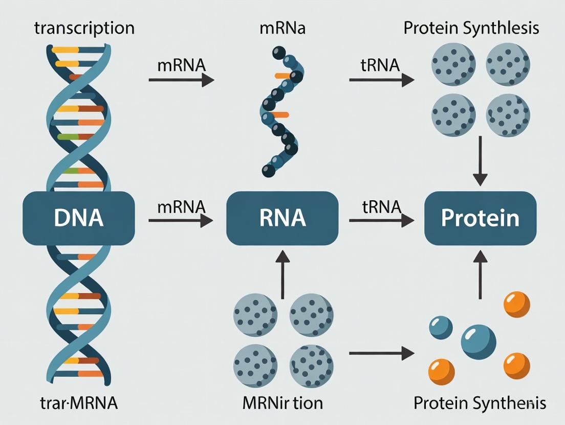
The unidirectional flow of genetic information from DNA to RNA to protein constitutes the central dogma of molecular biology. This process is orchestrated by a core set of molecular machines and informational intermediates. DNA-dependent RNA polymerases transcribe genes into messenger RNA (mRNA), which serves as a blueprint. This mRNA is decoded by the ribosome, a complex ribonucleoprotein comprising ribosomal RNA (rRNA) and proteins, with transfer RNA (tRNA) acting as the adaptor molecule that translates nucleotide triplets into amino acids. This whitepaper provides an in-depth technical analysis of these key players, focusing on their structure, function, quantitative dynamics, and experimental interrogation, framed within contemporary research aimed at understanding and therapeutic manipulation of this fundamental pathway.
Molecular Players: Structure, Function, and Quantitative Data
DNA-Dependent RNA Polymerases
RNA polymerases (RNAPs) are multisubunit enzymes that synthesize RNA transcripts complementary to a DNA template.
- Prokaryotes (e.g., E. coli): A single ~465 kDa RNAP core enzyme (α₂ββ'ω) requires a σ factor for promoter-specific initiation.
- Eukaryotes: Three major polymerases.
- RNA Polymerase II (Pol II), responsible for mRNA and most non-coding RNA synthesis, is a ~550 kDa, 12-subunit complex. Its C-terminal domain (CTD) heptapeptide repeats (YSPTSPS) undergo dynamic phosphorylation to regulate transcription initiation, elongation, and RNA processing.
Table 1: Key RNA Polymerase Types and Characteristics
| Polymerase Type | Organism | Primary Transcripts | Core Subunits | Approx. Mass (kDa) | Key Regulatory Feature |
|---|---|---|---|---|---|
| RNAP Core + σ70 | Prokaryote | mRNA, rRNA, tRNA | α₂, β, β', ω, σ | ~465 | σ factor for promoter recognition |
| RNA Polymerase I | Eukaryote | 28S, 18S, 5.8S rRNA | 14 subunits (RPA1,2, etc.) | ~590 | Localized in nucleolus |
| RNA Polymerase II | Eukaryote | mRNA, miRNA, snRNA | 12 subunits (RPB1-12) | ~550 | CTD phosphorylation cycle |
| RNA Polymerase III | Eukaryote | tRNA, 5S rRNA, other small RNAs | 17 subunits (RPC1-10, etc.) | ~700 | TFIIIB complex recruitment |
RNA Species: mRNA, tRNA, rRNA
Table 2: Characteristics of Principal RNA Species
| RNA Species | Primary Function | Key Structural Features | Avg. Length (nt) | Relative Cellular Abundance (%)* |
|---|---|---|---|---|
| mRNA | Protein-coding template | 5' cap, ORF, poly(A) tail, cis-regulatory elements | 500 - 10,000+ | ~2-5% |
| tRNA | Amino acid adaptor | Cloverleaf secondary; L-shaped 3D structure; anticodon loop | 76-90 | ~10-15% |
| rRNA | Catalytic & scaffold core of ribosome | Complex 2° & 3° structure; multiple functional domains | 120 - 5,000+ | ~80-85% |
*Percentages are approximate and vary by cell type and state.
The Ribosome
The ribosome is a two-subunit ribozyme that catalyzes peptide bond formation.
- Prokaryotic (70S): Composed of a large 50S subunit (23S & 5S rRNA + 33 proteins) and a small 30S subunit (16S rRNA + 21 proteins).
- Eukaryotic (80S): Composed of a large 60S subunit (28S, 5.8S, 5S rRNA + ~47 proteins) and a small 40S subunit (18S rRNA + ~33 proteins).
Table 3: Ribosome Composition Across Domains
| Ribosome (Sed. Coef.) | Large Subunit (LSU) | Small Subunit (SSU) | Key Functional Sites |
|---|---|---|---|
| Prokaryotic (70S) | 50S (23S, 5S rRNA, 33 proteins) | 30S (16S rRNA, 21 proteins) | A, P, E sites; Peptidyl Transferase Center (23S rRNA) |
| Eukaryotic Cytosolic (80S) | 60S (28S, 5.8S, 5S rRNA, ~47 proteins) | 40S (18S rRNA, ~33 proteins) | Similar to prokaryotic, with additional initiation factors |
Experimental Protocols
Protocol: Quantitative RT-PCR (qRT-PCR) for mRNA Analysis
Purpose: To quantify the expression level of specific mRNA transcripts. Methodology:
- RNA Extraction: Isolate total RNA using guanidinium thiocyanate-phenol-chloroform extraction (e.g., TRIzol).
- DNase Treatment: Treat RNA with DNase I to remove genomic DNA contamination.
- Reverse Transcription (RT): Synthesize cDNA using reverse transcriptase (e.g., M-MLV RT) and oligo(dT) or gene-specific primers.
- Quantitative PCR (qPCR): Perform real-time PCR using cDNA template, gene-specific primers, and a fluorescent reporter (SYBR Green or TaqMan probe).
- SYBR Green: Binds double-stranded DNA, emitting fluorescence.
- TaqMan Probe: Sequence-specific oligonucleotide with 5' fluorophore and 3' quencher; cleavage during amplification releases fluorescence.
- Data Analysis: Calculate relative expression using the ΔΔCt method, normalizing to housekeeping genes (e.g., GAPDH, ACTB).
Protocol: Ribosome Profiling (Ribo-seq)
Purpose: To map the positions of actively translating ribosomes on mRNA at nucleotide resolution. Methodology:
- Cell Lysis & Nuclease Footprinting: Rapidly lyse cells. Treat lysate with RNase I to digest mRNA regions not protected by bound ribosomes.
- Ribosome Isolation: Purify monosome complexes by sucrose density gradient centrifugation or size-exclusion chromatography.
- RNA Extraction & Size Selection: Recover protected ~30 nt mRNA "footprint" fragments.
- Library Construction: Dephosphorylate, ligate adaptors, reverse transcribe, and amplify footprints for deep sequencing.
- Bioinformatics: Map sequenced reads to the genome/transcriptome to determine ribosome positions and quantify translational efficiency.
Visualizations
Diagram 1: Central Dogma Flow from DNA to Protein
Diagram 2: Eukaryotic Transcription Initiation by RNA Pol II
Diagram 3: Ribosome Translocation Cycle During Elongation
The Scientist's Toolkit: Research Reagent Solutions
Table 4: Essential Reagents for DNA→RNA→Protein Research
| Reagent Category | Example Product/Kit | Primary Function in Research |
|---|---|---|
| RNA Polymerase Inhibitors | α-Amanitin (Pol II specific), Actinomycin D (general) | Mechanistic studies of transcription, blocking de novo RNA synthesis. |
| Reverse Transcriptases | SuperScript IV (Thermo Fisher), PrimeScript (Takara) | High-efficiency cDNA synthesis from RNA templates for downstream applications (qPCR, RNA-seq). |
| Ribosome Inhibitors | Cycloheximide (eukaryotic), Chloramphenicol (prokaryotic) | Arrest translating ribosomes on mRNA for ribosome profiling or translation inhibition studies. |
| In Vitro Translation Systems | Rabbit Reticulocyte Lysate, PURExpress (NEB) | Cell-free protein synthesis for functional studies, incorporation of modified amino acids. |
| Ribo-Seq Kits | ARTseq Ribosome Profiling Kit (Illumina) | Streamlined, optimized reagents for ribosome footprinting and sequencing library preparation. |
| tRNA Modifying Enzymes | Recombinant tRNA methyltransferases (e.g., TrmD) | Study of tRNA modification impact on structure, stability, and translational fidelity. |
| Cryo-EM Reagents | Graphene Oxide Grids, Gold Foils, Vitrification Robots | Sample preparation for high-resolution structural determination of large complexes like ribosomes and RNAPs. |
The flow of genetic information from DNA to RNA to protein is not a linear, invariant pipeline. It is a highly regulated process where control points determine which genes are expressed, at what level, and in which cell type. This regulation ensures cellular differentiation, adaptation, and homeostasis. Promoters, enhancers, and epigenetic modifications constitute the primary cis-regulatory and chromatin-based machinery that controls the first critical step: transcription initiation. Disruptions in this regulatory landscape are hallmarks of diseases like cancer and neurodegeneration, making its understanding paramount for therapeutic intervention.
Core Regulatory Elements & Mechanisms
Promoters: The Transcription Start Site Platform
Promoters are cis-acting DNA sequences immediately upstream of the transcription start site (TSS). They serve as the binding platform for RNA polymerase II (Pol II) and its associated general transcription factors (GTFs).
- Core Promoter Elements: Include the TATA box (bound by TBP), Initiator (Inr), and downstream promoter element (DPE). Their composition influences transcription efficiency and directionality.
- Quantitative Metrics: Promoter strength is often quantified by reporter assays (e.g., luciferase), with activity varying over several orders of magnitude (10- to 1000-fold differences). Mutations in promoter elements can reduce transcription by >80%.
Enhancers: The Long-Range Transcriptional Activators
Enhancers are distal cis-regulatory elements (located from several kb to >1 Mb from the TSS) that dramatically increase transcription rates. They function independently of orientation and position.
- Key Characteristics: Defined by specific chromatin signatures (see Table 1), they are bound by sequence-specific transcription factors (TFs) and co-activators (e.g., p300/CBP).
- Looping Mechanism: Enhancers physically contact promoters via chromatin looping, facilitated by cohesin and mediator complexes, bringing their bound activators into proximity with the promoter.
Epigenetic Modifications: The Chromatin Gatekeepers
Epigenetic modifications are heritable chemical marks on DNA or histones that regulate chromatin accessibility without altering the DNA sequence.
- DNA Methylation: The addition of a methyl group to cytosine (5mC), typically in CpG dinucleotides, associated with transcriptional repression.
- Histone Modifications: Post-translational modifications (e.g., acetylation, methylation, phosphorylation) on histone tails. These marks are read by specialized proteins to influence chromatin state (see Table 1).
Table 1: Key Chromatin Features of Regulatory Elements
| Feature | Active Promoter | Active Enhancer | Repressed/Inactive State |
|---|---|---|---|
| DNA Methylation | Low (Hypomethylated) | Low (Hypomethylated) | High (Hypermethylated) |
| Histone H3K4 Methylation | High H3K4me3 | High H3K4me1 | Low |
| Histone H3K27 Methylation | Low | Low | High H3K27me3 (Polycomb) |
| Histone Acetylation | High (e.g., H3K27ac) | High (e.g., H3K27ac) | Low |
| Chromatin Accessibility | High (DNase I hypersensitive) | High (DNase I hypersensitive) | Low (Closed) |
| Primary Assays | ChIP-seq (Pol II, H3K4me3), ATAC-seq | ChIP-seq (H3K27ac, p300), STARR-seq | ChIP-seq (H3K9me3, H3K27me3), DNAme-seq |
Table 2: Common Epigenetic Modifications and Their Functional Impact
| Modification | Catalytic Writer | Functional Outcome | Associated Genomic Region |
|---|---|---|---|
| H3K4me3 | MLL/COMPASS complexes | Transcription initiation | Active promoters |
| H3K27ac | p300/CBP | Transcriptional activation | Active enhancers & promoters |
| H3K36me3 | SETD2 | Transcription elongation | Gene bodies of active genes |
| H3K9me3 | SUV39H1/2 | Heterochromatin formation, repression | Repetitive regions, silenced genes |
| H3K27me3 | EZH2 (PRC2) | Facultative heterochromatin, repression | Developmentally regulated genes |
| DNA 5mC | DNMT3A/B, DNMT1 | Transcriptional repression, X-inactivation | CpG islands, repetitive elements |
Key Experimental Protocols
Mapping Chromatin Accessibility: ATAC-seq (Assay for Transposase-Accessible Chromatin)
Purpose: Identify genome-wide regions of open chromatin. Protocol Summary:
- Nuclei Isolation: Lyse cells with a gentle detergent to isolate intact nuclei.
- Tagmentation: Treat nuclei with the engineered Tn5 transposase. Tn5 simultaneously cuts open chromatin regions and inserts sequencing adapters.
- DNA Purification: Purify the tagmented DNA.
- PCR Amplification & Sequencing: Amplify the fragments with barcoded primers and perform high-throughput sequencing.
- Analysis: Align sequences to a reference genome; peaks correspond to accessible regions (promoters, enhancers).
Profiling Histone Modifications: Chromatin Immunoprecipitation Sequencing (ChIP-seq)
Purpose: Determine the genome-wide binding sites of a specific protein (e.g., TF) or histone modification. Protocol Summary:
- Crosslinking: Treat cells with formaldehyde to crosslink proteins to DNA.
- Chromatin Shearing: Sonicate or enzymatically digest chromatin to fragments of 200-500 bp.
- Immunoprecipitation: Incubate with an antibody specific to the target protein/modification. Capture antibody-bound complexes.
- Reverse Crosslinking & Purification: Reverse crosslinks and purify the associated DNA.
- Library Prep & Sequencing: Construct a sequencing library from the immunoprecipitated DNA.
- Analysis: Map reads to reference genome; significant peaks indicate binding/enrichment sites.
Measuring Enhancer-Promoter Interactions: Chromatin Conformation Capture (3C-based methods)
Purpose: Detect physical looping interactions between genomic loci (e.g., enhancer-promoter). Protocol Summary (Hi-ChIP variant):
- Crosslinking: Fix cells with formaldehyde.
- Chromatin Digestion: Restrict DNA with a frequent-cutter restriction enzyme (e.g., Mbol).
- Proximity Ligation: Under dilute conditions, ligate crosslinked DNA ends, joining spatially proximal fragments.
- Chromatin Immunoprecipitation: Perform ChIP (as in 4.2) for a protein of interest (e.g., H3K27ac, cohesin) to enrich for interacting fragments in regulatory regions.
- Library Prep & Sequencing: Process the DNA for paired-end sequencing.
- Analysis: Paired reads mapping to different restriction fragments identify long-range interactions.
Visualizations
Title: Enhancer-Promoter Looping Drives Transcription Initiation
Title: ChIP-seq Experimental Workflow
The Scientist's Toolkit: Key Research Reagents
Table 3: Essential Reagents for Gene Regulation Studies
| Reagent / Tool | Function / Application | Example |
|---|---|---|
| Tagmentase (Tn5) | Engineered transposase for simultaneous fragmentation and adapter tagging in ATAC-seq. | Illumina Nextera Tn5 |
| ChIP-Grade Antibodies | High-specificity, validated antibodies for immunoprecipitation of histone marks or TFs. | Anti-H3K27ac, Anti-RNA Pol II (CST/Abcam) |
| HDAC/DNMT Inhibitors | Small molecule inhibitors to perturb epigenetic states and study function. | Trichostatin A (HDACi), 5-Azacytidine (DNMTi) |
| dCas9-Epigenetic Effectors | CRISPR-dCas9 fused to epigenetic "writers" or "erasers" for locus-specific editing. | dCas9-p300 (activator), dCas9-KRAB (repressor) |
| Proximity Ligation Kits | Optimized reagents for 3C, Hi-C, and HiChIP experiments. | Arima Hi-C Kit, Proximo Hi-C Kit |
| Bisulfite Conversion Kit | Chemical conversion of unmethylated cytosine to uracil for DNA methylation analysis. | EZ DNA Methylation Kit (Zymo Research) |
The faithful and regulated conversion of genetic information from DNA to functional protein is a cornerstone of molecular biology. This "DNA to RNA to protein" paradigm, while conceptually linear, involves a series of intricate and highly regulated post-transcriptional RNA processing steps. For protein-coding genes, the primary transcript—pre-messenger RNA (pre-mRNA)—is biologically inert. It must undergo a precise suite of modifications to become a mature mRNA capable of nuclear export, translation, and regulation of its eventual decay. This whitepaper provides an in-depth technical guide to the four core nuclear mRNA processing events: 5' capping, splicing, editing, and 3' polyadenylation. These processes are not merely constitutive maturation steps but are critical control points for regulating gene expression, expanding proteomic diversity, and ensuring cellular homeostasis. Dysregulation in RNA processing is implicated in numerous diseases, making its machinery a compelling target for therapeutic intervention in oncology, neurology, and genetic disorders.
The 5' Cap: A Multifunctional Landmark
The 5' cap is a modified guanine nucleotide added co-transcriptionally to the first nucleotide of the nascent pre-mRNA.
Chemical Structure & Synthesis: Capping occurs via three enzymatic steps:
- RNA 5' Triphosphatase removes the terminal γ-phosphate from the 5' triphosphate of the pre-mRNA.
- Guanylyltransferase catalyzes the transfer of GMP from GTP to the resulting 5' diphosphate, forming a 5'-5' triphosphate linkage (GpppN).
- (Guanine-N7)-Methyltransferase adds a methyl group to the N7 position of the guanine, forming the canonical Cap-0 structure (m⁷GpppN).
Further methylation of the ribose 2'-O position of the first (and sometimes second) transcribed nucleotide by 2'-O-Methyltransferase generates Cap-1 and Cap-2, which are critical for distinguishing "self" from "non-self" RNA in the innate immune response.
Core Functions:
- Translation Initiation: The cap is recognized by the eukaryotic initiation factor 4F (eIF4F) complex, which recruits the 43S pre-initiation complex.
- mRNA Stability: Protects the 5' end from 5'→3' exonucleolytic degradation.
- Nuclear Export: Facilitates via interactions with the cap-binding complex (CBC) and subsequently with eIF4E.
- Immune Recognition: Cap-1 structure prevents recognition by innate immune sensors like RIG-I.
Quantitative Data: 5' Capping
| Parameter | Value / Description | Experimental Note |
|---|---|---|
| Addition Timing | Occurs after ~20-30 nucleotides are synthesized by Pol II | Measured by GRO-seq/NET-seq |
| Cap Structure | m⁷G(5')ppp(5')N (Cap-0); m⁷G(5')ppp(5')Nmp (Cap-1) | Defined by mass spectrometry |
| eIF4E Binding Affinity (Kd) | ~0.1 - 1 µM for m⁷GpppG cap analog | Measured by fluorescence polarization/ITC |
| Impact on mRNA Half-life | Can increase stability by >10-fold | Compared uncapped vs. capped RNA in vivo |
Experimental Protocol: In Vitro Capping Assay
Purpose: To assess the enzymatic activity of capping enzymes or to produce capped RNA for downstream applications.
Materials:
- Substrate: In vitro transcribed RNA with a 5' triphosphate.
- Enzymes: Recombinant capping enzyme (e.g., vaccinia virus capping enzyme) or cellular enzyme complex.
- Buffer: 50 mM Tris-HCl (pH 8.0), 5 mM DTT, 1 mM MgCl₂, 0.1 mM S-adenosyl methionine (SAM, for methylation step).
- Labeled Precursor: [α-³²P]GTP or [³H-methyl]SAM.
- Equipment: Heat block, gel electrophoresis apparatus, phosphorimager.
Procedure:
- Assemble a 20 µL reaction containing: 1 µg of RNA substrate, 1x reaction buffer, 5 µCi [α-³²P]GTP, 2.5 mM unlabeled GTP, and 1 µL of capping enzyme.
- Incubate at 37°C for 1 hour.
- Stop the reaction by adding 5 µL of 50 mM EDTA.
- Purify the RNA via phenol-chloroform extraction and ethanol precipitation.
- Resuspend the RNA and analyze by denaturing urea-PAGE (6-8%). The capped RNA will have a characteristic mobility shift. Autoradiography will visualize the radiolabeled cap.
- For methylation assay: Use unlabeled GTP and include 5 µCi [³H-methyl]SAM in the reaction. Analyze by filter binding or chromatography.
Diagram Title: Enzymatic Steps of 5' mRNA Capping
Pre-mRNA Splicing: Intron Removal and Exon Joining
Splicing is the precise removal of non-coding introns and ligation of coding exons. It is catalyzed by the spliceosome, a dynamic megadalton ribonucleoprotein complex.
The Spliceosome Cycle: The major U2-dependent spliceosome assembly occurs via ordered recruitment of small nuclear ribonucleoprotein particles (snRNPs: U1, U2, U4/U6, U5) and numerous proteins.
- Commitment (E Complex): U1 snRNP binds the 5' splice site (5'ss), and splicing factors (e.g., SF1, U2AF) bind the branch point (BP) and 3' splice site/polypyrimidine tract (3'ss).
- Pre-spliceosome (A Complex): U2 snRNP stably binds the BP, displacing SF1.
- Pre-catalytic B Complex: The U4/U6•U5 tri-snRNP joins, forming a pre-catalytic complex.
- Catalytic Activation: Extensive RNA-RNA rearrangements (U1 and U4 release) and protein remodeling lead to the formation of the activated B*act complex, which catalyzes the first transesterification reaction. The 2'OH of the branch point adenosine attacks the 5'ss, forming a free 5' exon and a lariat-intron-3' exon intermediate.
- Catalytic Step II (C Complex): Rearrangement positions the 5' exon for the second transesterification, where its 3'OH attacks the 3'ss, ligating the exons and releasing the intron lariat.
Alternative Splicing (AS): The selection of different splice sites generates multiple mRNA isoforms from a single gene, vastly expanding proteomic diversity. Major types include cassette exon skipping, alternative 5'/3' splice sites, mutually exclusive exons, and intron retention. AS is regulated by cis-acting RNA elements (enhancers/silencers) and trans-acting RNA-binding proteins (e.g., SR proteins, hnRNPs).
Quantitative Data: Pre-mRNA Splicing
| Parameter | Value / Description | Experimental Note |
|---|---|---|
| Human Gene % with Introns | ~95% of multi-exon genes | Genomic annotation (GENCODE) |
| Spliceosome Size | ~3-5 MDa (major U2-type) | Mass spectrometry, cryo-EM |
| Splicing Reaction Rate in vitro | ~1-2 min⁻¹ (for a single round) | Pre-mRNA substrate assays |
| Human Transcripts with AS | >95% of multi-exon genes | RNA-seq analysis (long-read) |
| Disease-Linked Splicing Mutations | >30% of human genetic disorders | ClinVar database analysis |
Experimental Protocol: Minigene Splicing Assay
Purpose: To test the impact of sequence variants or regulatory factors on splicing patterns.
Materials:
- Minigene Construct: A plasmid containing a genomic region of interest (exon(s) with flanking introns) cloned between two constitutive exons from a different gene (e.g., β-globin).
- Cells: Mammalian cell line (HEK293, HeLa).
- Transfection Reagent: Lipofectamine or PEI.
- RNA Isolation: TRIzol reagent, DNase I.
- RT-PCR: Reverse transcriptase, gene-specific or vector primers, PCR mix.
- Analysis: Agarose or capillary electrophoresis (Bioanalyzer).
Procedure:
- Transfect the minigene plasmid into cells (24-well plate format) using standard protocols.
- After 24-48 hours, harvest cells and isolate total RNA using TRIzol, treating with DNase I to remove plasmid DNA.
- Perform reverse transcription (RT) using an oligo(dT) or a primer specific to the downstream constitutive exon.
- Amplify the spliced products by PCR using primers in the flanking constitutive exons. Use a high-fidelity polymerase and cycle number within the linear range.
- Resolve PCR products by agarose gel electrophoresis or capillary electrophoresis. Bands corresponding to different isoforms (e.g., included exon vs. skipped exon) will be visible.
- Quantify band intensity using densitometry software. The percentage spliced in (PSI or Ψ) is calculated as: (Intensity of isoform with exon inclusion) / (Total intensity of all isoforms) x 100.
Diagram Title: Major Spliceosome Assembly and Catalytic Cycle
RNA Editing: Sequence Alteration Post-Transcription
RNA editing enzymatically alters the nucleotide sequence of an RNA molecule, creating a product that differs from its DNA template.
Major Types:
- A-to-I Editing: Catalyzed by ADAR (Adenosine Deaminases Acting on RNA) enzymes, which convert adenosine (A) to inosine (I) within double-stranded RNA regions. Inosine is read as guanosine (G) by the translation and splicing machinery. This can recode codons, create/abolish splice sites, or alter miRNA target sites. Important in neurobiology (e.g., editing of glutamate receptor GluA2 subunit).
- C-to-U Editing: Catalyzed by APOBEC (Apolipoprotein B mRNA Editing Catalytic Polypeptide-like) family enzymes, such as APOBEC1. Converts cytidine (C) to uridine (U). The classic example is editing of APOB mRNA in the intestine, creating a premature stop codon and a truncated protein (APOB48).
- Other Types: Include insertional editing in kinetoplastid mitochondria.
Quantitative Data: RNA Editing
| Parameter | Value / Description | Experimental Note |
|---|---|---|
| A-to-I Sites in Human Transcriptome | >4.5 million (Alu-rich); ~thousands in coding regions | REDIportal database |
| ADAR1/ADAR2 Knockout Phenotype | Embryonic lethality (ADAR1); seizures, death (ADAR2) | Mouse models |
| Editing Efficiency at Key Sites (e.g., GluA2 Q/R site) | ~99-100% | RNA-seq, Sanger sequencing |
| APOBEC1 Target Specificity | Requires mooring sequence 3' of edited C | In vitro editing assays |
Experimental Protocol: Detection of A-to-I RNA Editing by PCR and Restriction Digest (RFLP)
Purpose: To assess editing levels at a specific known site.
Materials:
- RNA Sample: Total RNA from tissue or cells.
- cDNA Synthesis Kit.
- PCR Primers: Flanking the editing site.
- Restriction Enzyme: An enzyme whose site is created or destroyed by the A-to-I (G) change. E.g., BbvCI site (CCTCAGC) is destroyed by A-to-I editing (becomes CCTIAGC, which is not recognized).
- Equipment: Thermocycler, agarose gel apparatus.
Procedure:
- Synthesize cDNA from DNase-treated RNA.
- PCR amplify the region of interest using high-fidelity polymerase.
- Purify the PCR product.
- Digest half of the purified product with the diagnostic restriction enzyme (e.g., BbvCI) in a 20 µL reaction for 2 hours.
- Run digested and undigested samples side-by-side on a high-percentage agarose gel (2.5-3%).
- Interpretation: The unedited sequence (A) will be cut, yielding two smaller bands. The edited sequence (I, read as G) will resist cutting, yielding one full-length band. The relative intensity of the bands quantifies the editing percentage.
3' End Processing: Cleavage and Polyadenylation
The 3' end of most eukaryotic mRNAs is generated by endonucleolytic cleavage followed by the addition of a poly(A) tail, a ~200-250 nucleotide homopolymer of adenosine.
Mechanism: The reaction requires recognition of conserved cis-acting elements on the pre-mRNA by a multi-subunit Cleavage and Polyadenylation Complex (CPC).
- Core Signals:
- Poly(A) Signal (PAS): AAUAAA (or a close variant) located 10-35 nucleotides upstream of the cleavage site (CS).
- Cleavage Site (CS): A CA dinucleotide (most common).
- Downstream Sequence Element (DSE): A U/GU-rich region located ~20-40 nucleotides downstream of the CS.
- Complex Assembly & Cleavage: CPSF (Cleavage and Polyadenylation Specificity Factor) binds the PAS. CstF (Cleavage Stimulation Factor) binds the DSE. CFI, CFII, and other factors assemble, leading to endonucleolytic cleavage at the CS.
- Poly(A) Addition: After cleavage, Poly(A) Polymerase (PAP) adds ~200-250 A residues in a processive manner, using ATP as a substrate. The initial phase is regulated by Nuclear Poly(A) Binding Protein (PABPN1), which stimulates PAP processivity and signals tail length control.
Functions:
- Translation: Enhances translation initiation via PABPC1 binding to the tail and interacting with eIF4G.
- Stability: Protects the mRNA from 3'→5' exonucleolytic decay.
- Export: The poly(A) tail and its associated proteins are part of the mRNA export competency signal.
Quantitative Data: Polyadenylation
| Parameter | Value / Description | Experimental Note |
|---|---|---|
| Canonical Poly(A) Signal | AAUAAA (approx. 60% of human genes) | Genomic analysis (PolyA_DB) |
| Average Poly(A) Tail Length (Human) | ~200-250 nucleotides in nucleus; dynamic in cytoplasm | PAT-seq, Nanopore sequencing |
| Cleavage Complex Proteins | >20 core subunits (CPSF, CstF, CFI/II) | Affinity purification/MS |
| Impact on mRNA Half-life | Poly(A)-deficient mRNA degraded in minutes | Transcriptional pulse-chase |
Experimental Protocol: Mapping Polyadenylation Sites by 3' RACE (Rapid Amplification of cDNA Ends)
Purpose: To identify the precise cleavage and polyadenylation site(s) used for a transcript.
Materials:
- RNA: High-quality, DNase-treated total RNA.
- Adaptor Oligos: A modified oligo(dT) primer with a known adapter sequence at its 5' end (e.g., QT primer: 5'-GCCACGCGTCGACTAGTAC(T)₁₇-3').
- Reverse Transcriptase: RNase H⁻ for first-strand synthesis.
- PCR Components: Gene-specific forward primer (GSP1) located upstream of the predicted poly(A) site, adapter-specific reverse primer, PCR mix.
- Nested PCR (optional): Nested gene-specific primer (GSP2) and adapter primer for increased specificity.
- Cloning & Sequencing: or direct Sanger/next-generation sequencing of PCR product.
Procedure:
- Synthesize first-strand cDNA using the QT primer and total RNA.
- Perform a first-round PCR using GSP1 and the adapter-specific primer.
- (Optional) Perform a second, nested PCR using GSP2 and a nested adapter primer, using a dilution of the first PCR product as template.
- Gel-purify the PCR product(s). Multiple bands may indicate alternative polyadenylation.
- Clone the product into a sequencing vector or purify for direct sequencing.
- Sequence the product. The junction between the gene-specific sequence and the poly(A) tail (or adapter sequence that replaced it) identifies the cleavage site.
Diagram Title: 3' End Cleavage and Polyadenylation Pathway
The Scientist's Toolkit: Key Research Reagent Solutions
| Reagent / Material | Primary Function | Example Use Case |
|---|---|---|
| Vaccinia Capping System | Recombinant enzyme complex to add Cap-0 to in vitro transcribed RNA. | Production of translationally competent or highly stable synthetic mRNA for transfection or therapeutic studies. |
| Spliceostatin A / Pladienolide B | Small molecule inhibitors of the SF3b complex within U2 snRNP. | Chemical probing of spliceosome function; inhibiting splicing as an anti-cancer strategy. |
| Anti-m³G Cap Antibody | High-affinity antibody specific for the N7-methylguanosine cap. | Immunoprecipitation of capped RNAs (e.g., for transcriptome-wide cap analysis). |
| Recombinant ADAR1/ADAR2 | Purified editing enzymes. | In vitro editing assays; development of RNA editing therapeutics (e.g., directed editing with guide RNAs). |
| 3'-Deoxyadenosine (Cordycepin) | Adenosine analog that terminates poly(A) tail elongation. | Inhibition of polyadenylation in cell culture to study mRNA metabolism. |
| Poly(A) Polymerase (E. coli or Yeast) | Enzyme to add homopolymeric A tails to RNA in vitro. | Adding poly(A) tails to synthetic RNAs; 3' end labeling of RNA. |
| α-Amanitin | RNA polymerase II-specific inhibitor. | Arresting transcription to study co-transcriptional processing events (e.g., ChIP-seq of processing factors). |
| LOCK-ANTI-oligo(dT) Probes | DNA probes that block oligo(dT) priming of abundant poly(A)+ RNA. | Enriching for non-polyadenylated or partially degraded transcripts in RNA-seq. |
RNA processing is not a series of isolated events but a highly coordinated and often interdependent network. Capping influences splicing efficiency; splicing can affect polyadenylation site choice; editing can alter splice sites. This complexity provides a rich layer of gene regulation that is essential for development, differentiation, and cellular response. From a translational research perspective, each step represents a node of vulnerability for disease and a potential target for intervention. Small molecules modulating splicing (e.g., for Spinal Muscular Atrophy, cancer), antisense oligonucleotides to redirect splicing or block editing, and the engineering of synthetic 5' and 3' ends for mRNA vaccines and therapeutics are all direct applications rooted in the fundamental biochemistry outlined in this guide. A deep understanding of these mechanisms is therefore indispensable for researchers and drug developers aiming to manipulate the flow of genetic information for diagnostic and therapeutic benefit.
Within the central dogma of molecular biology, the flow of information from DNA to RNA to protein is governed by the genetic code. This universal, yet nuanced, triplet code is deciphered during translation by the ribosome and transfer RNAs (tRNAs). This whitepaper delves into three critical, interconnected aspects of this decoding process: the non-random Codon Usage across genomes, the Wobble Hypothesis that explains tRNA degeneracy, and the strict maintenance of Reading Frames. Understanding these mechanisms is fundamental for research in synthetic biology, gene therapy, and the development of novel therapeutics targeting translation.
Codon Usage and Optimization
The genetic code is degenerate, with 61 sense codons specifying 20 standard amino acids. Synonymous codons are not used with equal frequency; this bias is termed codon usage bias. It varies significantly between organisms, across genes within a genome, and even along the length of a single gene.
Quantitative Data: Example Codon Usage Frequencies Table 1: Comparative Codon Usage Frequencies (per 1000 codons) in Model Organisms for the Amino Acid Leucine (Leu)
| Codon | E. coli | S. cerevisiae | H. sapiens | Amino Acid |
|---|---|---|---|---|
| UUA | 13.6 | 27.9 | 7.5 | Leu |
| UUG | 13.2 | 30.6 | 12.6 | Leu |
| CUU | 11.3 | 12.0 | 13.2 | Leu |
| CUC | 10.2 | 6.1 | 19.6 | Leu |
| CUA | 4.3 | 13.6 | 7.2 | Leu |
| CUG | 51.2 | 10.4 | 39.6 | Leu |
Key Drivers of Bias:
- tRNA Abundance: Highly expressed genes tend to use codons matched by abundant tRNAs, optimizing translational speed and accuracy.
- Mutation Pressure: Genomic GC content influences codon third-base composition.
- Natural Selection: Fine-tunes translation kinetics, co-translational folding, and mRNA stability.
Experimental Protocol: Analyzing Codon Usage
- Method: In silico Codon Usage Analysis.
- Procedure:
- Obtain the coding sequence (CDS) of interest from a database (e.g., NCBI GenBank).
- Use bioinformatics tools (e.g., CodonW, EMBOSS cusp) to calculate parameters like Relative Synonymous Codon Usage (RSCU) and the Codon Adaptation Index (CAI).
- Compare the gene's codon frequencies to a reference table for the host organism.
- For heterologous expression, use algorithms (e.g., IDT's OptimumGene, Twist Bioscience's optimization) to redesign the gene using host-preferred codons while avoiding problematic motifs (e.g., repetitive sequences, restriction sites).
- Validation: Synthesize the optimized gene, clone into an expression vector, and compare protein yield and kinetics to the wild-type sequence.
The Wobble Hypothesis
Proposed by Francis Crick, this hypothesis explains how a limited number of tRNAs can recognize multiple synonymous codons. Flexibility ("wobble") exists in the base pairing between the 5' base of the anticodon (position 1) and the 3' base of the codon (position 3).
Key Wobble Pairing Rules: Table 2: Standard Wobble Base-Pairing Rules
| Anticodon 5' Base (Position 1) | Can Pair with Codon 3' Base (Position 3) |
|---|---|
| G | U or C |
| U | A or G |
| I (Inosine, a modified base) | U, C, or A |
| C | G only |
| A | U only |
This modified base inosine (I) is critical for expanding decoding capacity. Wobble interactions reduce the cellular requirement for tRNA genes but can influence decoding speed and accuracy.
Experimental Protocol: Detecting tRNA Modification & Wobble Function
- Method: Mass Spectrometry (MS) Analysis of tRNA Nucleosides.
- Procedure:
- tRNA Purification: Isolate total tRNA from cells using phenol-chloroform extraction and anion-exchange chromatography or commercial kits.
- Nuclease Digestion: Digest purified tRNA to individual nucleosides using a combination of nuclease P1, snake venom phosphodiesterase, and alkaline phosphatase.
- LC-MS/MS Analysis: Separate the nucleoside mixture via Liquid Chromatography (LC) and analyze with tandem Mass Spectrometry (MS/MS).
- Identification & Quantification: Identify modified nucleosides (like inosine, pseudouridine, etc.) by comparing their mass/charge ratios and retention times to known standards. Quantify their relative abundance.
- Functional Assay: Combine with a reporter assay where a synonymous codon pair, predicted to be read by a single wobble tRNA, is mutated in a reporter gene. Correlate changes in translation efficiency (e.g., luciferase output) with the abundance of the specific modified tRNA.
Wobble Analysis: tRNA Modification Detection Workflow
Reading Frame Maintenance
The correct translation of a nucleotide sequence into a polypeptide is entirely dependent on the ribosome establishing and maintaining a single, uninterrupted reading frame. The reading frame is defined by the start codon (AUG) and is read in consecutive, non-overlapping triplets. A shift of one or two bases (+1 or +2 frameshift) completely alters the downstream amino acid sequence, usually leading to a nonfunctional or truncated protein.
Mechanisms of Maintenance:
- Ribosomal Precision: The ribosome's architecture ensures precise mRNA translocation by exactly three nucleotides.
- tRNA-mRNA Interactions: Correct codon-anticodon pairing stabilizes the complex.
- Restorative Frameshifting: In rare cases, programmed frameshifts (e.g., in viruses like HIV) are required for synthesis of alternative proteins. These are directed by specific mRNA cis-elements (slippery sequences, pseudoknots).
Experimental Protocol: Assaying Frameshift Mutagenesis
- Method: Dual-Luciferase Reporter Assay for Frameshift Efficiency.
- Procedure:
- Construct Design: Clone a sequence of interest (e.g., a putative slippery sequence) between the coding sequences for Renilla and firefly luciferase in a dual-reporter vector. The firefly luciferase must be placed in a different reading frame relative to the Renilla.
- Test & Control: Create a control construct where both luciferases are in-frame.
- Transfection: Transfert constructs into target cells.
- Measurement: Lyse cells and measure luminescence from each luciferase sequentially using a dual-luciferase assay kit.
- Calculation: The ratio of firefly to Renilla luminescence indicates frameshift efficiency. Normalize test ratios to the in-frame control.
Three Possible mRNA Reading Frames
The Scientist's Toolkit: Key Research Reagent Solutions
Table 3: Essential Reagents for Genetic Code Research
| Item | Function/Application | Example Vendor/Catalog |
|---|---|---|
| Codon-Optimized Gene Fragments | For synthetic gene construction with host-specific codon bias to maximize heterologous expression. | Twist Bioscience, IDT gBlocks, GenScript. |
| Dual-Luciferase Reporter Assay Systems | Quantitatively measure translational efficiency, frameshifting, or readthrough events. | Promega Dual-Luciferase Reporter (DLR) Assay. |
| In vitro Translation Kits | Cell-free systems to study translation mechanics, codon effects, and protein synthesis. | PURExpress (NEB), Flexi Rabbit Reticulocyte System (Promega). |
| tRNA Modification Analysis Kits | For extraction, purification, and initial analysis of modified tRNA nucleosides. | ChargeSwitch Total tRNA Isolation Kit (Thermo Fisher). |
| Ribosome Profiling (Ribo-Seq) Kits | Genome-wide mapping of translated reading frames and ribosome occupancy at codon resolution. | ARTseq/TruSeq Ribo Profile (Illumina-based). |
| Anti-Puromycin Antibodies | Detect newly synthesized polypeptides via puromycin incorporation (e.g., in SUnSET assays). | Kerafast, Merck Millipore. |
| Start & Stop Codon Suppressor tRNAs | For incorporation of unnatural amino acids or studying translation termination. | Chemical aminoacylated tRNAs (e.g., from Chemgenes). |
The flow of information from gene to protein is not a simple one-to-one cipher. It is dynamically regulated by the interplay of genomic codon bias, the biophysical rules of wobble pairing, and the absolute necessity of reading frame fidelity. Disruptions in these processes are linked to disease, while their manipulation offers powerful therapeutic avenues—from optimizing biologic drug production to designing small molecules that target frameshifting in pathogens. Continued research into these foundational mechanisms, powered by modern tools like ribosome profiling and quantitative mass spectrometry, remains crucial for advancing biomedicine and synthetic biology.
From Theory to Bench: Cutting-Edge Techniques for Tracking Genetic Information Flow
The central dogma of molecular biology outlines the unidirectional flow of genetic information from DNA to RNA to protein. Historically, studying this cascade has been limited by technological constraints that obscure heterogeneity, isoform complexity, and cellular context. Advanced sequencing technologies—long-read, single-cell, and spatial transcriptomics—now enable a high-resolution, multi-dimensional dissection of this flow. This guide details these technologies, providing a technical foundation for researchers interrogating gene expression regulation, RNA processing, and its ultimate phenotypic manifestation in physiology and disease.
Long-Read Sequencing Technologies
Core Principles and Platforms
Long-read sequencing, or third-generation sequencing, generates reads spanning thousands to millions of base pairs, enabling the direct interrogation of complex genomic regions, full-length RNA transcripts, and epigenetic modifications.
Key Platform Comparison: Table 1: Comparison of Major Long-Read Sequencing Platforms
| Platform | Technology | Avg. Read Length | Accuracy (Raw %) | Primary Application in Transcriptomics |
|---|---|---|---|---|
| PacBio (HiFi) | Circular Consensus Sequencing (CCS) | 10-25 kb | >99.9% | Full-length isoform sequencing, allele-specific expression, fusion detection |
| Oxford Nanopore (ONT) | Nanopore sensing | 10 kb - 2 Mb+ | ~96-98% (with Q20+ kits) | Direct RNA-seq, real-time sequencing, detection of RNA modifications |
Experimental Protocol: Full-Length Isoform Sequencing (Iso-Seq)
Objective: To obtain complete, unambiguously spliced cDNA sequences without assembly.
Detailed Methodology:
- RNA Extraction & QC: Isolate high-quality total RNA (RIN > 8.5) using a column-based or TRIzol method.
- cDNA Synthesis: Use a template-switching reverse transcriptase (e.g., Clontech SMARTer) to add universal adapters to the 5' end of first-strand cDNA.
- PCR Amplification: Amplify full-length cDNA with primers matching the adapters. Optimize cycle number to minimize PCR bias.
- Size Selection: Perform BluePippin or SageELF size selection to enrich for cDNAs >1 kb.
- SMRTbell Library Prep: Ligate hairpin adapters to both ends of the double-stranded cDNA to create a circularized SMRTbell template.
- Sequencing: Load onto a PacBio Sequel IIe/Revio system. Use the CCS mode where the polymerase repeatedly traverses the circular template, generating multiple subreads that are computationally polished into a single high-fidelity (HiFi) read.
- Bioinformatics Analysis: Process with the SMRT Link Iso-Seq pipeline: (1) Circular Consensus Calling, (2) Full-Length Read Identification (identification of 5' and 3' adapters and poly-A tail), (3) Clustering of identical transcripts to generate consensus isoforms, and (4) Alignment to the reference genome/transcriptome.
Iso-Seq Workflow for Full-Length Transcripts
Single-Cell RNA Sequencing (scRNA-seq)
Core Principles
scRNA-seq profiles the transcriptome of individual cells, uncovering cellular heterogeneity, developmental trajectories, and rare cell states within a tissue, directly linking genotypic information to cellular phenotype.
Key Quantitative Metrics: Table 2: Metrics and Performance of Common scRNA-seq Methods
| Method | Cells per Run | Cell Throughput | Sensitivity (Genes/Cell) | Key Feature |
|---|---|---|---|---|
| 10x Genomics Chromium | 500 - 10,000 | High | ~1,000-5,000 | Droplet-based, high throughput, robust |
| Smart-seq2 | 96 - 384 | Low | ~5,000-8,000 | Plate-based, full-length, high sensitivity |
| Seq-Well | ~10,000 | High | ~500-2,000 | Nanowell-based, cost-effective for many cells |
Experimental Protocol: Droplet-Based scRNA-seq (10x Genomics)
Objective: To profile gene expression from thousands of individual cells in parallel.
Detailed Methodology:
- Single-Cell Suspension Preparation: Dissociate tissue to a single-cell suspension. Achieve >90% viability. Remove cell clumps with a 40µm flow cell strainer. Count cells accurately.
- Gel Bead-in-emulsion (GEM) Generation: Load a Chromium chip with the cell suspension, Master Mix (with barcoded gel beads), and partitioning oil. The microfluidic system creates oil-separated aqueous droplets (GEMs), each containing a single cell, a single barcoded bead, and RT reagents.
- Reverse Transcription within GEMs: Cells are lysed within droplets. Poly-adenylated mRNA hybridizes to the bead's oligo-dT primers, which contain a cell-specific barcode and a Unique Molecular Identifier (UMI). Reverse transcription occurs inside each droplet, creating barcoded cDNA.
- Break Emulsion & cDNA Amplification: Droplets are broken, and pooled cDNA is purified and PCR-amplified.
- Library Construction: The amplified cDNA is fragmented, end-repaired, A-tailed, and ligated to sample index adapters via a second, shorter PCR.
- Sequencing: Libraries are sequenced on an Illumina platform (e.g., NovaSeq). A typical run uses paired-end sequencing: Read 1 for the cell barcode and UMI, Read 2 for the cDNA insert.
- Bioinformatics Analysis: Process with Cell Ranger (10x) or similar: (1) Demultiplexing by sample index, (2) Barcode/UMI processing, (3) Alignment to a reference genome, (4) Gene counting (aggregating reads with the same cell barcode, UMI, and gene), and (5) Downstream analysis (clustering, differential expression, trajectory inference).
Droplet-Based scRNA-seq Workflow
Spatial Transcriptomics
Core Principles
Spatial transcriptomics maps gene expression data directly onto tissue morphology, preserving the crucial spatial context of the DNA→RNA→protein flow within a tissue architecture.
Technology Comparison: Table 3: Comparison of Spatial Transcriptomics Methods
| Method | Resolution | Throughput (Genes) | Technology Basis | Preserves Morphology? |
|---|---|---|---|---|
| 10x Visium | 55 µm spots | Whole Transcriptome | Arrayed, barcoded oligo capture | Yes (H&E guided) |
| Nanostring GeoMx DSP | ~1-10 µm (ROI) | Whole Transcriptome/Protein | Photocleavable oligos, digital counting | Yes (imaging guided) |
| MERFISH / seqFISH | Subcellular | 100 - 10,000+ genes | In situ hybridization, imaging | Yes |
Experimental Protocol: Array-Based Capture (10x Visium)
Objective: To obtain whole-transcriptome data annotated with spatial coordinates from a tissue section.
Detailed Methodology:
- Tissue Preparation: Fresh-frozen tissue is sectioned at 10 µm thickness onto a Visium Gene Expression Slide. Each slide contains four 6.5x6.5 mm capture areas, each with ~5000 barcoded spots. Tissue is fixed in methanol and stained with H&E for imaging.
- Permeabilization Optimization: A critical step. Tissue is treated with a permeabilization enzyme to allow mRNA to diffuse from the tissue and bind to spatially barcoded capture probes on the slide. Optimization of time/enzyme concentration is required for each tissue type.
- Reverse Transcription On-Slide: mRNA hybridizes to slide-bound oligos containing a spatial barcode, a UMI, and an oligo-dT sequence. In situ reverse transcription creates barcoded cDNA.
- cDNA Harvest & Library Prep: cDNA is released from the slide and collected. A second-strand synthesis is performed, followed by denaturation and amplification to create a sequencing library with Illumina adapters and sample indices.
- Sequencing & Data Integration: Libraries are sequenced on an Illumina platform. The spaceranger pipeline aligns reads, assigns them to spatial barcodes, and generates a gene-spatial barcode matrix. This matrix is then overlaid onto the H&E image for visualization.
Spatial Transcriptomics Array Workflow
The Scientist's Toolkit: Key Research Reagent Solutions
Table 4: Essential Reagents and Kits for Advanced Sequencing
| Item / Kit Name | Provider | Primary Function |
|---|---|---|
| PacBio SMRTbell Prep Kit 3.0 | PacBio | Library preparation for long-read sequencing, converts dsDNA/cDNA to SMRTbell templates. |
| 10x Genomics Chromium Next GEM Chip K | 10x Genomics | Microfluidic chip for partitioning single cells and reagents into nanoliter-scale droplets (GEMs). |
| Chromium Next GEM Single Cell 3' Reagent Kits v3.1 | 10x Genomics | Contains all enzymes, beads, and buffers for GEM-RT, cDNA amplification, and library construction for 3' scRNA-seq. |
| Visium Spatial Gene Expression Reagent Kit | 10x Genomics | Contains slides and all reagents for tissue permeabilization, on-slide reverse transcription, and cDNA harvest for spatial mapping. |
| SMART-Seq v4 Ultra Low Input RNA Kit | Takara Bio | For plate-based, full-length scRNA-seq with high sensitivity from ultra-low input (1-1000 cells). |
| SQK-RNA004 | Oxford Nanopore | Kit for direct cDNA or direct RNA sequencing on Nanopore platforms, preserving native RNA modifications. |
| Dynabeads MyOne SILANE | Thermo Fisher | Magnetic beads used for SPRI-based clean-up and size selection in multiple NGS library prep protocols. |
| NovaSeq 6000 S4 Reagent Kit (300 cycles) | Illumina | Flow cell and chemistry for high-output, paired-end sequencing on the Illumina NovaSeq system. |
The convergence of long-read, single-cell, and spatial technologies provides an unprecedented, multi-layered view of genetic information flow. Long-read sequencing resolves molecular isoforms, single-cell profiling deconvolves cellular heterogeneity, and spatial mapping restores tissue-level context. Together, they form a powerful toolkit for researchers and drug developers aiming to understand disease mechanisms, identify novel biomarkers, and validate therapeutic targets with precise cellular and spatial resolution. Future integration with proteomics and live-cell imaging will further close the loop between genotype and phenotype.
The quantification of gene expression is a cornerstone of modern molecular biology, providing critical insights into the flow of genetic information from DNA to RNA to protein. This process, central to understanding cellular function, development, and disease, can be precisely measured using high-throughput transcriptomic platforms. Each major technology—RNA sequencing (RNA-Seq), quantitative polymerase chain reaction (qPCR), and the NanoString nCounter system—offers distinct advantages in sensitivity, throughput, and application. This technical guide provides an in-depth comparison of these platforms, framed within the broader research thesis of elucidating the dynamics of genetic information flow. Accurate quantification of RNA intermediates is essential for constructing predictive models of gene regulatory networks and protein output, which are fundamental to basic research and therapeutic development.
Quantitative Polymerase Chain Reaction (qPCR)
qPCR is the gold standard for targeted, sensitive quantification of specific RNA transcripts. It involves reverse transcribing RNA into complementary DNA (cDNA), followed by amplification with sequence-specific primers and fluorescent detection in real time.
Key Experimental Protocol (One-Step RT-qPCR):
- RNA Isolation & QC: Extract total RNA using silica-membrane columns or magnetic beads. Assess integrity via RIN (RNA Integrity Number) on a bioanalyzer and quantify by spectrophotometry (A260/A280).
- Reaction Setup: Combine in each well: 10-100 ng total RNA, gene-specific forward and reverse primers (200-500 nM each), a fluorescent DNA-binding dye (e.g., SYBR Green) or a sequence-specific probe (e.g., TaqMan), reverse transcriptase, hot-start DNA polymerase, dNTPs, and reaction buffer.
- Thermocycling & Detection: Run on a real-time thermocycler.
- Reverse Transcription: 50°C for 10-30 minutes.
- Enzyme Activation: 95°C for 2-5 minutes.
- Amplification (40-50 cycles): Denature at 95°C for 15 sec, anneal/extend at 60°C for 1 minute. Fluorescence is measured at the end of each extension phase.
- Data Analysis: Determine the cycle threshold (Ct) for each sample. Use a standard curve of known template concentrations or the ΔΔCt method for relative quantification to a reference gene.
RNA Sequencing (RNA-Seq)
RNA-Seq provides a comprehensive, unbiased profile of the transcriptome. It involves converting a population of RNA into a library of cDNA fragments, which are then sequenced en masse using high-throughput platforms.
Key Experimental Protocol (Illumina Poly-A Selection Workflow):
- RNA Isolation & QC: As for qPCR, with stringent requirement for high RIN (>8).
- Library Preparation:
- mRNA Enrichment: Use oligo(dT) magnetic beads to capture polyadenylated transcripts.
- Fragmentation: Heat or enzyme-based cleavage of RNA/cDNA to ~200-300 bp fragments.
- cDNA Synthesis: First-strand synthesis with random hexamers and reverse transcriptase, followed by second-strand synthesis.
- Adapter Ligation: Blunt-end repair, A-tailing, and ligation of platform-specific sequencing adapters containing unique dual indices (UDIs) for sample multiplexing.
- PCR Amplification: Enrich adapter-ligated fragments (typically 10-15 cycles).
- Library QC: Size selection via SPRI beads and quantification via qPCR.
- Sequencing: Pool libraries and load onto flow cell for cluster generation and sequencing-by-synthesis on platforms like NovaSeq or NextSeq (e.g., 150 bp paired-end reads).
- Data Analysis: Primary analysis involves demultiplexing, read alignment (e.g., to GRCh38 using STAR), and gene/transcript quantification (e.g., using featureCounts or Salmon). Differential expression is analyzed with tools like DESeq2 or edgeR.
NanoString nCounter Platform
The NanoString nCounter system offers direct, digital counting of RNA molecules without amplification or reverse transcription, minimizing bias. It uses sequence-specific fluorescent barcodes for multiplexed detection.
Key Experimental Protocol:
- Sample Preparation: Isolate total RNA (as above). No fragmentation or conversion to cDNA is required.
- Hybridization: Mix 100-300 ng of total RNA with a Reporter CodeSet (target-specific probes carrying a fluorescent barcode) and a Capture CodeSet (target-specific probes conjugated to biotin) in a single tube. Incubate at 65°C for 12-24 hours to allow specific probe-target hybridization.
- Purification & Immobilization: Load the reaction onto the nCounter Prep Station, which uses capillary electrophoresis to bind biotinylated complexes to a streptavidin-coated cartridge. Excess probes are washed away, and complexes are aligned in a linear fashion.
- Data Acquisition: The cartridge is scanned in the nCounter Digital Analyzer, which images the immobilized fluorescent barcodes at single-molecule resolution. Each barcode's count is directly proportional to the abundance of the target RNA in the original sample.
- Data Analysis: Raw counts are normalized using internal positive controls and housekeeping genes, followed by differential expression analysis with tools like nSolver or ROSALIND.
Quantitative Data Comparison Table
Table 1: Core Technical Specifications of Major Gene Expression Platforms
| Feature | qPCR (SYBR Green) | RNA-Seq (Illumina, Standard mRNA-Seq) | NanoString nCounter (Gene Expression) |
|---|---|---|---|
| Throughput (Targets/Sample) | Low (1-10s, typically) | Very High (All expressed transcripts, ~20,000 genes) | Medium-High (Customizable up to ~800 targets per panel) |
| Sensitivity (Limit of Detection) | Very High (1-10 copies) | High (Varies with sequencing depth) | High (~0.1-0.5 fM) |
| Dynamic Range | High (>7-8 log10) | Very High (>5-6 log10) | High (>4 log10) |
| Technical Reproducibility (%CV) | Excellent (<5%) | Good (10-20%) | Excellent (<5%) |
| Required RNA Input | Low (10 pg - 100 ng) | Medium-High (10 ng - 1 µg) | Medium (50 - 300 ng) |
| Amplification Bias | Yes (Exponential PCR) | Yes (PCR during library prep) | No (Amplification-free) |
| Primary Output Data | Cycle Threshold (Ct) | Sequence Read Counts (FASTQ) | Digital Barcode Counts |
| Turnaround Time (Hands-on) | Fast (Hours) | Slow (Days to Weeks) | Medium (1-2 Days) |
| Cost per Sample (Relative) | $ | $$$$ | $$-$$$ |
| Key Application | Targeted validation, high-precision low-plex | Discovery, splicing, novel transcripts, allelic expression | Targeted multiplex panels, degraded/FFPE samples |
Visualization of Methodologies and Data Flow
Title: Comparative Workflows of Three Gene Expression Platforms
Title: Quantifying RNA Within the Central Dogma Framework
The Scientist's Toolkit: Key Research Reagents & Materials
Table 2: Essential Reagent Solutions for Featured Experiments
| Item | Platform(s) | Function & Brief Explanation |
|---|---|---|
| DNase/RNase-free Water | All | Solvent for all reactions; eliminates nuclease contamination that degrades RNA or cDNA. |
| RNase Inhibitors | qPCR, RNA-Seq | Protects RNA templates from degradation during reverse transcription and library prep steps. |
| Oligo(dT) Magnetic Beads | RNA-Seq (Poly-A+) | Selectively binds poly-adenylated mRNA from total RNA, enriching for coding transcripts. |
| Random Hexamer Primers | qPCR, RNA-Seq | Binds randomly to RNA to prime first-strand cDNA synthesis, ensuring full transcript coverage. |
| dNTP Mix | qPCR, RNA-Seq | Provides the nucleotides (dATP, dCTP, dGTP, dTTP) as building blocks for DNA polymerization. |
| Hot-Start DNA Polymerase | qPCR, RNA-Seq | Remains inactive until a high-temperature step, preventing non-specific primer binding and amplification. |
| SYBR Green I Dye | qPCR (Intercalating) | Binds double-stranded DNA and fluoresces, providing a universal signal for real-time PCR quantification. |
| TaqMan Hydrolysis Probe | qPCR (Sequence-Specific) | Oligonucleotide with fluorophore/quencher; cleaved during amplification for target-specific signal. |
| Next-Gen Sequencing Adapters (UDI) | RNA-Seq | Short DNA sequences ligated to fragments; contain primer sites for cluster generation and unique sample indices. |
| SPRI (Solid Phase Reversible Immobilization) Beads | RNA-Seq | Magnetic beads that bind DNA by size for post-library prep cleanup and size selection. |
| nCounter Reporter & Capture CodeSet | NanoString | Custom panel of target-specific DNA probes with fluorescent barcodes (Reporter) and biotin handles (Capture). |
| Streptavidin Cartridge | NanoString | Solid surface that immobilizes biotinylated probe-target complexes for digital imaging and counting. |
The flow of genetic information from DNA to RNA to protein is a dynamic, regulated process. While genomics and transcriptomics provide foundational insights, they often fail to predict the functional proteome due to extensive post-transcriptional and translational control. This whitepaper details three core technological pillars—Mass Spectrometry-based Proteomics, Ribo-Sequencing (Ribo-Seq), and Puromycin-based Labeling—that enable researchers to directly quantify and analyze the translational output and its regulation. Integrating these methods is critical for a complete understanding of gene expression in health, disease, and in response to therapeutic intervention.
Core Methodologies: Principles and Applications
Mass Spectrometry (MS)-Based Proteomics
MS proteomics provides the definitive analysis of the proteome, identifying and quantifying thousands of proteins in a complex sample.
Key Principles:
- Bottom-Up Proteomics: Proteins are enzymatically digested into peptides, which are separated by liquid chromatography (LC), ionized, and analyzed by mass-to-charge (m/z) ratio in the mass spectrometer.
- Quantification: Achieved via label-free methods (comparative peak intensity) or isotopic labeling (e.g., TMT, SILAC).
- Data Acquisition: Tandem MS (MS/MS) fragments selected peptides to generate spectra matched to protein sequence databases.
Primary Application: Global protein identification, quantification, and characterization of post-translational modifications (PTMs).
Ribo-Sequencing (Ribo-Seq)
Ribo-Seq maps the precise positions of translating ribosomes on mRNAs genome-wide, providing a snapshot of translation in action.
Key Principles:
- Ribosomes are enzymatically halted and protected ~30 nucleotides of mRNA from nuclease digestion.
- This protected mRNA "footprint" is purified, sequenced, and mapped to the transcriptome.
- The periodic distribution of reads reveals the triplet reading frame and quantifies translational efficiency (TE = Ribo-Seq reads / mRNA-Seq reads).
Primary Application: Discovering translated open reading frames (including uORFs), measuring ribosome density, and identifying sites of translational pausing.
Puromycin-Based Labeling
Puromycin, a structural analog of aminoacyl-tRNA, incorporates into the growing polypeptide chain, causing premature chain termination. This property is harnessed for pulse-labeling of nascent chains.
Key Principles:
- Puro-PLA (Puromycylation-based Proximity Ligation Assay): Uses anti-puromycin antibodies to visualize nascent proteins in situ.
- PUNCH-P (Puromycin-associated Nascent Chain Proteomics): Biotinylated puromycin analogs (e.g., O-propargyl-puromycin) enable affinity purification and MS analysis of newly synthesized proteins.
- FUNCAT (Fluorescent Non-Canonical Amino Acid Tagging): Often combined, using methionine/puromycin analogs for click-chemistry-based detection.
Primary Application: Acute measurement of global or localized protein synthesis rates, often with high spatial resolution in cells and tissues.
Detailed Experimental Protocols
Protocol 1: TMT-Based Quantitative Mass Spectrometry Proteomics
- Sample Lysis & Protein Extraction: Lyse cells/tissue in RIPA buffer with protease/phosphatase inhibitors. Quantify protein via BCA assay.
- Digestion: Reduce (DTT), alkylate (iodoacetamide), and digest proteins with trypsin (1:50 w/w) overnight at 37°C.
- TMT Labeling: Desalt peptides. Label peptides from different conditions with unique TMT isobaric tags (e.g., TMT16-plex) for 1 hour at room temperature. Quench reaction with hydroxylamine.
- Pooling & Fractionation: Combine all TMT-labeled samples. Fractionate using high-pH reversed-phase HPLC to reduce complexity.
- LC-MS/MS Analysis: Analyze fractions on a nanoLC system coupled to an Orbitrap Eclipse Tribrid MS.
- Chromatography: 120-min gradient (3-25% ACN) on a C18 column.
- MS1: 120,000 resolution, 350-1500 m/z.
- MS2 (Selection): Cycle time 1s, MS2 fragmentation by CID at 35% NCE, detection in the ion trap.
- MS3 (Reporter Ion Quantification): Multi-notch synchronized precursor selection (SPS) of top 10 MS2 fragments, fragmented by HCD at 65% NCE, detected in the Orbitrap at 50,000 resolution.
- Data Analysis: Search data (e.g., using SequestHT in Proteome Discoverer 3.0) against a UniProt database. Apply filters: 1% FDR at PSM and protein levels. Normalize TMT reporter ion intensities across channels.
Protocol 2: Ribo-Sequencing (Adapted from McGlincy & Ingolia, 2017)
- Ribosome Arrest & Lysis: Treat cells with 100 µg/mL cycloheximide (CHX) for 2 min. Wash and lyse in polysome lysis buffer (PLB: 20 mM Tris pH 7.4, 150 mM NaCl, 5 mM MgCl₂, 1% Triton X-100, 1mM DTT, 100 µg/mL CHX, RNase inhibitors).
- Nuclease Digestion: Digest lysate with 750 U/mL RNase I for 45 min at RT. Quench with SUPERase•In RNase Inhibitor.
- Monoosome Purification: Layer lysate on a 1 M sucrose cushion (in PLB). Ultracentrifuge at 70,000 rpm (TLA-110 rotor) for 4h at 4°C. Resuspend ribosome pellet in TRIzol.
- Footprint Isolation: Extract RNA. Size-select ~30 nt ribosome-protected fragments (RPFs) on a 15% urea-PAGE gel.
- Library Preparation: Dephosphorylate RPFs. Ligate pre-adenylated 3' adapter. Reverse transcribe. Circularize cDNA. PCR amplify with unique dual indices.
- Sequencing & Analysis: Sequence on Illumina NextSeq 75bp single-end. Align reads to rRNA/tRNA sequences and remove matches. Map remaining reads to the transcriptome (e.g., using STAR). Analyze periodicity and quantify reads in coding sequences.
Protocol 3: Puromycin Click Chemistry (PUNCH-P) for Nascent Proteomics
- Pulse Labeling: Incubate live cells with 1 µM O-propargyl-puromycin (OP-Puro) for 10-30 min at 37°C.
- Cell Lysis & Click Reaction: Lyse cells in RIPA buffer. Perform copper-catalyzed azide-alkyne cycloaddition (CuAAC) reaction on clarified lysate: Incubate with 50 µM biotin-azide, 1 mM CuSO₄, 1 mM THPTA ligand, and 2.5 mM sodium ascorbate for 1h at RT.
- Streptavidin Purification: Incubate reaction with streptavidin magnetic beads overnight at 4°C. Wash beads stringently (SDS, urea, high-salt buffers).
- On-Bead Digestion & MS Prep: Reduce, alkylate, and digest proteins on beads with trypsin. Elute peptides and acidify.
- LC-MS/MS Analysis: Analyze by LC-MS/MS (as in Protocol 1, but label-free). Identify nascent proteins enriched in OP-Puro samples vs. no-puromycin controls.
Table 1: Comparative Analysis of Translation Profiling Methods
| Feature | Mass Spectrometry Proteomics | Ribo-Sequencing (Ribo-Seq) | Puromycin Labeling (PUNCH-P/FUNCAT) |
|---|---|---|---|
| Primary Measured Entity | Mature proteins/peptides | Ribosome-protected mRNA footprints | Newly synthesized polypeptides (nascent chains) |
| Temporal Resolution | Minutes to hours (steady-state) | ~1-2 minutes (acute, with CHX) | <10 minutes (acute pulse) |
| Throughput | High (multiplexing with TMT) | Medium (multiple samples per seq run) | Low to Medium (depends on MS setup) |
| Key Quantitative Output | Protein abundance, PTMs | Ribosome density, footprint reads, Translational Efficiency (TE) | Relative synthesis rate, nascent proteome |
| Spatial Resolution | None (bulk lysate) / Limited (fractionation) | None (bulk lysate) | High (possible with imaging, e.g., Puro-PLA) |
| Identifies Novel ORFs | Indirect (if novel peptide detected) | Direct (from footprint patterns) | Indirect (if novel peptide detected) |
| Major Limitations | Cost, dynamic range, indirect kinetics | Complex protocol, nuclease biases, RNA-seq dependency | Puromycin toxicity, requires click chemistry, background |
Table 2: Representative Quantitative Output from Integrated Study (Hypothetical Data)
| Gene | mRNA-seq (FPKM) | Ribo-Seq (FPKM) | Translational Efficiency (TE) | MS Protein (Log2 Intensity) | Puromycin Nascent (Fold Change vs. Ctrl) | Interpretation |
|---|---|---|---|---|---|---|
| MYC | 150.2 | 4500.5 | 30.0 | 12.8 | 8.5 | High translation, rapid synthesis |
| ACTB | 500.1 | 6000.2 | 12.0 | 15.2 | 1.2 | High mRNA, efficient but stable protein |
| p53 | 50.5 | 100.1 | 2.0 | 9.5 | 3.5 | Low TE, but synthesis induced by stress |
| Novel_uORF | 10.2 | 25.5 | 2.5 | N/A | N/A | Actively translated upstream ORF |
Visualization of Workflows and Relationships
Title: Central Dogma Analysis Technologies
Title: Core Experimental Workflows
The Scientist's Toolkit: Essential Research Reagents & Solutions
Table 3: Key Reagents for Translation Analysis
| Reagent / Kit | Primary Function | Key Consideration |
|---|---|---|
| Cycloheximide (CHX) | Arrests translating ribosomes during Ribo-Seq lysis. | Use high purity; toxic. Critical for snapshot. |
| RNase I | Digests mRNA not protected by ribosomes to generate footprints. | Requires optimization of concentration/time. |
| O-Propargyl-Puromycin (OP-Puro) | Click-chemistry compatible analog for labeling nascent chains. | Pulse concentration/time varies by cell type. |
| Tandem Mass Tag (TMT) 16-plex | Isobaric labels for multiplexed quantitative MS of up to 16 samples. | Requires high-resolution MS3 for accuracy. |
| SuperScript IV Reverse Transcriptase | High-efficiency, robust reverse transcription for Ribo-Seq library prep. | Essential for low-input RPF cDNA synthesis. |
| Streptavidin Magnetic Beads | Captures biotinylated nascent proteins after puromycin click reaction. | Stringent washing is critical to reduce background. |
| Ribo-Zero rRNA Depletion Kit | (Alternative to gel size-selection) Removes rRNA from RPF prep. | Can simplify but may lose some small footprints. |
| Protease/Phosphatase Inhibitor Cocktail | Preserves protein integrity and PTMs during cell lysis for MS. | Must be added fresh to lysis buffers. |
| SILAC "Heavy" Amino Acids (Lys⁸/Arg¹⁰) | Metabolic labeling for MS quantification; alternative to TMT. | Requires complete cell passaging in heavy media. |
| Polyribosome Buffer (with CHX/DTT) | Maintains polysome integrity during lysis for Ribo-Seq or sucrose gradients. | Must be RNase-free and kept ice-cold. |
This whitepaper, framed within the broader thesis of DNA-to-RNA-to-protein flow of genetic information, details the use of CRISPR-based functional genomic screens to establish causal links between genetic sequences and cellular phenotypes. These screens systematically perturb gene elements—enhancers, promoters, open reading frames (ORFs)—and measure downstream molecular (RNA, protein) and cellular (proliferation, morphology) outcomes.
Core Principles and Quantitative Data
CRISPR screens leverage the Cas9 nuclease or catalytically dead Cas9 (dCas9) fused to effector domains to create genetic perturbations. The table below summarizes key CRISPR screening modalities and their primary applications in the genotype-to-phenotype pipeline.
Table 1: Modalities of CRISPR Screening for Genotype-Phenotype Investigation
| Modality | CRISPR System | Primary Perturbation | Typical Phenotypic Readout | Throughput (Typical Library Size) |
|---|---|---|---|---|
| Knockout | Cas9 | Indels causing frameshifts/NHEJ | Cell survival, drug resistance, fluorescence | Genome-wide (~60-80k sgRNAs) |
| Activation | dCas9-VPR | Transcriptional upregulation | Drug resistance, differentiation, reporter expression | Focused or genome-wide (~10-70k sgRNAs) |
| Interference | dCas9-KRAB | Transcriptional downregulation | Essentiality, synthetic lethality, signaling output | Focused or genome-wide (~10-70k sgRNAs) |
| Base Editing | dCas9-Cytidine/ Adenosine Deaminase | Point mutations (C>T or A>G) | Drug resistance, protein function alteration | Targeted (~1-10k sgRNAs) |
| Epigenetic | dCas9-p300/ DNMT3A | Histone acetylation / DNA methylation | Gene expression changes, cellular differentiation | Focused (~5-20k sgRNAs) |
| Imaging | dCas9-EGFP | Genomic locus labeling | Spatial genome organization (microscopy) | Targeted (10s-100s sgRNAs) |
Table 2: Representative Quantitative Outcomes from Published CRISPR Screens
| Study Focus | Screening Type | Key Hit Metric | Number of Significant Hits | Validation Rate (approx.) |
|---|---|---|---|---|
| Cancer essential genes | Knockout (Avana) | Gene effect score (Chronos) | ~2,000 pan-essential genes | >80% |
| Immuno-oncology targets | Knockout + Activation | Fold-change in sgRNA abundance | 50-150 hits per screen | 60-75% |
| SARS-CoV-2 host factors | Knockout | Log2 fold-change (infection vs control) | ~300 host dependency factors | ~70% |
| Enhancer mapping | CRISPRi | Log2 fold-change (phenotype) | Hundreds of functional enhancers | Varies by assay |
Detailed Experimental Protocols
Protocol 1: Pooled CRISPR-KO Screen for Essential Genes
Objective: Identify genes essential for cell proliferation. Workflow:
- Library Design: Select a genome-wide sgRNA library (e.g., Brunello, ~76k sgRNAs). Clone into lentiviral transfer plasmid.
- Virus Production: Produce lentivirus in HEK293T cells via transfection with packaging plasmids (psPAX2, pMD2.G).
- Cell Infection & Selection: Infect target cells at a low MOI (~0.3) to ensure single integration. Select with puromycin (2-5 µg/mL) for 5-7 days.
- Population Maintenance: Passage cells, maintaining >500x library representation at each step. Harvest initial reference sample (T0).
- Phenotype Propagation: Culture cells for ~14 population doublings. Harvest final sample (T_end).
- Genomic DNA (gDNA) Extraction & NGS Prep: Isolate gDNA (Qiagen Maxi Prep). Amplify integrated sgRNA cassettes via PCR with indexed primers.
- Sequencing & Analysis: Sequence on Illumina platform. Align reads to library reference. Use MAGeCK or similar tool to calculate sgRNA depletion and gene-level essentiality scores (e.g., negative binomial p-value, log2 fold-change).
Protocol 2: CRISPRi/dCas9-KRAB Screen for Transcriptional Repression
Objective: Identify regulatory elements (e.g., enhancers) controlling a gene of interest. Workflow:
- Cell Line Engineering: Stably express dCas9-KRAB in target cell line via lentiviral transduction and blasticidin selection.
- Library Design: Design tiling sgRNAs targeting non-coding regions (~5 sgRNAs per 500bp region).
- Virus Production & Infection: As in Protocol 1.
- Phenotype Assay: After selection, assay phenotype (e.g., FACS for reporter fluorescence, drug treatment).
- Cell Sorting & gDNA Extraction: Sort cells into phenotype bins (e.g., top/bottom 20% of fluorescence) or treat vs control. Extract gDNA from each bin.
- NGS & Analysis: Amplify and sequence sgRNAs. Compare sgRNA abundance between phenotype bins to identify regulatory elements whose perturbation alters expression.
Visualization of Workflows and Pathways
Title: Pooled CRISPR Screen Core Workflow
Title: Genetic Info Flow in CRISPR Screens
The Scientist's Toolkit
Table 3: Essential Research Reagent Solutions for CRISPR Screening
| Reagent / Material | Provider Examples | Function in Screen |
|---|---|---|
| Validated sgRNA Library (e.g., Brunello, Calabrese) | Addgene, Sigma-Aldrich | Pre-designed, QC'd pooled sgRNA clones for specific screening goals (genome-wide, focused). |
| Lentiviral Packaging Plasmids (psPAX2, pMD2.G) | Addgene | Second-generation system for producing recombinant lentivirus to deliver CRISPR components. |
| Lentiviral Transfer Plasmid (lentiCRISPRv2, lentiGuide-Puro) | Addgene | Backbone for cloning sgRNA library; contains sgRNA scaffold and selection marker (e.g., PuroR). |
| dCas9-KRAB / dCas9-VPR Expression Constructs | Addgene | For transcriptional repression (CRISPRi) or activation (CRISPRa) screens. |
| High-Titer Lentivirus Production System | Takara Bio, Thermo Fisher | Optimized transfection reagents and protocols for generating high-MOI virus pools. |
| Next-Generation Sequencing Kit (for sgRNA amplicons) | Illumina, New England Biolabs | Kits for preparing and barcoding PCR-amplified sgRNA sequences for multiplexed NGS. |
| Cell Line-Specific Culture & Transduction Media | Thermo Fisher, ATCC | Optimized media and transduction enhancers (e.g., Polybrene) for efficient gene delivery. |
| Bioinformatics Analysis Pipeline (MAGeCK, BAGEL2) | Open Source (GitHub) | Software for robust statistical identification of enriched/depleted sgRNAs and gene hits. |
| CRISPR Screening Positive Control sgRNAs | Horizon Discovery | sgRNAs targeting essential genes (e.g., RPA3) for assay quality control. |
| PCR Purification & Clean-Up Kits | Qiagen, Macherey-Nagel | For clean amplification of sgRNA inserts from genomic DNA prior to sequencing. |
The central dogma of molecular biology, describing the unidirectional flow of genetic information from DNA to RNA to protein, provides the foundational framework for modern therapeutic intervention. Disruptions in this flow—through genetic mutations, aberrant expression, or dysregulated translation—underlie countless diseases. Contemporary drug discovery directly targets specific stages of this information cascade. This whitepaper details the applications of target validation, antisense oligonucleotides (ASOs), small interfering RNA (siRNA), and mRNA therapeutics, all of which are technologies designed to precisely interrogate and modulate the DNA-to-RNA-to-protein pathway for therapeutic benefit.
Target Validation: Establishing Causal Links in the Genetic Information Flow
Target validation is the critical process of establishing a causal relationship between a molecular target (e.g., a gene, RNA transcript, or protein) and a disease phenotype, confirming its role within the genetic information pathway.
Core Experimental Protocols:
CRISPR-Cas9 Knockout/Knockin:
- Protocol: Design single-guide RNAs (sgRNAs) targeting the gene of interest. Co-transfect with a Cas9 expression plasmid into relevant cell lines. For knockin, include a donor DNA template with homology arms. Validate edits via Sanger sequencing or next-generation sequencing (NGS). Phenotypic assays (e.g., proliferation, migration, specific pathway reporter assays) are then performed.
- Purpose: Permanently disrupt or alter the DNA sequence, testing the necessity of the gene at the origin of the information flow.
RNA Interference (siRNA/shRNA) Knockdown:
- Protocol: Transfert cells with synthetic siRNAs or lentiviral vectors expressing shRNAs against the target mRNA. Include non-targeting (scramble) controls. Assess knockdown efficiency at the mRNA (qRT-PCR) and protein (Western blot) levels 48-72 hours post-transfection, followed by phenotypic analysis.
- Purpose: Temporarily degrade specific mRNA transcripts, validating the target's role at the RNA stage without altering the genome.
Antisense Oligonucleotide (ASO) Knockdown:
- Protocol: Treat cells or in vivo models with gapmer ASOs (typically 16-20 nucleotides) complementary to the target pre-mRNA or mature mRNA. Use scrambled ASO controls. Measure mRNA reduction by qRT-PCR and protein by Western blot after 24-96 hours.
- Purpose: Induce RNase H1-mediated degradation of RNA-DNA heteroduplexes, validating the target at the RNA level.
Quantitative Data from Key Validation Studies:
Table 1: Comparative Output of Target Validation Techniques
| Technique | Target Stage | Efficacy Metric (Typical Range) | Duration of Effect | Primary Readout |
|---|---|---|---|---|
| CRISPR Knockout | DNA (Gene) | >95% editing efficiency | Permanent | Genotype, Phenotype |
| siRNA Knockdown | mRNA | 70-90% mRNA reduction | 5-7 days | mRNA/protein level, Phenotype |
| ASO Knockdown | mRNA/pre-mRNA | 60-85% mRNA reduction | 2-4 weeks (in vivo) | mRNA/protein level, Phenotype |
| CRISPRa/i | DNA (Promoter) | 5-50x gene expression modulation | Transient to Stable | mRNA level, Phenotype |
Target Validation within the Central Dogma
Oligonucleotide Therapeutics: ASOs and siRNA
These modalities target the RNA stage, preventing the flow of information to protein.
Antisense Oligonucleotides (ASOs):
- Mechanism: Single-stranded DNA/RNA hybrids (typically 16-20mer) that bind to complementary RNA via Watson-Crick base pairing.
- Key Modifications: Phosphorothioate (PS) backbone for nuclease resistance and protein binding; 2'-O-Methoxyethyl (2'-MOE) or Locked Nucleic Acid (LNA) for enhanced affinity and stability.
- Action: 1. RNase H1-mediated degradation (Gapmers: central DNA block flanked by modified nucleotides). 2. Steric blockade of splicing (Splice-switching ASOs) or translation.
Small Interfering RNA (siRNA):
- Mechanism: Double-stranded RNA (typically 21-23bp) where the guide strand is loaded into the RNA-induced silencing complex (RISC).
- Key Modifications: Extensive 2'-modifications (e.g., 2'-F, 2'-O-Me) on passenger and guide strands; PS linkages; GalNAc conjugation for hepatocyte delivery.
- Action: RISC-mediated, sequence-specific cleavage and degradation of complementary mRNA via the Argonaute 2 (Ago2) protein.
Detailed Experimental Protocol for In Vitro siRNA/ASO Screening:
- Design: Design 3-5 siRNAs/ASOs per target using algorithms to minimize off-target effects. Include positive (essential gene) and negative (scramble, non-targeting) controls.
- Formulation: For transfection, dilute siRNAs/ASOs in buffer. Use lipid-based transfection reagent (e.g., Lipofectamine RNAiMAX for siRNA, Lipofectamine 3000 for ASOs) in serum-free Opti-MEM medium.
- Transfection: Reverse transfect cells in 96-well plates. For siRNA: 5-50 nM final concentration; for ASOs: 10-100 nM. Incubate complex/cell mixture for 48-72 hours.
- Viability Assay: Perform CellTiter-Glo luminescent assay to measure ATP content as a proxy for cell viability/cytotoxicity.
- Efficacy Validation: Harvest RNA for qRT-PCR using TaqMan assays. Normalize to housekeeping genes (GAPDH, HPRT1). Calculate % target mRNA remaining vs. scramble control.
- Hit Selection: Select leads with >70% knockdown and <20% reduction in cell viability.
The Scientist's Toolkit: Key Reagent Solutions
Table 2: Essential Reagents for Oligonucleotide Research
| Reagent/Material | Function/Description | Example Vendor/Product |
|---|---|---|
| Modified Oligonucleotides | Chemically synthesized siRNA or ASO with PS, 2'-MOE, LNA modifications for stability & activity. | Integrated DNA Technologies (IDT), Horizon Discovery |
| Lipid Transfection Reagent | Forms cationic complexes with anionic oligonucleotides for cellular delivery in vitro. | Thermo Fisher (Lipofectamine RNAiMAX), Mirus Bio (TransIT-X2) |
| GalNAc Conjugation Kit | For synthesizing siRNA conjugates for targeted liver delivery in vivo. | Thermo Fisher Click Chemistry Tools |
| RNase H1 Enzyme | For in vitro assays to validate gapmer ASO mechanism of action. | New England Biolabs (NEB) |
| TaqMan Gene Expression Assays | Sequence-specific probes for precise quantification of mRNA knockdown by qRT-PCR. | Thermo Fisher (Applied Biosystems) |
| RISC Immunoprecipitation Kit | Isolate RISC complexes to confirm siRNA loading and identify off-target mRNA interactions. | Abcam (anti-Ago2 antibodies) |
mRNA Therapeutics
mRNA therapeutics intervene by introducing exogenous mRNA to direct the de novo synthesis of proteins, effectively adding a new stream of information into the cytoplasmic translation machinery.
Core Principles and Workflow:
- mRNA Design: Sequence optimization (codon usage, GC content), 5' cap1 structure (CleanCap), 5' and 3' untranslated regions (UTRs) for stability/translation, modified nucleosides (N1-methylpseudouridine) to reduce innate immune recognition, and a poly(A) tail.
- Delivery: Formulation in lipid nanoparticles (LNPs) containing ionizable cationic lipids, phospholipids, cholesterol, and PEG-lipids for encapsulation, cellular uptake, and endosomal escape.
- Action: Delivered mRNA is translated in the cytoplasm to produce intracellular, secreted, or membrane-bound therapeutic proteins (e.g., vaccines, monoclonal antibodies, enzyme replacements).
Quantitative Data on mRNA Therapeutic Platforms:
Table 3: Key Characteristics of mRNA Therapeutic Platforms
| Platform Feature | Vaccine (e.g., SARS-CoV-2) | Protein Replacement (e.g., PAH for PKU) | Cell Therapy (e.g., CAR-mRNA) |
|---|---|---|---|
| Protein Expression Onset | 2-6 hours post-transfection | 1-4 hours | 2-8 hours |
| Peak Protein Expression | 24-48 hours | 6-24 hours | 12-48 hours |
| Expression Duration | Days to weeks | 2-7 days (requires redosing) | 3-7 days (transient) |
| Key LNP Component | ALC-0315 (Moderna), SM-102 (Pfizer) | Proprietary ionizable lipids | Customized for cell types (e.g., T-cells) |
| Primary Mechanism | Adaptive immune activation | Metabolic enzyme supplementation | Transient cell engineering |
Experimental Protocol for In Vitro mRNA Transfection and Analysis:
- mRNA Preparation: Thaw modified mRNA stock on ice. Dilute in nuclease-free buffer.
- LNP Formulation (Microfluidics): Prepare an aqueous phase (mRNA in citrate buffer, pH 4.0) and an organic phase (ionizable lipid, phospholipid, cholesterol, PEG-lipid in ethanol). Use a microfluidic device to mix rapidly at a controlled ratio (e.g., 3:1 aqueous:organic). Dialyze against PBS to remove ethanol and raise pH.
- Cell Transfection: Plate cells 24h prior. Add LNP-mRNA complexes at an mRNA dose of 0.1-1 µg/well in a 24-well plate. Incubate for 24-72 hours.
- Analysis:
- Expression: Harvest supernatant or lysates. Use ELISA or MSD assay for secreted/intracellular protein quantitation.
- Immunogenicity: Measure IFN-α/β levels in supernatant via ELISA.
mRNA Therapeutic Mechanism of Action
The strategic modulation of the genetic information flow from DNA to RNA to protein represents the cornerstone of next-generation therapeutics. Target validation technologies like CRISPR and RNAi allow for the precise deconvolution of this pathway in disease. Building on this understanding, ASO, siRNA, and mRNA platforms offer a direct, sequence-specific toolkit to inhibit, correct, or supplement gene expression. The continued integration of advanced chemistry, delivery technologies, and insights from fundamental molecular biology is driving the clinical translation of these transformative modalities, enabling the treatment of previously undruggable targets across a vast spectrum of diseases.
Navigating Experimental Pitfalls: Ensuring Fidelity in Gene Expression Workflows
Within the central dogma of molecular biology—the DNA to RNA to protein flow of genetic information—RNA serves as the critical, yet labile, intermediary. Accurate analysis of RNA is therefore paramount for interpreting gene expression and regulatory networks. However, experimental RNA data is frequently confounded by technical artifacts, primarily degradation, contamination, and reverse transcription (RT) biases. These artifacts can skew quantification, lead to false conclusions, and compromise the integrity of downstream research and drug development pipelines. This whitepaper provides an in-depth technical guide to identifying, mitigating, and correcting for these pervasive challenges.
RNA Degradation: The Ubiquitous Challenge
RNA degradation is the enzymatic breakdown of RNA molecules, primarily by ribonucleases (RNases). Its extent directly impacts the accuracy of expression profiling, as it preferentially affects longer transcripts and alters the representation of transcript regions.
- Endogenous RNases: Released during cell lysis if protocols are not rapid or inhibitory.
- Exogenous RNases: Ubiquitous contaminants from skin, dust, or laboratory surfaces.
- Metal-Ion Catalyzed Hydrolysis: Can occur in certain buffer conditions.
- Physical Shearing: From vigorous pipetting or vortexing.
Quantitative Impact Assessment
The RNA Integrity Number (RIN), generated by microfluidic capillary electrophoresis (e.g., Agilent Bioanalyzer), is the gold standard metric.
Table 1: Correlation Between RIN Values and Downstream Application Suitability
| RIN Value | Integrity Level | Implications for Downstream Applications |
|---|---|---|
| 10.0 - 9.0 | High/Intact | Ideal for all applications, including long-read RNA-seq and full-length cDNA library prep. |
| 8.9 - 7.0 | Good | Suitable for standard RNA-seq, qPCR, and microarrays; 3' bias may be detectable. |
| 6.9 - 5.0 | Moderate | Use with caution; only robust for 3'-biased assays (e.g., 3' RNA-seq, targeted qPCR). Significant bias expected. |
| < 5.0 | Degraded | Not reliable for quantitative work; consider alternative samples or assay types. |
Protocol: Assessment of RNA Integrity
Materials: RNA sample, Agilent RNA 6000 Nano Kit, Bioanalyzer instrument. Procedure:
- Prepare an RNA gel matrix and dye mixture according to the kit protocol.
- Prime the microfluidic chip using the provided syringe station.
- Load 1 µL of marker into the appropriate wells, followed by 1 µL of each RNA sample and ladder.
- Pipette-mix the sample and ladder wells.
- Vortex the chip for 1 minute at 2400 rpm.
- Run the chip in the Bioanalyzer within 5 minutes.
- Analyze the electrophoretogram: sharp 18S and 28S ribosomal peaks (2:1 ratio for mammalian RNA) and a high RIN algorithm score indicate integrity.
Contamination: Genomic DNA and Beyond
Contaminants introduce non-target signals, confounding data interpretation.
- Genomic DNA (gDNA): The most common contaminant. Causes false-positive signals in qPCR and spurious reads in RNA-seq that map to intronic/non-genic regions.
- Protein/Phenol Carryover: Inhibits enzymatic reactions in RT and PCR.
- Cross-Contamination: Between samples during processing.
Protocol: DNase I Treatment for gDNA Removal
Materials: Purified RNA, RNase-free DNase I, 10x DNase Buffer, EDTA. Procedure:
- Combine in a nuclease-free tube: 1-5 µg RNA, 1 µL 10x DNase Buffer, 1 µL DNase I (1 U/µL), Nuclease-free water to 10 µL.
- Mix gently and incubate at 25°C for 15 minutes.
- Add 1 µL of 25 mM EDTA (to chelate Mg2+ and inactivate DNase I).
- Incubate at 65°C for 10 minutes.
- Proceed to reverse transcription or store at -80°C.
Validation: Perform a no-reverse-transcriptase (-RT) control qPCR assay targeting a non-transcribed region or an intron-spanning amplicon. A Cq value >5 cycles later than the +RT sample indicates effective gDNA removal.
Reverse Transcription Biases: The Hidden Variable
The RT step, where RNA is copied into cDNA, is a major source of quantitative and qualitative bias, directly affecting the faithful representation of the transcriptome.
- Priming Bias:
- Oligo(dT) Priming: Favors polyadenylated RNA 3' ends; underrepresents non-poly(A) RNA and degraded samples.
- Random Hexamer Priming: Can prime anywhere on RNA, but efficiency varies by sequence and secondary structure, leading to uneven coverage.
- Sequence/Secondary Structure Bias: Stable RNA secondary structures can cause RTase pausing or premature dissociation, leading to drop-offs and underrepresentation of certain regions.
- Enzyme Processivity: Different reverse transcriptases have varying fidelity, thermostability, and ability to read through secondary structures.
Table 2: Comparison of Common Reverse Transcription Strategies
| Priming Method | Principle | Advantages | Disadvantages | Best For |
|---|---|---|---|---|
| Oligo(dT) | Binds poly(A) tail. | Selective for mRNA; simple. | 3'-biased; misses non-poly(A) RNA (e.g., some lncRNAs); poor for degraded RNA. | Standard mRNA profiling, 3' RNA-seq. |
| Random Hexamers | Binds random complementary sequences. | Whole-transcriptome, includes non-coding RNA; works with degraded RNA. | Can prime on rRNAs; variable priming efficiency; biased genomic background. | Total RNA analysis, degraded samples. |
| Gene-Specific | Binds specific target sequence. | Highly specific, high efficiency for target. | Multiplexing limited; not for global profiling. | Targeted qPCR assays. |
| Mixed (dT + Random) | Combination of above. | Balances coverage and sensitivity. | Optimization required; complex bias profile. | General-purpose full-transcriptome. |
Protocol: Evaluating RT Bias with ERCC RNA Spike-Ins
Materials: ERCC ExFold RNA Spike-In Mix (known molar concentrations), chosen reverse transcriptase and priming kit. Procedure:
- Spike a constant amount (e.g., 1 µL of 1:1000 dilution) of ERCC mix into equal aliquots of your RNA sample before RT.
- Perform separate RT reactions using the different priming methods/enzymes you wish to compare.
- Perform qPCR for a panel of endogenous genes and several ERCC spike-in transcripts across a range of abundances.
- Analyze: Compare the Cq values of the same spike-in between different RT methods. Consistent recovery indicates lower bias. Deviations in the expected ratios of high-to-low abundance spikes reveal dynamic range compression or bias.
The Scientist's Toolkit: Research Reagent Solutions
Table 3: Essential Reagents for Mitigating RNA Artifacts
| Reagent/Category | Example Product(s) | Primary Function & Rationale |
|---|---|---|
| RNase Inhibitors | Murine RNase Inhibitor, Recombinant RNasin | Binds and inhibits a broad spectrum of RNases, protecting RNA during extraction and reverse transcription. |
| DNA Removal | DNase I, RNase-free; gDNA removal columns | Enzymatically digests or physically traps gDNA contaminants during or after RNA purification. |
| RNA Stabilizers | RNAlater, PAXgene Tubes | Immediately denatures RNases upon contact with tissue/cells, preserving in vivo transcriptome profiles. |
| Integrity Assessment | Agilent Bioanalyzer RNA kits, TapeStation | Provides quantitative (RIN) and qualitative (electropherogram) assessment of RNA degradation. |
| High-Fidelity RT Enzymes | SuperScript IV, Maxima H Minus | Engineered for high thermostability, processivity, and reduced secondary-structure bias for more complete cDNA synthesis. |
| Standardized Spike-Ins | ERCC ExFold RNA Spike-Ins, SIRVs | External RNA controls of known concentration/sequence to quantify technical variation, bias, and detection limits. |
| Magnetic Bead Cleanup | SPRI/AMPure beads | Size-selective cleanup to remove primers, enzymes, salts, and fragmented nucleic acids post-reaction. |
Visualizing Workflows and Biases
Diagram 1: RNA Analysis Workflow & Key Checkpoints
Diagram 2: Key Sources of Reverse Transcription Bias
Faithful interrogation of the RNA layer of the central dogma requires vigilant management of degradation, contamination, and RT bias. These artifacts are not merely nuisances but systematic technical variables that can distort biological interpretation. By implementing rigorous quality control (RIN assessment, -RT controls), utilizing strategic reagents (RNase inhibitors, high-fidelity enzymes), and employing standardized spike-ins for bias detection, researchers can significantly improve the accuracy and reproducibility of their RNA data. This rigor is non-negotiable for foundational research and is critical for the development of robust biomarkers and therapeutics based on gene expression signatures.
Optimizing Conditions for High-Yield, High-Quality RNA and Protein Isolation
The central dogma of molecular biology, describing the precise flow of genetic information from DNA to RNA to protein, forms the foundational framework for modern biological research. Investigations into gene expression regulation, proteomic responses, and cellular signaling cascades rely entirely on the integrity of the analyzed molecules. Consequently, the simultaneous isolation of high-quality RNA and protein from a single biological sample is not merely a technical procedure but a critical prerequisite for robust, correlative multi-omics data. This guide details optimized protocols to co-isolate these analytes, ensuring that downstream applications—from quantitative PCR and RNA sequencing to western blotting and mass spectrometry—accurately reflect the in vivo state of the genetic information pipeline.
Core Principles & Challenges
The primary challenge in co-isolation is managing the incompatibility of standard isolation methods: RNA requires an RNase-free environment, often employing guanidinium thiocyanate, while protein isolation frequently uses denaturing detergents like SDS. The key is to rapidly inactivate all enzymatic activity (RNases, DNases, and proteases) immediately upon cell lysis and then partition the lysate for parallel processing.
Table 1: Comparison of Co-Isolation Methodologies
| Method/Kit | Principle | Avg. RNA Yield (µg/10^6 cells) | Avg. Protein Yield (mg/10^6 cells) | RNA Integrity (RIN) | Protein Integrity (SDS-PAGE) | Best For |
|---|---|---|---|---|---|---|
| Tri-Reagent/Monophasic Lysis | Phenol-guanidinium based, phase separation | 8-15 | 0.5-1.5 | 8.5-10 | Good, but may require cleanup | High-yield total RNA & total protein |
| Column-Based Co-Purification | Lysate filtering, sequential elution | 5-10 | 0.2-0.8 | 9.0-10 | Excellent, compatible with MS | High-quality RNA for NGS; intact proteins |
| Magnetic Bead Separation | Bead-based binding of RNA, protein from supernatant | 4-8 | 0.5-2.0 | 8.0-9.5 | Variable, depends on protocol | Automated, high-throughput processing |
Optimized Detailed Protocol: Monophasic Lysis with Phase Separation
This classic method offers high yield and cost-effectiveness.
Reagents & Equipment:
- Monophasic lysis reagent (e.g., TRIzol, QIAzol).
- Chloroform.
- Isopropanol (for RNA), 100% Ethanol (for DNA optional), Acetone (for protein).
- RNase-free water, 0.1% SDS DEPC-treated water.
- Benchtop centrifuge capable of 12,000 x g, pre-cooled to 4°C.
- RNase-free tubes and pipette tips.
Procedure: A. Lysis and Phase Separation:
- Lyse cells or homogenize tissue directly in the monophasic reagent (e.g., 1 mL per 50-100 mg tissue). Immediate and thorough lysis is critical.
- Incubate 5 min at RT for complete dissociation.
- Add 0.2 mL chloroform per 1 mL of lysate. Cap tube securely.
- Vortex vigorously for 15 seconds. Incubate at RT for 2-3 min.
- Centrifuge at 12,000 x g for 15 min at 4°C. The mixture separates into three phases: a colorless upper aqueous (RNA), interphase (DNA), and red lower organic (protein).
B. RNA Isolation from Aqueous Phase:
- Transfer the aqueous phase (≈50% of original volume) to a new RNase-free tube.
- Add an equal volume of 100% isopropanol. Mix by inversion. Incubate at RT for 10 min.
- Centrifuge at 12,000 x g for 10 min at 4°C. Discard supernatant.
- Wash pellet with 75% ethanol (in DEPC-water). Vortex, centrifuge at 7,500 x g for 5 min.
- Air-dry pellet for 5-10 min. Do not over-dry.
- Resuspend in RNase-free water or 0.1% SDS DEPC-water. Determine purity (A260/A280 ≈ 2.0) and integrity (RIN > 8.5).
C. Protein Isolation from Organic Phase:
- Transfer the organic phase and interphase to a new tube. Note: If DNA is needed, precipitate from interphase with ethanol.
- Precipitate proteins by adding 1.5 volumes of 100% acetone. Mix by inversion.
- Incubate at -20°C for at least 1 hour (or overnight for maximum yield).
- Centrifuge at 12,000 x g for 10 min at 4°C. Discard supernatant.
- Wash protein pellet twice with 0.3 M guanidine hydrochloride in 95% ethanol.
- Wash pellet once with 100% ethanol. Centrifuge briefly.
- Air-dry pellet for 5-10 min.
- Solubilize pellet in 1% SDS or appropriate buffer (e.g., RIPA) using gentle heating (50°C) and pipetting. Quantify by BCA or Bradford assay.
Visualizing the Workflow and Central Dogma Context
Diagram Title: Co-Isolation Workflow for RNA and Protein
Diagram Title: Co-Isolation's Role in Central Dogma Research
The Scientist's Toolkit: Essential Reagents & Materials
Table 2: Key Research Reagent Solutions for Co-Isolation
| Reagent/Material | Function & Rationale |
|---|---|
| Monophasic Lysis Reagent (e.g., TRIzol) | Contains phenol and guanidine isothiocyanate. Simultaneously denatures proteins and inhibits RNases/DNases, enabling stabilization of all biomolecules upon initial contact. |
| RNase Decontamination Solution | Used to treat surfaces and equipment. Critical for preventing exogenous RNase contamination, which can degrade RNA samples post-isolation. |
| RNase-Free Water (0.1% DEPC-treated) | Solvent for resuspending RNA pellets. The DEPC treatment inactivates any RNases present in the water. The 0.1% SDS variant helps solubilize RNA and inhibit RNases. |
| Protein Solubilization Buffer (e.g., 1% SDS or RIPA) | Used to dissolve the precipitated protein pellet. Must be compatible with downstream assays (e.g., avoid SDS for certain enzyme assays, use it for western blotting). |
| Phase Lock Gel Tubes | Optional but highly recommended. A dense inert gel barrier that sits between the organic and aqueous phases after centrifugation, preventing interphase carryover during pipetting, increasing purity and yield. |
| Magnetic Bead-Based Kits (e.g., RNA-protein co-purification kits) | Enable automation and high-throughput processing. Beads selectively bind RNA, allowing protein to be purified from the supernatant via precipitation, streamlining the workflow. |
Troubleshooting Low Translation Efficiency and Protein Yield in Heterologous Systems
Within the broader thesis investigating the fidelity and efficiency of the central dogma—DNA to RNA to protein—in complex biological systems, the challenge of heterologous protein expression stands as a critical bottleneck. This guide provides a systematic, technical approach to diagnosing and resolving low translation efficiency and poor protein yield in heterologous hosts such as E. coli, yeast, insect, and mammalian cell systems.
Foundational Analysis: Pinpointing the Bottleneck
The first step is to determine whether the limitation lies at the transcriptional or translational level. Key quantitative metrics must be collected.
Table 1: Diagnostic Assays for Bottleneck Identification
| Assay | Target | Method | Interpretation of Low Yield |
|---|---|---|---|
| qRT-PCR | mRNA abundance | Quantitate transcript copy number per cell. | Low mRNA suggests transcriptional issue (promoter strength, mRNA stability). |
| Northern Blot | mRNA integrity & size | Electrophoretic separation and probe hybridization. | Degraded or truncated mRNA indicates stability/processing problems. |
| Ribosome Profiling | Ribosome occupancy on mRNA | Deep sequencing of ribosome-protected mRNA footprints. | Low ribosome occupancy indicates direct translation initiation/elongation defects. |
| Polysome Profiling | Active translation complexes | Sucrose gradient centrifugation to separate polysomes. | mRNA shift to monosomes/free fractions confirms translational defect. |
Experimental Protocol: Polysome Profiling
- Cell Treatment: Rapidly chill culture cycloheximide (100 µg/mL) to freeze ribosomes.
- Lysis: Lyse cells in hypotonic buffer with RNase inhibitors.
- Centrifugation: Layer lysate on a 10-50% linear sucrose density gradient.
- Ultracentrifugation: Centrifuge at 35,000 rpm for 3 hours (4°C) in a swing-bucket rotor.
- Fractionation & Analysis: Puncture tube bottom, collect fractions via density gradient fractionator, monitoring A254. High A254 in heavy fractions indicates robust polysome formation.
Diagram Title: Diagnostic Workflow for Expression Bottlenecks
Key Optimization Strategies and Protocols
A. Optimizing Transcriptional & mRNA Stability Elements
Protocol: mRNA Half-Life Determination via Transcriptional Pulse-Chase
- Use a tightly regulated inducible promoter (e.g., T7, Tet-On).
- Pulse: Induce transcription for a short, defined period (e.g., 10 min).
- Chase: Add transcription inhibitor (e.g., rifampicin for prokaryotes, actinomycin D for eukaryotes).
- Time Points: Collect samples at t=0, 2, 5, 10, 20, 40 min post-inhibition.
- Analysis: Quantitate target mRNA via qRT-PCR, normalize to stable control, plot log(amount) vs. time to calculate half-life.
B. Enhancing Translation Initiation
The ribosome binding site (RBS) strength is paramount in prokaryotes. Use computational design (e.g., RBS Calculator) and screen libraries.
Table 2: Optimization Targets and Solutions
| Target Factor | Proposed Solution | Key Reagent/Kit | Expected Outcome |
|---|---|---|---|
| Weak RBS/5' UTR | Synthetic RBS library screening | Commercial or custom cloning kits (e.g., NEB Golden Gate). | Increased initiation rate. |
| Rare Codon Clusters | Host-optimized gene synthesis or tRNA supplementation | Plasmid-based tRNA supplements (e.g., pRARE for E. coli). | Improved elongation, reduced ribosome stalling. |
| Protein Misfolding | Co-expression of chaperones, use of fusion tags | Chaperone plasmids (GroEL/ES, DnaK/J), solubility tags (MBP, SUMO). | Increased soluble fraction. |
| Host Cell Stress | Use of engineered strains, cultivation optimization | Strains for disulfide bond formation (SHuffle), protease-deficient (BL21(DE3)). | Enhanced cell viability and product stability. |
Diagram Title: Central Dogma Flow and Key Optimization Levers
The Scientist's Toolkit: Research Reagent Solutions
Table 3: Essential Reagents for Troubleshooting Expression
| Reagent/Tool | Category | Primary Function | Example Product/Strain |
|---|---|---|---|
| T7 RNA Polymerase Strains | Expression Host | Drives high-level transcription from T7 promoters. | E. coli BL21(DE3), Rosetta(DE3). |
| Protease-Deficient Strains | Expression Host | Minimizes target protein degradation. | E. coli BL21 (lon-/ompT-). |
| tRNA Supplement Plasmids | Translation Aid | Supplies rare tRNAs for non-optimal codons. | pRARE (Merck), pRIG (Addgene). |
| Chaperone Co-expression Vectors | Folding Aid | Enhances proper folding of complex proteins. | pG-KJE8 (DnaK/DnaJ/GrpE), pGro7 (GroEL/ES). |
| Solubility Enhancement Tags | Fusion Partner | Increases solubility and aids purification. | MBP (maltose-binding protein), SUMO (Small Ubiquitin-like Modifier). |
| Ribosome Profiling Kit | Diagnostic Tool | Captures and sequences ribosome-protected mRNA fragments. | ARTseq/TruSeq Ribo Profile kits. |
| mRNA Stability Assay Kits | Diagnostic Tool | Quantitates mRNA decay rates post-transcriptional inhibition. | Actinomycin D chase assay kits. |
| Anti-Translation Inhibitors | Experimental Control | Arrests translation for polysome profiling. | Cycloheximide (eukaryotes), Chloramphenicol (prokaryotes). |
Integrated Workflow for Systematic Improvement
Protocol: High-Throughput RBS/5' UTR Screening in Microplates
- Library Construction: Clone target ORF downstream of a diverse 5' UTR library (e.g., using degenerate primers) into an expression vector.
- Transformation: Transform library into expression host, ensuring high coverage (>10x library diversity).
- Cultivation & Induction: Grow clones in 96-deep well plates, induce expression under standardized conditions.
- High-Throughput Yield Quantification:
- Option A (Lysozyme/SDS Lysis): Lyse cells chemically, clarify, use SDS-PAGE with fluorescent staining and plate-based gel imaging.
- Option B (Split-GFP/AlphaScreen): Fuse target to reporter fragment; measure complementation via fluorescence or luminescence.
- Validation: Isolate top-performing clones, sequence 5' UTR, and validate in shake-flask culture.
Addressing low yields in heterologous systems requires a methodical dissection of the central dogma. By quantitatively diagnosing the bottleneck and iteratively applying targeted optimizations—from transcript engineering to translational tuning and post-translational folding support—researchers can systematically restore robust protein expression, advancing both fundamental genetic information flow studies and applied biopharmaceutical development.
Addressing Discrepancies Between mRNA Abundance and Protein Output
The canonical flow of genetic information from DNA to RNA to protein, as outlined by the Central Dogma, forms the bedrock of molecular biology. However, a critical complication in this linear model is the frequent and often substantial disconnect between messenger RNA (mRNA) abundance and the final output of functional protein. This discrepancy is not an anomaly but a fundamental regulatory layer, where post-transcriptional and post-translational controls fine-tune gene expression. For researchers and drug development professionals, understanding and quantifying these mechanisms is essential for accurate biomarker identification, target validation, and therapeutic intervention.
Core Biological Mechanisms of Discrepancy
The relationship between mRNA and protein levels is modulated by a series of interconnected biological processes.
2.1 Transcriptional & Post-Transcriptional Regulation
- Alternative Splicing: Generates multiple mRNA isoforms from a single gene, not all of which are translated efficiently or into stable proteins.
- mRNA Stability & Decay: mRNA half-lives vary dramatically (minutes to over 24 hours), influenced by cis-elements (e.g., AU-rich elements) and trans-acting factors (e.g., RNA-binding proteins, miRNAs).
- Translation Initiation & Elongation: The rate-limiting step, controlled by the 5' cap, 5' UTR structure, initiation factors (eIFs), and codon optimality. Rare codons can slow ribosome elongation.
2.2 Post-Translational Regulation
- Protein Folding & Maturation: Requires chaperones; misfolded proteins are targeted for degradation.
- Protein Stability & Turnover: Regulated by degradation signals (degrons), post-translational modifications (e.g., ubiquitination), and proteasome/autophagy activity.
- Subcellular Localization & Sequestration: Alters functional availability and detection.
Diagram: Key Regulatory Nodes Between mRNA and Protein
Quantitative Landscape of mRNA-Protein Correlation
Recent multi-omics studies have systematically quantified the mRNA-protein relationship across different organisms and conditions. The correlation coefficients (Pearson's r) typically range from 0.4 to 0.8.
Table 1: Representative mRNA-Protein Correlation Coefficients from Recent Studies
| System / Cell Type | Study Focus | Avg. Correlation (r) | Key Influencing Factor Identified | Reference (Year) |
|---|---|---|---|---|
| Human Cell Lines (NCI-60) | Pan-cancer proteogenomics | 0.47 | Protein complex stability & degradation rates | (Li et al., 2023) |
| Saccharomyces cerevisiae | Response to stress | 0.58 - 0.76 | Transcriptional bursts & mRNA half-life | (Lahtvee et al., 2022) |
| Mouse Liver | Circadian rhythms | 0.41 | Phased translation of metabolic enzymes | (Robles et al., 2021) |
| Human Plasma | Biomarker discovery | < 0.30 | Extensive post-secretory processing | (Geyer et al., 2023) |
Table 2: Impact of mRNA and Protein Half-Lives on Output Discrepancy
| Feature | Typical Range | Consequence for Discrepancy |
|---|---|---|
| mRNA Half-life | 2 min - 24+ hours | Short half-life necessitates high transcription rates for steady protein output. |
| Protein Half-life | 2 min - weeks | Stable proteins accumulate beyond mRNA presence; unstable proteins require constant synthesis. |
| Differential Ratio | mRNA:Protein half-life ~1:10 to 1:1000 | Large ratios decouple temporal dynamics; protein levels lag and persist relative to mRNA. |
Experimental Methodologies for Investigation
Protocol: Parallel Multi-Omic Profiling (RNA-seq + Mass Spectrometry)
Objective: To measure genome-wide mRNA and protein abundances simultaneously from the same sample. Workflow Diagram:
Detailed Steps:
- Cell Lysis & Aliquot: Homogenize cells in a denaturing buffer (e.g., Guanidine-HCl). Immediately split the lysate into two aliquots for RNA and protein extraction.
- RNA-seq Library Prep (RNA Aliquot): Isolate total RNA using magnetic oligo-dT beads. Prepare sequencing libraries with strand-specific protocols. Sequence on an Illumina platform (≥ 30M reads/sample).
- Proteomic Sample Prep (Protein Aliquot): Digest proteins with trypsin after reduction/alkylation. Use Tandem Mass Tag (TMTpro 16plex) or label-free approaches for multiplexing. Desalt peptides with C18 stage tips.
- LC-MS/MS Analysis: Separate peptides on a 50cm C18 column using a nano-UPLC system. Analyze with a high-resolution tandem mass spectrometer (e.g., Orbitrap Exploris 480) in data-dependent acquisition (DDA) or data-independent acquisition (DIA) mode.
- Bioinformatic Integration: Map RNA-seq reads to a reference genome (STAR aligner). Quantify transcripts (e.g., with Salmon). Identify and quantify proteins from MS/MS spectra (using MaxQuant, DIA-NN, or Spectronaut). Perform correlation analysis (Spearman/Pearson) and regression modeling.
Protocol: Ribosome Profiling (Ribo-seq)
Objective: To map the exact positions of translating ribosomes, providing a snapshot of translation efficiency (TE = ribosome footprint density / mRNA abundance). Key Steps:
- Cell Harvest & Lysis: Rapidly freeze cells in liquid nitrogen. Lyse with a buffer containing cycloheximide to arrest ribosomes.
- Nuclease Digestion: Treat lysate with RNase I to digest mRNA regions not protected by ribosomes.
- Ribosome-Protected Fragment (RPF) Purification: Isolve ~28-30nt RNA fragments by size selection on a sucrose cushion or gel.
- Library Construction & Sequencing: Dephosphorylate, ligate adapters, reverse transcribe, and circularize RPFs for deep sequencing.
- Analysis: Align RPFs to the transcriptome. Calculate translational efficiency per gene by normalizing RPF counts to mRNA-seq counts from a parallel sample.
Protocol: Dynamic Pulse-Chase SILAC (pSILAC)
Objective: To measure de novo protein synthesis and degradation rates independently of mRNA levels. Key Steps:
- Metabolic Labeling: Grow cells in "light" medium with natural Lysine and Arginine. Switch one population to "medium" (Lys4, Arg6) and another to "heavy" (Lys8, Arg10) SILAC media.
- Time-Course Harvest: Harvest cells at multiple time points (e.g., 0, 30min, 2h, 8h, 24h) after the switch.
- Mixed Sample MS: Combine equal protein amounts from "medium" and "heavy" time points, with a common "light" spike-in standard for normalization. Process for LC-MS/MS.
- Kinetic Modeling: Calculate synthesis and degradation rates by modeling the incorporation of "medium"/"heavy" labels over time relative to the "light" standard.
The Scientist's Toolkit: Essential Research Reagents & Solutions
Table 3: Key Reagents for Investigating mRNA-Protein Discrepancies
| Reagent / Material | Function & Application | Key Consideration |
|---|---|---|
| Cycloheximide | Translation inhibitor; arrests ribosomes on mRNA for Ribo-seq and polysome profiling. | Use at low concentration (e.g., 100 µg/mL) for short durations to minimize stress responses. |
| Harvestastat / RNAlater | Nucleic acid stabilization solution; rapidly penetrates tissue to stabilize in vivo RNA/protein expression states. | Critical for preserving in vivo translational profiles during sample collection. |
| Tandem Mass Tag (TMTpro) 16plex | Isobaric chemical labels for multiplexed quantitative proteomics; allows parallel analysis of up to 16 conditions. | Requires high-resolution MS2 or MS3 for accurate quantification to overcome ratio compression. |
| DIA-NN Software | Data-Independent Acquisition (DIA) mass spectrometry data analysis; enables deep, reproducible proteome quantification without missing data. | Superior for large cohort studies where label-free DIA is preferred over TMT multiplexing. |
| Puromycin | Aminoacyl-tRNA analog; causes premature chain termination. Used in puromycin-associated nascent chain proteomics (PUNCH-P) to isolate newly synthesized proteins. | Can be conjugated to beads for pull-down or to a fluorophore for imaging (FUNCAT). |
| CRISPRi/a Screening Libraries | For genome-wide perturbation of non-coding regulatory elements (UTRs, promoters) to assess impact on protein output. | Enables functional mapping of cis-regulatory sequences affecting translation and stability. |
| Proteasome Inhibitors (MG-132, Bortezomib) | Inhibit the 26S proteasome; used to measure contribution of proteasomal degradation to protein turnover. | Distinguish proteasomal from lysosomal (autophagic) degradation (use chloroquine/leupeptin for latter). |
| Methoxyamine | Reagents for click chemistry (e.g., Click-iT AHA) to metabolically label and purify nascent proteins. | Requires a compatible detection reagent (e.g., alkyne-biotin for streptavidin pull-down). |
The discrepancy between mRNA and protein is a defining feature of complex gene regulation, not noise. For drug development, this underscores the necessity of directly measuring target protein dynamics, as mRNA levels can be poor surrogates. Emerging technologies like single-cell proteomics, spatial omics, and improved in vivo biosensors for protein turnover will further dissect this regulatory layer. Ultimately, integrating transcriptional, translational, and degradational kinetics into predictive mathematical models will be crucial for accurately engineering biological systems and developing effective therapeutics.
Best Practices for Experimental Design and Reproducibility in Omics Studies
Introduction: Within the Central Dogma Framework
The systematic study of biomolecules—genomics, transcriptomics, proteomics, and metabolomics—has revolutionized our understanding of the flow of genetic information from DNA to RNA to protein. However, the complexity and scale of omics data amplify the consequences of poor experimental design, making reproducibility a paramount challenge. This guide outlines best practices to ensure robust, reliable findings that accurately reflect biological mechanisms within the central dogma.
1. Foundational Experimental Design
- Hypothesis-Driven Design: Clearly define the biological question within the DNA→RNA→protein pathway (e.g., "Does knockdown of Transcription Factor X alter the proteome downstream of its known mRNA targets?").
Power Analysis and Sample Size: Conduct a priori power analysis using pilot data or published effect sizes to determine the minimum sample number needed to detect a biologically meaningful change.
Table 1: Example Sample Size Estimation for a Transcriptomics Study
Parameter Value Justification Primary Outcome Differentially expressed genes (DEGs) Focus on RNA-level output. Effect Size (Log2 Fold Change) 1.5 Based on prior qPCR validation of key targets. Desired Power (1-β) 0.8 Standard threshold to limit false negatives. Significance Level (α) 0.05 (adjusted) Account for multiple testing. Estimated Sample Size per Group n ≥ 6 Determined using RNA-seq power calculation tools (e.g., Scotty). Replication vs. Pseudoreplication: Biological replicates (samples from distinct biological units) are non-negotiable for inferring population-level effects. Technical replicates (repeated measurements of the same sample) control for assay noise but cannot substitute for biological replicates.
- Randomization & Blinding: Randomize sample processing order (e.g., RNA extraction, library prep) to avoid batch effects. When possible, blinding analysts to group assignment during data processing and analysis reduces unconscious bias.
2. Sample Preparation & Quality Control (QC)
Robust findings require high-quality input material that faithfully represents the in vivo molecular state.
- Standardized Protocols: Document and adhere to SOPs for sample collection, storage, and processing. For multi-omics integration, plan fractionation strategies that preserve molecules for downstream assays (e.g., PAXgene for simultaneous RNA/DNA, RIPA with inhibitors for protein/phosphoprotein).
- Rigorous QC Metrics:
- Genomics/DNA: Fragment analyzer for DNA integrity (DV200 > 50% for FFPE), Qubit for accurate quantification.
- Transcriptomics/RNA: RNA Integrity Number (RIN > 7 for standard RNA-seq), absence of genomic DNA contamination.
- Proteomics: Protein yield, purity (A260/A280), and visual confirmation of lack of degradation via SDS-PAGE.
QC Data Table: Record all QC data.
Table 2: Mandatory QC Checkpoints for Omics Studies
Omics Layer QC Metric Acceptance Threshold Tool/Method Genomics DNA Integrity Number (DIN) DIN ≥ 7 (for WGS) Genomic DNA ScreenTape Transcriptomics RNA Integrity Number (RIN) RIN ≥ 8 (optimal) Bioanalyzer/Tapestation Proteomics Protein Concentration Consistent yield across replicates BCA/LC-MS total ion count All Sample Contamination Absence of adapter/lane carryover FastQC, MultiQC
3. Data Generation & Process Controls
- Batch Design: Process samples in small, balanced batches that include representatives from all experimental groups. Include control samples (e.g., reference RNA, pooled quality control samples) in every batch to monitor technical variation.
- Negative & Positive Controls: Include negative controls (e.g., no-template, mock IP) to identify contamination or background signal. Use spike-in controls (e.g., SIRVs for RNA-seq, UPS2 for proteomics) for absolute quantification and to detect global technical biases.
4. Data Management & Computational Reproducibility
- Metadata Standards: Adhere to the FAIR (Findable, Accessible, Interoperable, Reusable) principles. Use community-standard metadata schemas (e.g., MIAME, MIAPE) and ontologies (e.g., GO, PSI-MS). A sample metadata table should detail every aspect from phenotype to processing date.
- Version Control & Code Sharing: Use Git for all analysis code. Share scripts (R, Python) in repositories like GitHub or GitLab, with a clear README and an explicit software environment (e.g., Docker container, Conda
environment.yml). - Pipeline Documentation: Record all software tools with exact version numbers and parameters. Where possible, use workflow managers (Nextflow, Snakemake).
5. Detailed Experimental Protocol: Integrated Multi-Omic Workflow
Protocol: Sequential RNA-seq and Proteomics from the Same Cellular Sample Aim: To correlate transcriptional changes with subsequent alterations in the proteome following a genetic perturbation.
- Cell Culture & Perturbation: Culture two biological cohorts (Control vs. Knockout) in triplicate (n=6 total). Apply perturbation for 24 hours.
- Cell Lysis & Fractionation: Lyse cells in TRIzol. Perform phase separation:
- Organic Phase: Store at -80°C for subsequent protein precipitation.
- Aqueous Phase: Proceed with RNA isolation.
- RNA-seq Library Prep (Aqueous Phase): a. Purify RNA from the aqueous phase using the Direct-zol RNA Miniprep kit, including on-column DNase I digestion. b. Assess RNA quality (RIN > 7) and quantity. c. Prepare libraries using the Illumina Stranded mRNA Prep kit. Use unique dual indices (UDIs) to prevent index hopping. d. Pool libraries equimolarly and sequence on an Illumina NovaSeq (2x150bp, 30M reads/sample minimum).
- Proteomics Sample Prep (Organic Phase): a. Precipitate proteins from the organic phase with isopropanol. Wash pellets 3x with 0.3M Guanidine HCl in 95% ethanol. b. Resolubilize and denature pellets in 8M Urea, 100mM Tris pH 8.5. c. Reduce (5mM DTT, 30min), alkylate (15mM IAA, 30min in dark), and digest with Lys-C/Trypsin (overnight, 37°C). d. Desalt peptides with C18 StageTips. Dry and resuspend in 0.1% Formic Acid for LC-MS/MS.
- LC-MS/MS Acquisition: a. Load 1μg of peptides onto a 25cm C18 column. b. Use a 120min gradient (3-30% ACN in 0.1% FA) on a nanoLC coupled to a Orbitrap Exploris 480. c. Acquire data in Data-Independent Acquisition (DIA) mode: MS1: 120k resolution, scan range 350-1200 m/z. MS2: 30k resolution, 28 variable windows.
- Data Analysis:
- RNA-seq: FastQC → Trim Galore! (adapter trim) → STAR (alignment to ref. genome) → featureCounts (quantification) → DESeq2 (DEG analysis).
- Proteomics: DIA-NN (library-free search against organism-specific database) → Normalization (median centering) → limma (differential expression).
The Scientist's Toolkit: Key Research Reagent Solutions
Table 3: Essential Reagents for Integrated Omics Studies
| Reagent/Kit | Function | Key Consideration |
|---|---|---|
| TRIzol / Qiazol | Simultaneous extraction of RNA, DNA, and protein from a single sample. | Enables sequential multi-omics from limited material; requires careful phase separation. |
| RNase Inhibitors (e.g., Protector) | Inactivate RNases during protein handling. | Critical when proceeding to proteomics after RNA isolation from the same lysate. |
| Universal Protein Standard 2 (UPS2) | A defined mix of 48 recombinant human proteins at known concentrations. | Spike-in control for LC-MS/MS for absolute quantification and inter-batch normalization. |
| Sequencing Spike-in Controls (e.g., ERCC, SIRVs) | Synthetic RNA sequences at known ratios. | Assess sensitivity, dynamic range, and technical performance of RNA-seq assay. |
| Unique Dual Index (UDI) Kits | Molecular barcodes for NGS library multiplexing. | Eliminates index-hopping crosstalk, essential for sample integrity in large pools. |
| Mass Spectrometry Grade Trypsin/Lys-C | High-purity enzymes for protein digestion. | Ensures complete, specific cleavage, minimizing missed cleavages for reliable peptide identification. |
Visualizations
Integrated Multi-Omic Experimental Workflow
Omics QC Checkpoints in Central Dogma Flow
Confirming the Pathway: Integrative and Comparative Analysis for Robust Findings
Within the central dogma of molecular biology—the DNA to RNA to protein flow of genetic information—each step introduces regulatory complexity. While RNA sequencing (RNA-Seq) provides a comprehensive snapshot of the transcriptome, mRNA levels often correlate poorly with functional protein abundance due to post-transcriptional regulation, translation efficiency, and protein turnover. This whitepaper details orthogonal validation methodologies, framing them as essential for rigorous research and therapeutic development, where functional outcomes are paramount.
The Validation Imperative: Bridging Transcriptome, Proteome, and Phenotype
Discrepancies between RNA and protein levels are well-documented. Validation is not merely confirmatory; it is a critical step to establish biological causality. Orthogonal methods, employing different physical or technical principles, strengthen conclusions by minimizing platform-specific artifacts.
- Technical: Platform sensitivities, sample preparation biases.
- Biological: Post-transcriptional regulation (miRNAs, RNA stability), translational control, post-translational modifications, protein degradation rates.
Core Methodological Frameworks
Quantitative Proteomics for Transcriptome Validation
Primary Technique: Mass Spectrometry (MS)-Based Proteomics.
- Data-Independent Acquisition (DIA-MS): Preferred for its reproducibility and comprehensive digitization of the proteome. Provides a permanent, searchable record of all peptide signals in a sample.
- Tandem Mass Tag (TMT) / Isobaric Tagging: Allows multiplexed (e.g., 11-plex) quantitative comparison across multiple conditions simultaneously, enhancing throughput and reducing run-to-run variability.
Experimental Protocol: Integrating RNA-Seq and DIA-MS
- Sample Preparation: Use the same biological sample aliquot split for RNA and protein extraction. For proteins: lyse, reduce, alkylate, and digest with trypsin.
- Peptide Library Generation (for DIA): Fractionate a pooled sample offline (e.g., high-pH reversed-phase) and analyze each fraction by Data-Dependent Acquisition (DDA) MS. Use software (Spectronaut, DIA-NN) to generate a spectral library.
- DIA-MS Acquisition: Analyze individual samples using a defined, wide isolation window scheme (e.g., 25-30 Da windows covering 400-1000 m/z). Each cycle fragments all peptides in the window.
- Data Analysis: Map DIA data against the spectral library for peptide/protein identification and quantification. Correlate with RNA-Seq TPM (Transcripts Per Million) or FPKM (Fragments Per Kilobase Million) values.
Functional Assays for Phenotypic Anchoring
A. Proximity-Based Functional Proteomics: PPI Validation
- Technique: Proximity-Dependent Biotinylation (e.g., BioID, TurboID).
- Protocol: Fuse a protein of interest (identified via RNA-Seq/proteomics) to a promiscuous biotin ligase. Express in cells and incubate with biotin. Biotinylated proximal proteins are streptavidin-captured and identified by MS. This validates predicted interactions from co-expression networks.
B. High-Content Phenotypic Screening
- Technique: RNAi/CRi knockdown or CRISPRa overexpression of target genes followed by high-content imaging.
- Protocol: (1) Prioritize gene list from RNA-Seq. (2) Perform targeted perturbation in relevant cell model. (3) Stain for relevant phenotypic markers (cytoskeleton, organelle markers). (4) Automated imaging and analysis (CellProfiler). Correlate gene expression changes with quantitative phenotypic scores.
C. Reporter Assays for Pathway Validation
- Technique: Luciferase-based or fluorescent transcriptional reporters.
- Protocol: Clone the putative regulatory element (e.g., promoter, enhancer) of a differentially expressed gene upstream of a firefly luciferase gene. Co-transfect with a control Renilla luciferase plasmid. Measure activity ratio to validate that RNA expression changes are driven by specific regulatory element activity.
Data Integration and Correlation Analysis
Statistical correlation (Spearman's rank is robust to outliers) is calculated between RNA and protein abundances. Critical Consideration: Account for the temporal disconnect; introduce a time-lag in correlation analyses for dynamic studies. Functional assay data (e.g., phenotypic score, interaction strength) can be correlated in a ternary analysis.
Table 1: Representative RNA-Protein Correlation Coefficients Across Systems
| Biological System / Condition | Median Spearman's ρ (RNA-Protein) | Key Influencing Factor | Reference Year |
|---|---|---|---|
| Human Cell Lines (Steady State) | 0.41 - 0.58 | Protein half-life, mRNA stability | 2020 |
| Mouse Liver (Circadian Rhythm) | 0.20 - 0.80 (time-lag dependent) | Phasing of transcription/translation | 2021 |
| Cancer vs. Normal Tissue | 0.35 (Cancer) vs 0.55 (Normal) | Increased translational dysregulation in disease | 2022 |
| Bacterial Stress Response | 0.60 - 0.85 | Tight coupling in rapid response systems | 2023 |
Table 2: Orthogonal Validation Success Rates for Hypothetical Drug Target Study
| Target Gene ID | RNA-Seq Log2FC | Proteomics Log2FC | BioID-Validated PPIs Changed? | Phenotypic Score Correlation | Orthogonal Validation Outcome |
|---|---|---|---|---|---|
| Gene A | +3.2 | +2.8 | Yes (3/5) | Strong (ρ=0.89) | High Confidence |
| Gene B | +2.5 | +0.9 | No (0/2) | Weak (ρ=0.21) | Low Confidence |
| Gene C | -1.8 | -1.7 | Yes (2/2) | Moderate (ρ=0.65) | High Confidence |
Visualizing the Workflow and Relationships
Diagram 1: Orthogonal Validation Workflow
Diagram 2: Central Dogma & Points of Validation
The Scientist's Toolkit: Research Reagent Solutions
Table 3: Essential Reagents and Kits for Orthogonal Validation
| Item Name | Vendor Examples | Primary Function in Validation |
|---|---|---|
| TMTpro 16plex | Thermo Fisher Scientific | Isobaric mass tags for multiplexed quantitative comparison of up to 16 samples in a single MS run. |
| Trypsin, MS-Grade | Promega, Thermo Fisher | High-purity protease for reproducible protein digestion into peptides for LC-MS/MS analysis. |
| Streptavidin Magnetic Beads | Pierce, New England Biolabs | Capture biotinylated proteins in BioID/TurboID experiments for interaction partner isolation. |
| TurboID Kit | Addgene, academic labs | All-in-one vector systems for proximity-dependent biotinylation in live cells. |
| Dual-Luciferase Reporter Assay System | Promega | Quantifies firefly luciferase (experimental) and Renilla luciferase (control) activity for promoter/enhancer validation. |
| CRISPRa/dCas9-VPR & sgRNA Libraries | Synthego, Horizon Discovery | For targeted gene activation to test phenotypic consequences of gene expression changes. |
| Cell Painting Kits | Revvity | Standardized fluorescent dye sets for high-content morphological profiling post-perturbation. |
| Spectronaut/Perseus/DIA-NN | Biognosys, Max Quant, open-source | Software for DIA-MS data analysis, proteomic statistics, and integration with transcriptomic data. |
Orthogonal validation, correlating RNA-Seq data with proteomics and functional assays, is non-negotiable for robust scientific conclusions within the DNA-RNA-protein paradigm. It moves research beyond correlation to causation, de-risking drug target identification and mechanistic studies. The integrated workflow—leveraging advanced mass spectrometry, proximity labeling, and high-content phenotyping—provides a multi-layered, systems-level understanding of biological function, ensuring that discoveries at the transcript level are meaningfully connected to the operative proteome and resulting phenotype.
This whitepaper examines comparative genomics and transcriptomics as essential disciplines for understanding the flow of genetic information from DNA to RNA to protein. By leveraging model organisms—from yeast (S. cerevisiae) and nematodes (C. elegans) to zebrafish (D. rerio) and mice (M. musculus)—researchers can decipher conserved genetic circuits, regulatory motifs, and post-transcriptional networks that govern cellular function in human cells. This comparative approach accelerates the identification of disease mechanisms and therapeutic targets.
Core Methodologies and Experimental Protocols
Comparative Genome Alignment and Analysis
Protocol: Whole-Genome Alignment Using Progressive Cactus
- Input Data: Prepare genome assemblies in FASTA format for multiple species (e.g., human, mouse, rat, dog).
- Alignment: Run the Progressive Cactus pipeline, which builds a phylogenetic guide tree and performs base-level alignment in a hierarchical manner.
- Extraction of Conserved Elements: Use the
halPhyloPTrain.pyandhalPhyloPtools to compute evolutionary conservation scores (PhyloP) and identify constrained genomic elements. - Variant Calling: Use
hal2mafto convert the HAL alignment to MAF (Multiple Alignment Format) for downstream single-nucleotide variant (SNV) and indel analysis.
Cross-Species Transcriptomics (RNA-Seq)
Protocol: Differential Expression Analysis Across Species
- Sample Preparation & Sequencing: Isolate RNA from homologous tissues (e.g., liver) across model organisms and humans. Perform paired-end 150bp sequencing on an Illumina platform to a depth of 30-40 million reads per sample.
- Pseudo-alignment and Quantification: For each species, use a tailored approach:
- For well-annotated models: Align reads to the respective reference genome (GRCm39 for mouse, GRCz11 for zebrafish) using STAR aligner.
- For cross-species comparison: Use kallisto in
--pseudobammode with a composite reference containing all species' cDNA sequences to obtain cross-mapped counts.
- Conserved Differential Expression: Perform differential expression analysis within each species using DESeq2. Identify orthologs via Ensembl Compara. Apply rank-rank hypergeometric overlap (RRHO) analysis to detect conserved expression patterns across species pairs.
Quantitative Data Synthesis
Table 1: Genomic Conservation Metrics Across Key Model Organisms and Humans
| Organism | Genome Size (Gb) | Protein-Coding Genes | % 1-to-1 Orthologs with Human | Average Nucleotide Identity in Conserved Regions (%) | Divergence Time from Human (Million Years) |
|---|---|---|---|---|---|
| Human | 3.2 | ~19,500 | 100% | 100% | 0 |
| Mouse | 2.7 | ~21,500 | 80% | 85% | ~90 |
| Zebrafish | 1.4 | ~25,500 | 70%* | 71% | ~450 |
| C. elegans | 0.1 | ~20,000 | 40%* | ~50 | ~600 |
| S. cerevisiae | 0.012 | ~6,000 | 20%* | ~35 | ~1,000 |
Note: *Many genes have a one-to-many orthology relationship due to whole-genome duplications.
Table 2: Conserved Transcriptomic Responses to Hypoxia in Liver Tissue
| Gene Ortholog Group | Human (Log2FC) | Mouse (Log2FC) | Zebrafish (Log2FC) | Adjusted P-value (Conserved) | Putative Conserved Function |
|---|---|---|---|---|---|
| HIF1A | +3.2 | +2.9 | +2.5 | 1.2e-10 | Master hypoxia regulator |
| VEGFA | +4.1 | +3.8 | +3.0 | 5.4e-12 | Angiogenesis |
| BNIP3 | +5.2 | +4.7 | +3.8 | 2.3e-14 | Autophagy & Apoptosis |
| PDK1 | +2.8 | +2.5 | +1.9 | 3.1e-08 | Metabolic reprogramming |
Visualizing Conserved Pathways and Workflows
Title: Conserved Genetic Information Flow Pathway
Title: Cross-Species Transcriptomics Workflow
The Scientist's Toolkit: Key Research Reagent Solutions
| Reagent / Material | Function in Comparative Genomics/Transcriptomics | Example Product/Provider |
|---|---|---|
| Cross-Reactive Antibodies | Immunodetection of conserved protein epitopes across species for validating translation of conserved transcripts. | Cell Signaling Technology's Phospho-Histone H3 (Ser10) Antibody (works in human, mouse, rat, zebrafish). |
| Ultra II FS DNA Library Prep Kit | High-fidelity library preparation for whole-genome sequencing to generate accurate genomic data for alignment. | New England Biolabs (NEB) #E7805. |
| NEBNext Poly(A) mRNA Magnetic Kit | Isolation of poly-adenylated RNA from total RNA for standard mRNA-seq across eukaryotes. | New England Biolabs (NEB) #E7490. |
| RiboMinus Eukaryote Kit v2 | Depletion of ribosomal RNA for total RNA-seq, crucial for non-model organisms or samples with low poly-A RNA. | Thermo Fisher Scientific #A15020. |
| Dual-Luciferase Reporter Assay System | Functional testing of conserved non-coding regulatory elements (e.g., promoters, enhancers) in cell lines from different species. | Promega #E1910. |
| Clontech In-Fusion HD Cloning Kit | Seamless cloning of orthologous gene sequences or regulatory regions into various vectors for functional comparison. | Takara Bio #638909. |
| Species-Specific siRNA/mRNA | Knockdown or overexpression of orthologous genes in respective model organism cell lines to assess conserved function. | Horizon Discovery (siGENOME); TriLink BioTechnologies (CleanCap mRNA). |
Benchmarking Tools and Pipelines for RNA-Seq and Proteomics Data Analysis
In the central dogma of molecular biology, genetic information flows from DNA to RNA to proteins. Understanding this flow at a systems level is fundamental to modern biological research and therapeutic development. RNA-Seq and quantitative proteomics are the primary technologies for measuring the transcriptome and proteome, respectively. Benchmarking the computational tools and integrated pipelines that analyze this data is critical for ensuring accurate biological interpretation and translational success. This whitepaper provides a technical guide to current benchmarking strategies, protocols, and resources for these omics technologies.
The Imperative for Benchmarking in Multi-Omics Research
Discrepancies between mRNA and protein abundances—due to post-transcriptional regulation, translation efficiency, and protein degradation—highlight the complexity of the genetic information flow. Robust, benchmarked computational methods are required to reliably quantify these molecules and integrate the data to uncover true biological signals amidst technical noise. Systematic benchmarking evaluates tools on defined datasets with known ground truth or validated outcomes, providing empirical evidence for selection and guiding future tool development.
Core Benchmarking Strategies and Metrics
RNA-Seq Analysis Benchmarking
Benchmarking focuses on key steps: read alignment, transcript quantification, differential expression analysis, and isoform detection.
Common Metrics:
- Accuracy/Precision/Recall (F1-score): For event detection (e.g., differential expression).
- Correlation with ground truth: e.g., qPCR, spike-in controls (e.g., ERCC, SIRV).
- Reproducibility: Consistency across technical replicates.
- Computational Resource Use: CPU time, memory footprint, I/O.
Key Benchmarking Studies & Resources:
- SEQC/MAQC-III and IV Consortia: Provide extensive RNA-seq reference datasets with validated qPCR and microarray benchmarks.
- Simulated Data: Tools like
Polyester(R) andRSEM-simgenerate reads from a known transcriptome, offering perfect ground truth for alignment and quantification. - Reference Datasets: The
LexogenSIRV spike-in controls (known isoform sequences) are gold standards for isoform quantification and differential expression benchmarking.
Proteomics Data Analysis Benchmarking
Benchmarking targets: peptide-spectrum matching (PSM), protein inference, label-free or labelled quantification, and post-translational modification (PTM) detection.
Common Metrics:
- False Discovery Rate (FDR) calibration: Comparison of reported vs. actual FDR using decoy databases.
- Quantitative Accuracy: Precision (coefficient of variation) and accuracy (deviation from known ratios) using defined protein mixtures (e.g.,
UPS1/2standards,ProteomeToolssynthetic peptides). - Sensitivity/Depth: Number of true identifications at a given FDR threshold.
Key Benchmarking Resources:
- Complex Standard Mixtures:
UPS1(48 human proteins) in a S. cerevisiae background for detection sensitivity. - Controlled Ratio Mixtures:
SPIKE-INexperiments with known fold-change ratios (e.g., 1:1, 2:1, 5:1). - Public Repositories:
PRIDEandCPTACprovide well-characterized benchmark datasets, such as the CPTAC Interlaboratory Study datasets.
Integrated DNA->RNA->Protein Pipeline Benchmarking
True systems biology requires integrating data across omics layers. Benchmarking integrated pipelines is challenging due to the lack of comprehensive ground-truth datasets. Current strategies use:
- Synthetic Multi-Omics Data: Simulated datasets with pre-defined correlations.
- Spike-in Controlled Experiments: Applying RNA and protein spike-ins to the same sample.
- Consortium-Generated Gold Standards: Efforts like the
SEQCandCPTACconsortia generate matched transcriptomic, proteomic, and genomic data from well-characterized reference samples (e.g.,Hela,HCC1395cell lines).
Experimental Protocols for Generating Benchmark Data
Protocol 1: Generating a Spike-In Controlled RNA-Seq Benchmark Dataset
Objective: Assess differential expression tool performance with known fold-changes.
Materials (Research Reagent Solutions):
- SIRV Spike-In Mix (Lexogen): Contains 92 synthetic RNA isoforms in known molar concentrations, divided into sets with defined log2-ratios (e.g., Set A vs. Set B). Provides ground truth for isoform-level analysis.
- ERCC ExFold RNA Spike-In Mix (Thermo Fisher): 92 synthetic transcripts with known concentration ratios between two mixes. Provides ground truth for transcript-level differential expression.
- High-Quality Total RNA: From a well-characterized cell line (e.g., HEK293).
- RNA-Seq Library Prep Kit: e.g., TruSeq Stranded mRNA (Illumina) or NEBNext Ultra II (NEB).
Methodology:
- Spike-in Addition: Split the high-quality total RNA into two aliquots (Condition A and B). To Condition A, add a defined volume of SIRV/ERCC Mix 1. To Condition B, add the same volume of SIRV/ERCC Mix 2.
- Library Preparation: Perform RNA-seq library construction on both spiked samples in parallel, using identical protocols and reagents to minimize batch effects.
- Sequencing: Pool libraries and sequence on an Illumina platform to a sufficient depth (e.g., 30-50M paired-end reads per sample).
- Ground Truth Table: Create a tab-delimited file listing every spike-in transcript ID, its known concentration in each condition, and the resulting expected log2(fold-change).
Protocol 2: Generating a Controlled-Proteome Benchmark Dataset for Quantification
Objective: Assess quantitative proteomics pipeline accuracy and dynamic range.
Materials (Research Reagent Solutions):
- UPS1 Protein Standard (Sigma-Aldrich): 48 recombinant human proteins at defined concentrations. Spiked into a complex background (e.g., S. cerevisiae lysate) to test detection sensitivity and quantitative accuracy.
- ProteomeTools 2.0 Synthetic Peptides (JPT/Thermo Fisher): >330,000 tryptic peptides representing the human proteome. Ideal for benchmarking DIA/SWATH and library generation.
- HeLa Cell Protein Digest (Pierce): Provides a consistent, complex background matrix.
- TMT or TMTpro Isobaric Label Reagents (Thermo Fisher): For multiplexed ratio experiments.
Methodology:
- Sample Preparation: Create a series of samples where the UPS1 standard is spiked into a constant amount of HeLa digest at varying, known ratios (e.g., 1:1, 2:1, 5:1, 10:1 across different TMT channels).
- Multiplexing: Label each sample with a different isobaric tag (TMT channel) following manufacturer protocol.
- Pooling & Fractionation: Combine the labeled samples into a single pool. Perform basic pH reverse-phase fractionation to increase proteome coverage.
- LC-MS/MS Analysis: Analyze each fraction on a high-resolution tandem mass spectrometer.
- Ground Truth Table: Create a file listing each UPS1 protein, its known spiked-in amount in each TMT channel, and the expected reporter ion ratio relative to the reference channel.
Table 1: Benchmarking Metrics for Key RNA-Seq Quantification Tools (Representative Data)
| Tool | Alignment-Based | Pseudoalignment | Correlation with qPCR (r) | Runtime (min) | Memory (GB) | Best For |
|---|---|---|---|---|---|---|
| STAR | Yes | No | 0.85-0.92 | 15-30 | 28 | Spliced alignment, variant detection |
| HISAT2 | Yes | No | 0.83-0.90 | 20-40 | 8 | Memory-efficient alignment |
| Kallisto | No | Yes | 0.88-0.93 | 3-5 | 5 | Rapid transcript-level quantification |
| Salmon | No | Yes | 0.89-0.94 | 5-10 | 6 | Accurate quant, bias correction |
Table 2: Benchmarking Metrics for Proteomics Search Engines (CPTAC Study Summary)
| Search Engine | PSM FDR Accuracy | Protein ID Depth (HeLa, 1% FDR) | Quant. Precision (Median CV) | Key Strength |
|---|---|---|---|---|
| MaxQuant | High | ~10,000 | 8-12% | User-friendly, integrated workflow |
| MSFragger | High | ~10,500 | 7-11% | Ultra-fast open search, PTM discovery |
| Spectronaut | Very High | ~9,800 | 5-9% | Excellent DIA/SWATH performance |
| Proteome Discoverer | High | ~9,700 | 9-13% | Vendor integration, customizable |
Visualizing Workflows and Relationships
Title: RNA-Seq Benchmarking Workflow with Spike-Ins
Title: Central Dogma and Multi-Omics Integration
Title: Decision Logic for Selecting Tools to Benchmark
The Scientist's Toolkit: Essential Research Reagent Solutions
| Reagent/Resource | Vendor/Provider | Primary Function in Benchmarking |
|---|---|---|
| SIRV Spike-In Mixes | Lexogen | Provides known isoform sequences and ratios for RNA-seq tool validation, especially for isoform quantification and DE. |
| ERCC ExFold RNA Spike-Ins | Thermo Fisher Scientific | Defined mRNA controls with known fold-changes between mixes for assessing accuracy of differential expression pipelines. |
| UPS1 & UPS2 Protein Standards | Sigma-Aldrich | 48-49 human proteins at defined concentrations; spiked into complex backgrounds to test proteomics sensitivity and quantitative linearity. |
| TMTpro 16/18plex Isobaric Labels | Thermo Fisher Scientific | Enables multiplexed quantification of up to 18 samples simultaneously, critical for generating controlled ratio datasets with minimal missing values. |
| ProteomeTools 2.0 Peptide Library | JPT / Thermo Fisher | Synthetic tryptic peptide library representing human proteome; essential for benchmarking DIA/SWATH acquisition and spectral library generation. |
| HeLa & Yeast Standard Protein Digests | Pierce / Sigma | Well-characterized, consistent complex protein mixtures used as a background matrix in spike-in experiments. |
| SEQC/CPTAC Reference Datasets | GEO / PRIDE | Publicly available gold-standard multi-omics datasets from consortia, providing pre-validated benchmarks for integrated pipeline testing. |
Rigorous benchmarking of RNA-Seq and proteomics tools is non-negotiable for credible systems biology research into the flow of genetic information. The field is moving towards integrated, end-to-end pipeline assessments using well-characterized, multi-omics reference materials. By employing standardized spike-in protocols, consortium-generated gold standards, and clearly defined metrics as outlined herein, researchers can critically evaluate analytical workflows. This ensures that subsequent biological conclusions about the relationships between DNA, RNA, and protein are built upon a foundation of reliable computational analysis, ultimately accelerating robust discovery in basic research and drug development.
The validation of a novel therapeutic target is a cornerstone of modern drug discovery, demanding rigorous evidence across the DNA → RNA → protein axis. This case study outlines a systematic, technical framework for target validation, from initial human genetics through to functional protein characterization, all within the context of elucidating the flow of genetic information. We use the hypothetical gene PROT1, implicated in inflammatory disease via genome-wide association studies (GWAS), as a continuous example.
Phase 1: From Genomic Locus to Candidate Gene
Objective: Prioritize a causal gene from a disease-associated genomic locus identified by GWAS.
1.1. Data Integration and Bioinformatics Triage
- Method: Integrate GWAS summary statistics with functional genomic datasets from resources like ENCODE, GTEx, and single-cell ATAC-seq databases.
- Protocol: Use tools like FUMA or Open Targets Genetics. Overlap the GWAS locus (e.g., lead SNP and its linkage disequilibrium block) with:
- Promoter/Enhancer Marks: H3K4me3, H3K27ac ChIP-seq peaks.
- Chromatin Interaction Maps: Hi-C or promoter capture Hi-C data to link regulatory elements to gene promoters.
- Expression Quantitative Trait Loci (eQTL/pQTL): Data linking SNP genotypes to mRNA (PROT1) or protein levels in relevant tissues.
Quantitative Data Table: PROT1 Locus Prioritization
| Data Type | Source | Relevant Tissue/Cell | Association (p-value/β) | Interpretation |
|---|---|---|---|---|
| GWAS Lead SNP | IBD Consortium | Whole Blood | rs12345, p=5.2x10^-9 | Significant disease association |
| Chromatin State | ENCODE | Monocytes | H3K27ac peak at locus | Active enhancer element |
| Hi-C Interaction | Promoter Capture Hi-C | Macrophages | Interacts with PROT1 promoter | Physical gene linkage |
| cis-eQTL | GTEx v9 | Whole Blood | rs12345, p=1.8x10^-6, β=0.3 | Risk allele increases PROT1 mRNA |
1.2. Candidate Gene Selection Logic
Phase 2: RNA-Level Validation and Modulation
Objective: Establish disease-relevant expression patterns and probe gene function via transcript manipulation.
2.1. Expression Profiling
- Protocol (qRT-PCR): Isolate RNA from patient-derived monocytes (cases vs. controls). Perform reverse transcription. Use TaqMan assays specific for PROT1. Normalize to housekeeping genes (GAPDH, ACTB). Analyze via ΔΔCt method.
2.2. Functional Knockdown/CRISPRi
- Protocol (siRNA Knockdown in Cell Line): Culture THP-1 macrophages. Transferd with 50nM PROT1-specific siRNA or non-targeting control using lipid-based reagent. Incubate 72h. Validate knockdown via qRT-PCR (>70% efficiency) and proceed to functional assays (e.g., cytokine release).
Quantitative Data Table: PROT1 Transcript Validation
| Experiment | Condition | Mean PROT1 mRNA (Relative) | P-value | Functional Readout (e.g., IL-1β) |
|---|---|---|---|---|
| Patient qRT-PCR | Healthy Controls (n=20) | 1.0 ± 0.2 | -- | -- |
| Patient qRT-PCR | Active Disease (n=20) | 2.8 ± 0.4 | 3.1x10^-7 | -- |
| siRNA Knockdown | Control siRNA | 1.0 ± 0.15 | -- | 450 pg/mL ± 32 |
| siRNA Knockdown | PROT1 siRNA | 0.25 ± 0.08 | 2.4x10^-6 | 180 pg/mL ± 25 |
Phase 3: Protein-Level Characterization and Pathway Mapping
Objective: Characterize the protein, its interactors, and its role in a disease-relevant signaling pathway.
3.1. Protein Detection and Localization
- Protocol (Western Blot): Lyse cells in RIPA buffer. Separate 30μg protein via SDS-PAGE. Transfer to PVDF membrane. Incubate with anti-PROT1 primary antibody (1:1000, overnight, 4°C) and HRP-conjugated secondary antibody (1:5000, 1h). Develop with ECL. Use β-actin as loading control.
- Protocol (Immunofluorescence): Seed cells on coverslips. Fix with 4% PFA, permeabilize with 0.1% Triton X-100. Block with 5% BSA. Incubate with anti-PROT1 antibody, then fluorescent secondary. Image with confocal microscopy.
3.2. Pathway Mapping via Co-Immunoprecipitation (Co-IP)
- Protocol: Lyse cells in mild NP-40 buffer. Incubate 500μg lysate with 2μg anti-PROT1 antibody (or IgG control) for 2h at 4°C. Add Protein A/G beads for 1h. Wash beads 3x. Elute proteins in Laemmli buffer. Analyze by Western blot for hypothesized interactors (e.g., components of the NF-κB pathway).
PROT1 Inflammatory Signaling Pathway
Phase 4: Functional Validation and Druggability Assessment
Objective: Establish direct causal link between target activity and disease phenotype, and assess amenability to inhibition.
4.1. Phenotypic Rescue with Genetic Tools
- Protocol (CRISPR-Cas9 Knockout): Transferd cells with plasmids expressing Cas9 and a gRNA targeting PROT1 exon 2. Single-cell clone and validate frameshift by sequencing and Western blot. Subject KO clones to disease-relevant stimulation (e.g., LPS) and measure cytokine output.
4.2. Pharmacological Inhibition
- Protocol (Dose-Response with Tool Compound): Treat primary human macrophages with a putative PROT1 small-molecule inhibitor (Compound X) across a 10-point dilution series (1nM – 30μM) for 1h prior to LPS stimulation. After 24h, measure cytokine release (ELISA) and cell viability (MTT assay). Calculate IC50 and CC50.
Quantitative Data Table: Functional and Druggability Assessment
| Assay | Condition | Key Metric | Value | Conclusion |
|---|---|---|---|---|
| CRISPR-KO Phenotype | WT + LPS | IL-6 Secretion | 1200 pg/mL ± 105 | PROT1 is required for |
| CRISPR-KO Phenotype | PROT1 KO + LPS | IL-6 Secretion | 310 pg/mL ± 45 | maximal cytokine response |
| Compound X Efficacy | Inhibitor + LPS | IC50 (IL-1β) | 150 nM | Potent inhibitor |
| Compound X Toxicity | Inhibitor (72h) | CC50 (Viability) | >20 μM | High therapeutic index |
The Scientist's Toolkit: Key Research Reagent Solutions
| Reagent/Material | Function in Validation Pipeline |
|---|---|
| GWAS Summary Statistics | Provides the initial genetic association linking locus to disease. |
| eQTL/pQTL Datasets (GTEx, UK Biobank) | Links genetic variant to molecular trait (RNA/Protein), supporting causality. |
| ChIP-seq Grade Antibodies | For mapping histone modifications (H3K27ac) to identify regulatory elements. |
| TaqMan Gene Expression Assays | For precise, specific quantification of PROT1 mRNA levels in patient samples. |
| Validated siRNA/sgRNA | For specific knockdown or knockout of PROT1 to establish functional necessity. |
| Anti-PROT1 Antibody (Validated) | Essential for protein detection (Western, IF), localization, and Co-IP studies. |
| Protein A/G Magnetic Beads | For efficient immunoprecipitation of PROT1 and its protein interactors. |
| Recombinant Cytokines/TLR Ligands | To stimulate the disease-relevant pathway (e.g., LPS) in cellular models. |
| Electrochemiluminescence (ECL) Reagent | For sensitive detection of proteins on Western blots. |
| Selective PROT1 Tool Compound | Pharmacological probe to test druggability and establish target engagement. |
Conclusion This multi-phase framework demonstrates a systematic approach to target validation, traversing the central dogma from genetic association to protein function. Quantitative data integration, rigorous experimental perturbation at each level (DNA, RNA, protein), and pathway elucidation are critical to de-risking novel targets like PROT1 for therapeutic development.
The Role of Multi-Omics Integration in Understanding Regulatory Networks
Understanding the flow of genetic information from DNA to RNA to protein has moved beyond linear, single-layer analysis. The central dogma is now recognized as a dense, interconnected regulatory network. Multi-omics integration is the critical framework for elucidating these networks, providing a systems-level view of cellular function, disease mechanisms, and therapeutic targets. This technical guide details the methodologies, data integration strategies, and analytical tools required to map these networks within the context of DNA→RNA→Protein research.
The Multi-Omics Data Landscape
Multi-omics approaches measure multiple molecular layers simultaneously. Key datasets include:
- Genomics/Epigenomics: DNA sequence, chromatin accessibility (ATAC-seq), histone modifications (ChIP-seq), DNA methylation.
- Transcriptomics: RNA abundance (bulk/single-cell RNA-seq), RNA isoforms, non-coding RNAs.
- Proteomics: Protein abundance (mass spectrometry), post-translational modifications.
- Metabolomics: Abundance of small-molecule metabolites.
The integration of these layers reveals how genetic and epigenetic variation regulates transcript abundance, which in turn dictates protein levels and ultimately metabolic activity.
Core Integration Methodologies & Protocols
A. Vertical Integration (Multi-Layer Profiling on the Same Sample)
This gold-standard approach minimizes biological noise by analyzing multiple omics layers from the same cell population.
Protocol: Coordinated DNA-RNA-Protein Extraction from Primary Cells
- Cell Lysis: Lyse 1-5x10^6 cells in a commercial dual-purpose lysis buffer (e.g., AllPrep kit from Qiagen). Vortex vigorously.
- Phase Separation: Transfer lysate to a DNA/RNA/protein separation column. Centrifuge. DNA and RNA bind to the silica membrane; proteins and metabolites flow through.
- DNA/RNA Elution: Wash columns. Elute DNA and RNA separately using dedicated buffers.
- Protein Precipitation: Add ice-cold acetone to the flow-through fraction. Incubate at -20°C for 2 hours. Centrifuge at 15,000g for 20 min. Wash pellet with cold 80% acetone. Air-dry and resuspend in urea buffer.
- Downstream Processing: DNA for WGS/ATAC-seq; RNA for RNA-seq; proteins for tryptic digestion and LC-MS/MS.
Protocol: Single-Cell Multi-Omics (CITE-seq)
- Cell Staining: Incubate a single-cell suspension with a panel of ~100 DNA-barcoded antibodies targeting surface proteins (TotalSeq from BioLegend).
- Cell Partitioning: Load stained cells, barcoded oligo-dT beads, and reagents into a microfluidic device (10x Genomics Chromium).
- mRNA Capture & Library Prep: Perform GEM-RT. Generate separate sequencing libraries for: a) Transcriptome: from poly-A captured mRNA, b) Surface Protein: from antibody-derived tags (ADTs).
- Sequencing & Analysis: Sequence libraries. Align mRNA reads to transcriptome and ADT reads to a tag reference. Analyze paired transcript and protein expression per cell.
B. Horizontal Integration (Cross-Sample Correlation)
This method integrates large, disparate datasets (e.g., a cohort's genomics with a separate cell line's proteomics) using statistical and machine learning models.
Methodology: Multi-Omic Factor Analysis (MOFA)
- Data Input: Prepare matrices for each omics dataset (e.g., genotypes, RNA counts, protein intensities) across matched or related samples. Handle missing values via imputation.
- Model Training: Apply a Bayesian framework to decompose the variation in each data view into a set of common Latent Factors.
- Interpretation: Analyze factor loadings to identify which features (e.g., SNPs, genes, proteins) drive each factor. Correlate factors with sample phenotypes (e.g., disease state).
Quantitative Data Synthesis
Table 1: Common Multi-Omics Integration Tools & Their Applications
| Tool Name | Integration Type | Core Algorithm | Primary Output |
|---|---|---|---|
| MOFA+ | Horizontal | Bayesian Factor Analysis | Latent factors explaining variance across omics layers. |
| Seurat (v5+) | Vertical (Single-Cell) | Canonical Correlation Analysis (CCA), Weighted Nearest Neighbors | Integrated single-cell multi-omics clusters and joint embeddings. |
| Arboreto | Horizontal | GRN Inference | Gene Regulatory Networks (GRNs) from transcriptomics + prior info (ATAC-seq). |
| LIMMA | Differential Analysis | Linear Models | Lists of differentially expressed/abundant features across conditions per omics layer. |
Table 2: Key Metrics from a Hypothetical Multi-Omics Study on Drug Response
| Omics Layer | Measurement | Control Mean | Treated Mean | P-value | Integrated Inference |
|---|---|---|---|---|---|
| Epigenomics | Chromatin Accessibility at Gene X promoter | 120 ATAC-seq reads | 450 ATAC-seq reads | 1.2e-08 | Drug activates Gene X promoter. |
| Transcriptomics | Gene X mRNA Expression | 15.5 TPM | 62.3 TPM | 3.5e-10 | Increased transcription confirmed. |
| Proteomics | Protein X Abundance | 1,200 ppm | 4,800 ppm | 7.8e-07 | mRNA increase translates to protein. |
| Metabolomics | Downstream Metabolite M | 5.0 µM | 0.8 µM | 2.1e-05 | Protein X enzyme activity depletes M. |
Visualizing Regulatory Networks
Title: Multi-Omics Feedback in Gene Regulation
Title: Vertical Multi-Omics Experimental Workflow
The Scientist's Toolkit: Research Reagent Solutions
Table 3: Essential Reagents & Kits for Multi-Omics Experiments
| Item Name (Example) | Vendor | Function in Multi-Omics Workflow |
|---|---|---|
| AllPrep DNA/RNA/Protein Mini Kit | Qiagen | Simultaneous, column-based purification of genomic DNA, total RNA, and proteins from a single biological sample. |
| TotalSeq Antibodies | BioLegend | DNA-barcoded antibodies for CITE-seq, enabling concurrent protein surface marker detection and transcriptome sequencing. |
| Chromium Single Cell Multiome ATAC + Gene Exp. | 10x Genomics | Microfluidic kit for simultaneous profiling of chromatin accessibility (ATAC-seq) and gene expression (RNA-seq) in the same single nucleus. |
| TMTpro 16plex Isobaric Label Reagents | Thermo Fisher | Tandem mass tags for multiplexing up to 16 proteomic samples in a single LC-MS/MS run, enhancing throughput and quantitation. |
| Nextera XT DNA Library Prep Kit | Illumina | Rapid preparation of sequencing-ready libraries from low-input DNA, suitable for ATAC-seq and other epigenomic applications. |
| TruSeq Stranded mRNA Library Prep Kit | Illumina | Gold-standard library preparation for whole transcriptome RNA sequencing from purified mRNA. |
Conclusion
The linear flow from DNA to RNA to protein is governed by a complex, highly regulated network. Mastery of its foundational principles, coupled with modern methodological tools, is indispensable for rigorous biomedical research. Success requires not only technical proficiency but also systematic troubleshooting and robust, multi-layered validation to translate molecular observations into reliable biological insights. Future directions point towards the increasing integration of spatial context, real-time kinetics, and AI-driven predictive models of gene expression. For drug development, this refined understanding directly enables more precise targeting of pathogenic pathways, from nucleic acid-based therapies to small molecules, paving the way for a new generation of mechanism-driven therapeutics. Continued innovation in tracking and manipulating this central pathway will remain a cornerstone of biomedical advancement.