Chromosomal Landscape of NBS Genes in Plants: Distribution Patterns, Disease Resistance Implications, and Genomic Analysis Methods
This article provides a comprehensive analysis of Nucleotide-Binding Site (NBS) gene distribution across plant chromosomes, tailored for researchers, scientists, and drug development professionals.
Chromosomal Landscape of NBS Genes in Plants: Distribution Patterns, Disease Resistance Implications, and Genomic Analysis Methods
Abstract
This article provides a comprehensive analysis of Nucleotide-Binding Site (NBS) gene distribution across plant chromosomes, tailored for researchers, scientists, and drug development professionals. We explore the foundational biology of NBS genes as key disease resistance (R-gene) components, detailing their genomic organization and evolutionary patterns. Methodological sections cover cutting-edge bioinformatics tools and sequencing techniques for NBS gene identification and mapping. We address common challenges in NBS gene annotation and analysis optimization, followed by comparative validation of distribution patterns across major crop species. The synthesis offers insights into breeding applications, synthetic biology, and the translational potential for novel disease resistance strategies in agriculture and biomedicine.
Understanding NBS Genes: The Genomic Architects of Plant Disease Resistance
Nucleotide-binding site (NBS) genes constitute the largest family of plant disease resistance (R) genes. They encode intracellular immune receptors that directly or indirectly recognize pathogen effectors, triggering a robust defense response. This technical guide defines their core structure, function, and classification. The analysis is framed within ongoing research on NBS gene distribution across plant chromosomes, a critical endeavor for understanding genome evolution, R-gene clustering, and for breeding durable resistant cultivars through marker-assisted selection.
Core Structure and Classification
NBS genes are characterized by a conserved NB-ARC domain (Nucleotide-Binding Adaptor shared by APAF-1, R proteins, and CED-4). They are primarily classified based on their N-terminal and C-terminal domains.
Table 1: Major Classes of NBS-Encoding R Genes
| Class | N-Terminal Domain | C-Terminal Domain | Representative Subfamily | Example R Gene |
|---|---|---|---|---|
| TNL | TIR (Toll/Interleukin-1 Receptor) | Leucine-Rich Repeat (LRR) | TIR-NBS-LRR | Arabidopsis RPS4 |
| CNL | Coiled-Coil (CC) | Leucine-Rich Repeat (LRR) | CC-NBS-LRR | Arabidopsis RPM1 |
| NL | (None) | Leucine-Rich Repeat (LRR) | NBS-LRR | Potato R1 |
| TN | TIR | (None) | TIR-NBS | Arabidopsis TN2 |
| CN | Coiled-Coil | (None) | CC-NBS | Rice RGU2 |
Table 2: Quantitative Distribution of NBS Genes in Model Plant Genomes
| Plant Species | Approx. Total NBS Genes | TNL Count | CNL Count | Other | Major Chromosomal Distribution Pattern |
|---|---|---|---|---|---|
| Arabidopsis thaliana | ~150 | ~55% | ~45% | Minimal | Dispersed, with clusters on Chr. 1, 3, 4, 5. |
| Oryza sativa (Rice) | ~500 | <1% | ~99% | Minimal | Large clusters on Chr. 6, 11, 12. |
| Zea mays (Maize) | ~120 | <1% | ~99% | Minimal | Clustered, often in telomeric regions. |
| Glycine max (Soybean) | ~400+ | ~30% | ~70% | Present | Dense clusters across all chromosomes. |
Key Signaling Pathways
Upon pathogen recognition, NBS proteins initiate defense signaling. TNLs and CNLs often converge on downstream hubs but utilize distinct upstream components.
Diagram 1: TNL and CNL Immune Signaling Pathways
Key Experimental Protocols
Genome-Wide Identification & Chromosomal Distribution Analysis
Objective: To catalog and map all NBS genes in a plant genome. Workflow:
- Sequence Retrieval: Download genome assembly (FASTA) and annotation (GFF3) files from Phytozome or NCBI.
- HMMER Search: Use Hidden Markov Model (HMM) profiles (PF00931 for NB-ARC) with
hmmsearch(e-value < 1e-5) against the proteome. - Domain Validation: Confirm hits using Pfam/InterProScan to check for full-length NB-ARC and presence of TIR, CC, LRR domains.
- Chromosomal Mapping: Parse GFF3 coordinates of validated genes. Use custom scripts (Python/R) to calculate physical positions and gene densities.
- Synteny & Cluster Analysis: Use MCScanX to identify tandem and segmental duplications. Define a cluster as ≥2 NBS genes within 200 kb.
Diagram 2: NBS Gene Identification & Mapping Workflow
Functional Validation via Transient Assay (Agroinfiltration)
Objective: To test if a candidate NBS gene confers recognition of a specific pathogen effector. Protocol:
- Cloning: Clone the candidate NBS gene into a binary expression vector (e.g., pCAMBIA1300 with 35S promoter).
- Strain Preparation: Transform the construct into Agrobacterium tumefaciens strain GV3101. Grow overnight, resuspend in infiltration buffer (10 mM MES, 10 mM MgCl₂, 150 µM acetosyringone, pH 5.6) to an OD₆₀₀ of 0.5.
- Co-infiltration: Mix Agrobacterium cultures expressing the NBS gene and the candidate effector gene (1:1 ratio). Infiltrate into leaves of a model plant (e.g., Nicotiana benthamiana).
- Phenotyping: Monitor for Hypersensitive Response (HR) – localized cell death – at 24-72 hours post-infiltration.
- Ion Leakage Quantification: To objectively measure HR, use a conductivity meter to assay ion leakage from leaf discs.
The Scientist's Toolkit: Key Research Reagent Solutions
Table 3: Essential Reagents for NBS Gene Research
| Reagent / Material | Function & Application |
|---|---|
| NB-ARC HMM Profile (PF00931) | Core bioinformatics tool for identifying NBS-like sequences in genomic data. |
| pCAMBIA Series Vectors | Plant binary vectors for stable transformation or transient expression of NBS gene constructs. |
| Agrobacterium tumefaciens GV3101 | Standard strain for delivering DNA constructs into plant cells via agroinfiltration. |
| Acetosyringone | Phenolic compound that induces Agrobacterium vir genes, critical for efficient T-DNA transfer. |
| Nicotiana benthamiana | Model plant for transient expression assays due to its susceptibility to agroinfiltration and clear HR readout. |
| Conductivity Meter | Quantitative measurement of ion leakage (electrolyte) as a proxy for HR-induced cell death. |
| Anti-GFP / HA / FLAG Antibodies | For detecting tagged NBS protein expression, localization, and protein-protein interaction studies. |
| CRISPR/Cas9 Kit (Plant-specific) | For generating knock-out mutants to study NBS gene function in planta. |
NBS Domain Structure and Functional Classification (TNL, CNL, RNL)
Within the broader research on NBS (Nucleotide-Binding Site) gene distribution across plant chromosomes, understanding their structural domains and functional classification is paramount. The chromosomal arrangement of these genes is not random but is intimately linked to their evolutionary trajectories and functional specializations. This guide provides a technical foundation for categorizing NBS genes—primarily into TNL, CNL, and RNL classes—enabling researchers to correlate genomic localization patterns with potential immune signaling functions.
Domain Architecture and Classification
Plant NBS-LRR (NLR) genes encode intracellular immune receptors. They are classified based on their N-terminal domains.
Table 1: Core Classification of Major NBS-LRR Families
| Class | N-Terminal Domain | Canonical Structure (N-to-C) | Representative Clade(s) | Primary Signaling Mechanism |
|---|---|---|---|---|
| TNL | Toll/Interleukin-1 Receptor (TIR) | TIR - NBS - LRR | TIR-NBS-LRR (TNL) | Often requires EDS1-PAD4/SAG101; promotes defense gene expression & HR. |
| CNL | Coiled-Coil (CC) | CC - NBS - LRR | CC-NBS-LRR (CNL) | Often activates calcium-permeable channels (e.g., NRG1, NIA) leading to HR. |
| RNL | RPW8-like CC (CCR) | CCR - NBS - LRR | ADR1, NRG1 | Acts as helper NLRs (hNLRs), amplifying signals from sensor CNLs/TNLs. |
Key Domains:
- N-Terminal Domain: Determines initial signaling partnerships (TIR or CC/CCR).
- NBS (NB-ARC) Domain: Binds ATP/ADP. Nucleotide-dependent conformational changes regulate activity.
- LRR Domain: Involved in effector recognition and auto-inhibition.
Detailed Functional Signaling Pathways
TNL Signaling Pathway
TNLs recognize pathogen effectors directly or indirectly, leading to TIR domain enzymatic activity. Recent studies confirm TIR domains are NADase enzymes, producing signaling molecules.
Experimental Protocol: TIR NADase Activity Assay (in vitro)
- Cloning & Purification: Clone the TIR domain (e.g., from AtRPP1) into an E. coli expression vector with a His-tag. Purify using Ni-NTA affinity chromatography.
- Reaction Setup: In a 50 µL reaction buffer (20 mM HEPES pH 7.5, 150 mM NaCl, 10 mM MgCl2), combine purified TIR protein (1-5 µM) with NAD+ substrate (100 µM). Incubate at 25°C for 30 min.
- Product Detection:
- HPLC/MS: Stop reaction, separate metabolites by HPLC, and identify ADPR, cADPR, or other variants via mass spectrometry.
- TLC: Resolve reaction products on polyethyleneimine-cellulose TLC plates with 0.5 M LiCl2/1 M formic acid as mobile phase. Visualize using UV shadowing.
- Validation: Use catalytically dead mutants (E→A in catalytic glutamates) as negative controls.
Diagram: TNL Immune Signaling Cascade
Title: TNL-EDS1-RNL immune signaling pathway
CNL and RNL Signaling Pathway
Sensor CNLs recognize effectors and often require helper RNLs (NRG1, ADR1) to execute a robust hypersensitive response (HR).
Experimental Protocol: HR Cell Death Reconstitution Assay (in Nicotiana benthamiana)
- Construct Preparation: Clone full-length genes for: a) Sensor CNL, b) Helper RNL (e.g., NRG1), c) Corresponding pathogen effector (or Avr gene) into binary vectors (e.g., pCambia) with distinct tags (HA, FLAG).
- Agroinfiltration: Transform each construct into Agrobacterium tumefaciens strain GV3101. Grow cultures, resuspend in infiltration buffer (10 mM MES, 10 mM MgCl2, 150 µM acetosyringone, pH 5.6). Mix bacterial suspensions (OD600 = 0.5 for each) as per experimental design:
- Test: Sensor CNL + Effector + Helper RNL.
- Controls: Each construct alone and pairwise combinations.
- Infiltration & Monitoring: Infiltrate mixes into leaves of 4-5 week old N. benthamiana plants. Monitor visually for HR cell death (collapsed, water-soaked tissue) at 24-72 hours post-infiltration.
- Quantification: Conduct ion conductivity assays on leaf discs to quantify cell death, or stain with trypan blue to visualize dead cells.
Diagram: CNL-RNL Cooperation in Immunity
Title: CNL and RNL cooperative cell death signaling
The Scientist's Toolkit: Research Reagent Solutions
Table 2: Essential Reagents for NBS-LRR Functional Studies
| Reagent / Material | Function & Application | Example / Note |
|---|---|---|
| pEAQ-HT Expression Vector | High-yield transient protein expression in N. benthamiana via agroinfiltration. | Contains silencing suppressor p19. |
| Gateway Cloning System | Enables rapid recombination-based cloning of NLR genes into multiple destination vectors. | LR Clonase II enzyme mix. |
| Anti-ATP/ADP Agarose Beads | Affinity purification to assess nucleotide-binding status of purified NBS domains. | Pull-down assay for NBS domain activity. |
| Fluorescent Dyes (e.g., Fluo-4 AM, PI) | Measure cytosolic Ca2+ flux (Fluo-4) or cell death permeability (Propidium Iodide, PI). | Used in plate reader or microscopy assays. |
| NAD+/NADH Assay Kit (Colorimetric) | Quantify NAD+ depletion in in vitro TIR domain enzymatic reactions. | Confirms TIR NADase activity. |
| EDS1/PAD4 Antibodies | Immunoprecipitation (IP) or western blot to probe TNL signaling complex formation. | Validate protein-protein interactions. |
| N. benthamiana eds1/pad4/nrg1 Mutant Lines | Genetic backgrounds to dissect specific signaling requirements for TNL/CNL pathways. | Essential for in planta complementation tests. |
| Firefly Luciferase Reporter under Defense Promoter | Quantify defense gene activation downstream of NLR signaling (e.g., PR1::LUC). | Luminescence as a quantitative readout. |
Chromosomal Distribution Context
The classification directly informs distribution studies. TNL and CNL genes often reside in complex, lineage-specific clusters on chromosomes, likely facilitating tandem duplication and neofunctionalization. RNLs (helper NLRs) are typically fewer in number, more conserved, and may be located separately from sensor clusters. Mapping the chromosomal positions of these structurally defined classes can reveal evolutionary pressures (e.g., balancing selection) and hotspot regions for NLR diversification, a core aim of the overarching thesis research.
The genomic arrangement of Nucleotide-Binding Site-Leucine-Rich Repeat (NBS-LRR) genes is a cornerstone of plant innate immunity research. These disease-resistance genes are not randomly scattered across chromosomes but follow distinct distribution patterns—clusters, tandems, and singletons—that have profound implications for genome evolution, adaptive responses, and breeding strategies. Understanding these patterns is critical for mapping and isolating novel R-genes and for engineering durable resistance in crops. This whitepaper provides a technical dissection of these chromosomal patterns, framed explicitly within contemporary plant NBS gene research.
Defining Distribution Patterns: Core Concepts
- Gene Cluster: A group of two or more paralogous genes (genes related by duplication within a genome) located within a defined chromosomal region, often spanning several hundred kilobases. Clusters may contain genes from different families but are functionally related (e.g., NBS-LRR genes). Homologous recombination and unequal crossing-over are key drivers.
- Tandem Array: A specific, tight arrangement where multiple genes of the same family are positioned in direct succession, head-to-tail, with little intervening sequence. This is a subset of clustering and is a primary mechanism for the rapid expansion of NBS-LRR gene families.
- Singleton Gene: A gene with no closely related paralogs within a ~1-10 Mb region. It exists in isolation, often representing ancient, highly conserved genes or recently transferred sequences.
Quantitative Analysis of NBS Gene Distribution in Model Plants
Recent genome-wide analyses reveal consistent patterns across plant species. The following table summarizes key quantitative findings.
Table 1: NBS-LRR Gene Distribution Patterns in Selected Plant Genomes
| Plant Species | Total NBS-LRR Genes | % in Clusters/Tandems | Average Cluster Size (Genes) | Largest Cluster | % as Singletons | Primary Chromosomal Hotspots | Key Reference (Example) |
|---|---|---|---|---|---|---|---|
| Arabidopsis thaliana | ~200 | ~75% | 4-5 | 15 genes | ~25% | Chromosomes 1, 3, 5 | (Meyers et al., 2003) |
| Oryza sativa (Rice) | ~500 | ~85% | 6-8 | >30 genes | ~15% | Chromosomes 11, 12 | (Zhou et al., 2004) |
| Zea mays (Maize) | ~150 | ~70% | 3-4 | 12 genes | ~30% | Chromosomes 2, 10 | (Xiao et al., 2021) |
| Glycine max (Soybean) | ~500 | ~80% | 5-7 | 25 genes | ~20% | Chromosomes 10, 13, 15 | (Kang et al., 2012) |
Note: Percentages are approximate and vary between annotation methods.
Experimental Protocols for Characterizing Distribution Patterns
Protocol: Genome-Wide Identification and Localization of NBS-LRR Genes
Objective: To identify all NBS-encoding genes within a genome and map their physical positions.
- Sequence Retrieval: Download the complete genome assembly (FASTA) and annotation (GFF3) files from a repository like Phytozome or NCBI.
- HMMER Search: Use HMMER (v3.3) with a curated Hidden Markov Model (HMM) profile (e.g., Pfam: PF00931 for NB-ARC domain) to scan the proteome.
- Command:
hmmsearch --domtblout nbs_results.txt NB-ARC.hmm protein_fasta.fa
- Command:
- Domain Validation: Filter hits with an E-value < 1e-5. Manually verify the presence of conserved kinase motifs (P-loop, RNBS-A-D) using multiple sequence alignment (e.g., with MAFFT).
- Chromosomal Mapping: Extract genomic coordinates from the corresponding gene models in the GFF3 file. Use a custom Python/R script to plot positions along chromosomes.
- Pattern Classification:
- Tandem/Cluster: Genes separated by ≤ 5 intervening non-NBS genes.
- Singleton: No other NBS gene within a 1 Mb window upstream or downstream.
Protocol: FluorescenceIn SituHybridization (FISH) for Physical Clustering Validation
Objective: To visually confirm the physical clustering of predicted NBS gene sequences on metaphase chromosomes.
- Probe Preparation: Clone a conserved NBS domain fragment (e.g., via PCR from genomic DNA) into a plasmid. Label the probe using Nick Translation with a fluorophore-conjugated nucleotide (e.g., Cy3-dUTP).
- Chromosome Spread Preparation: Treat root tips with colchicine to arrest cells in metaphase. Fix in 3:1 ethanol:acetic acid. Digest with pectinase/cellulase. Drop cells onto slides and air dry.
- Hybridization: Denature probe and chromosomal DNA together at 75°C for 5 min. Incubate in a humid chamber at 37°C overnight for hybridization.
- Washing and Detection: Wash slides in stringent buffer (2x SSC, 0.1% SDS at 42°C) to remove non-specific binding. Counterstain chromosomes with DAPI (4',6-diamidino-2-phenylindole).
- Imaging: Visualize using a fluorescence microscope equipped with appropriate filter sets for DAPI and Cy3. Co-localized signals on a chromosome arm indicate a physical cluster.
Visualizing Analysis Workflows and Genetic Relationships
Diagram 1: Computational & Experimental Workflow for NBS Gene Mapping
Diagram 2: Evolutionary Dynamics of NBS Gene Clusters
The Scientist's Toolkit: Research Reagent Solutions
Table 2: Essential Reagents and Tools for NBS Gene Distribution Research
| Item/Category | Specific Example/Kit | Function in Research |
|---|---|---|
| Domain Detection | HMMER Suite, Pfam NB-ARC HMM (PF00931) | Bioinformatics tool and profile for identifying NBS domain sequences in proteomes/genomes. |
| Sequence Alignment | MAFFT, Clustal Omega | Software for aligning protein sequences to confirm conserved motifs and classify subfamilies. |
| Genomic Database | Phytozome, Ensembl Plants | Curated repositories for plant genome assemblies, annotations, and comparative genomics data. |
| FISH Probe Labeling | Nick Translation Kit (e.g., Abbott Molecular) | Enzymatically incorporates fluorescently tagged nucleotides into DNA probes for in situ hybridization. |
| Chromosome Spread Enzymes | Pectinase (from Aspergillus niger), Cellulase (from Trichoderma viride) | Digest plant cell walls to prepare clean metaphase chromosome spreads for FISH. |
| Fluorophores | Cy3-dUTP, DAPI Counterstain | Cy3 provides a stable red-orange signal for probe detection. DAPI stains DNA to visualize chromosomes. |
| Visualization Software | Circos, IGV (Integrative Genomics Viewer) | Generates publication-quality circular and linear plots of gene positions along chromosomes. |
| PCR for Probes | High-Fidelity DNA Polymerase (e.g., Phusion) | Amplifies specific NBS gene fragments from genomic DNA with low error rates for probe generation. |
This whitepaper examines the evolutionary mechanisms of gene duplication, subsequent diversification, and the selection pressures that shape gene families, with a specific focus on Nucleotide-Binding Site (NBS) encoding genes in plants. Understanding these processes is critical for elucidating the uneven distribution of NBS disease-resistance genes across plant chromosomes, a core thesis in plant genomics and resistance breeding. These evolutionary dynamics directly influence the architecture of plant immune systems and offer targets for synthetic biology approaches in crop protection and drug discovery.
Gene Duplication: Mechanisms and Initial Expansion
Gene duplication is the primary source of raw genetic material for evolution. In the context of NBS genes, duplication events create copies that are liberated from conserved functional constraints.
Primary Duplication Mechanisms
- Whole Genome Duplication (WGD/Polyploidy): Creates complete copies of the genome, including all NBS genes. Subsequent diploidization and fractionation lead to selective gene loss, contributing to cluster disruption or formation.
- Tandem Duplication: Unequal crossing over during meiosis results in adjacent copies on the same chromosome. This is the primary driver of localized NBS gene clusters.
- Retrotransposition (Retroduplication): mRNA is reverse-transcribed and inserted into the genome, often into new chromosomal locations. These copies usually lack introns and promoters, leading to pseudogenization or neofunctionalization.
- DNA-Based Transposition/Replicative Transposition: DNA segments, potentially containing NBS genes, are copied and mobilized by transposable elements, facilitating dispersal.
Quantitative Data on Duplication Rates in Plant NBS Genes
Table 1: Prevalence of Duplication Mechanisms in Plant NBS-LRR Gene Families
| Plant Species | Estimated NBS-LRR Count | % from Tandem Duplication | % from WGD/Dispersed | % from Retrotransposition | Key Chromosomal Hotspots | Reference (Example) |
|---|---|---|---|---|---|---|
| Arabidopsis thaliana | ~200 | 70-80% | 15-20% | <5% | Chr. 1, 3, 5 | (Meyers et al., 2003) |
| Oryza sativa (Rice) | ~500 | ~60% | ~35% | ~5% | Chr. 6, 11, 12 | (Zhou et al., 2004) |
| Zea mays (Maize) | ~150 | ~50% | ~45% (Recent WGD) | ~5% | Chr. 2, 4, 10 | (Xiao et al., 2007) |
| Glycine max (Soybean) | ~500+ | ~40% | ~55% (Ancient & Recent WGD) | ~5% | Multiple, complex | (Schmutz et al., 2010) |
Diversification: Sequence and Functional Divergence
Post-duplication, gene copies undergo diversification through several molecular processes.
Key Diversification Processes
- Subfunctionalization: Partitioning of ancestral gene functions among duplicates (e.g., different expression patterns or protein interaction partners).
- Neofunctionalization: Acquisition of a novel function by one duplicate, such as recognition of a new pathogen effector (Avr protein).
- Positive Selection/Diversifying Selection: Particularly acts on the solvent-exposed residues of the LRR domain, altering effector-binding specificity.
- Birth-and-Death Evolution: New genes are created by duplication; some are maintained by selection, while others degenerate into pseudogenes or are deleted.
- Intergenic Recombination/Concerted Evolution: Unequal crossing-over and gene conversion homogenize sequences within clusters, or create novel chimeric genes with new specificities.
Experimental Protocol: Detecting Selection Pressures (dN/dS Analysis)
Objective: To calculate the ratio of non-synonymous (dN) to synonymous (dS) substitutions to identify selection pressures acting on duplicated NBS gene pairs or families.
Methodology:
- Gene Family Alignment: Identify paralogous NBS gene sequences from genome annotations. Isolate and align coding sequences (CDS) using codon-aware aligners (e.g., MAFFT, PRANK).
- Phylogeny Construction: Generate a maximum-likelihood tree from the aligned CDS using tools like IQ-TREE or RAxML.
- Pairwise dN/dS Calculation: Use CodeML from the PAML package or the
seqinrpackage in R.- Define foreground (e.g., specific duplicate clade) and background branches on the phylogeny.
- Run site models (M7 vs. M8) to detect sites under positive selection across the alignment.
- Run branch-site models to test if positive selection acts on specific lineages (e.g., after a duplication event).
- Interpretation: dN/dS >> 1 indicates positive/diversifying selection; dN/dS ≈ 1 indicates neutral evolution; dN/dS << 1 indicates purifying/negative selection.
Selection Pressures and Genomic Distribution
The distribution of NBS genes across chromosomes is non-random, shaped by balancing selection, frequency-dependent selection, and host-pathogen co-evolution.
Forces Shaping Distribution
- Balancing Selection: Maintains high allelic diversity at resistance loci over long evolutionary timescales (e.g., RPP8 locus in Arabidopsis).
- Purifying Selection: Acts on core NBS domain structures to maintain functional integrity.
- Adaptive Conflict: Trade-offs between optimizing different functions can drive subfunctionalization after duplication.
Experimental Protocol: Chromosomal Distribution & Cluster Analysis
Objective: To map NBS gene physical locations and define clusters to correlate with genomic features.
Methodology:
- NBS Gene Identification: Perform genome-wide HMMER searches using PFAM profiles (e.g., PF00931 for NB-ARC) against the target plant proteome/genome.
- Physical Mapping: Extract chromosomal coordinates for identified genes from the genome annotation (GFF3 file).
- Cluster Definition: Define a cluster using criteria (e.g., ≥2 NBS genes within a 200 kb genomic window with ≤1 non-NBS gene intervening).
- Data Integration & Visualization: Overlay NBS cluster maps with genomic features (recombination rate, transposable element density, synteny blocks) using Circos or karyoploteR in R. Perform statistical tests (e.g., permutation) to assess significance of co-localization.
Quantitative Data on Selection and Distribution
Table 2: Selection Pressures and Distribution Features in Model Plant NBS Genes
| Genomic Feature/Measure | Arabidopsis thaliana | Oryza sativa (Indica) | Implications for Distribution |
|---|---|---|---|
| Avg. dN/dS in LRR domain | 1.2 - 2.5 (Paralogs) | 1.5 - 3.0 (Paralogs) | Strong positive selection for diversification |
| Avg. dN/dS in NB-ARC domain | 0.1 - 0.3 | 0.15 - 0.35 | Strong purifying selection for conserved function |
| % NBS in Clustered Arrangement | ~75% | ~65% | Tandem duplication is dominant force |
| Correlation with Low-Recomb. Regions | Moderate | Strong | Clusters often in pericentromeric regions |
| Common Associated TEs | Helitrons, Copia LTR | Gypsy LTR, MULEs | TEs facilitate non-homologous dispersal |
Visualizing Evolutionary Pathways and Workflows
Evolutionary Fate of Duplicated NBS Genes
NBS Gene Evolutionary Analysis Workflow
The Scientist's Toolkit: Research Reagent Solutions
Table 3: Essential Reagents and Resources for NBS Gene Evolution Studies
| Item | Function/Application | Example/Supplier |
|---|---|---|
| PFAM HMM Profiles | Hidden Markov Models for identifying NBS (NB-ARC, TIR, LRR) domains in protein sequences. | PF00931 (NB-ARC), PF01582 (TIR), PF13855 (LRR). |
| PAML (CodeML) Software | Statistical package for phylogenetic analysis by maximum likelihood, used for dN/dS calculation. | http://abacus.gene.ucl.ac.uk/software/paml.html |
| Plant Genomic DNA Kits | High-molecular-weight DNA extraction for long-read sequencing (PacBio, Nanopore) to resolve complex clusters. | Qiagen Genomic-tip, CTAB-based protocols. |
| cDNA Synthesis & RT-PCR Kits | For expression analysis of NBS paralogs to assess subfunctionalization (tissue-specific, induced). | SuperScript IV Reverse Transcriptase (Thermo Fisher). |
| Gateway Cloning System | Modular cloning for functional validation of duplicated NBS genes via agroinfiltration (e.g., in N. benthamiana). | Thermo Fisher Scientific. |
| Effector & Avirulence (Avr) Proteins | Recombinant proteins to test recognition specificity of diversified NBS-LRR proteins. | Often produced in E. coli or via cell-free systems. |
| CRISPR-Cas9 Editing Systems | For targeted mutagenesis or deletion of specific NBS gene copies to assess functional redundancy/novelty. | Custom gRNAs targeting variable regions. |
| Genome Browser & Database | Integrated platform for visualizing gene clusters, synteny, and associated genomic features. | Phytozome, Ensembl Plants, JBrowse. |
The evolutionary trajectory of NBS genes—from duplication through diversification under varying selection pressures—provides the mechanistic foundation for their complex, non-random chromosomal distribution. This understanding, derived from integrated bioinformatic and experimental protocols, is paramount for advancing the thesis on NBS gene architecture. It enables researchers to decipher patterns of disease resistance evolution and informs strategic manipulation of these genes for developing durable crop protection strategies and novel therapeutic targets.
Correlation Between NBS Gene Density and Genomic Features (Centromeres, Telomeres, Recombination Hotspots)
Thesis Context: This whitepaper provides a technical guide within the context of broader thesis research aimed at elucidating the patterns and evolutionary forces shaping the non-random distribution of Nucleotide-Binding Site Leucine-Rich Repeat (NBS-LRR) genes across plant chromosomes. Understanding this distribution is critical for leveraging natural variation in disease resistance.
NBS-LRR genes constitute the largest family of plant disease resistance (R) genes. Their genomic distribution is not random but shows significant correlations with specific chromosomal landmarks. This guide synthesizes current research on the relationship between NBS gene density and core genomic features: gene-poor, heterochromatic centromeres; telomeres; and recombination hotspots. This spatial patterning has profound implications for R-gene evolution, breeding, and synthetic biology approaches in crop improvement.
Recent analyses across multiple plant genomes reveal consistent patterns of NBS gene distribution relative to genomic features. The following tables summarize key quantitative findings.
Table 1: NBS Gene Density Relative to Chromosomal Zones
| Genomic Zone | Average NBS Gene Density (genes/Mb) | Characteristic Recombination Rate | Typical Chromatin State | Example Plant (Reference) |
|---|---|---|---|---|
| Pericentromere | 0.5 - 2.0 | Very Low (≤ 0.5 cM/Mb) | Heterochromatic | Arabidopsis thaliana, Oryza sativa |
| Distal Chromosome Arms | 10.0 - 25.0 | High (≥ 5 cM/Mb) | Euchromatic | Glycine max, Solanum lycopersicum |
| Subtelomeric Region | 15.0 - 30.0 | Moderate to High | Euchromatic with repetitive elements | Zea mays, Hordeum vulgare |
| Recombination Hotspot | Often 1.5-3x higher than surrounding arm | Very High (Peak) | Open, accessible chromatin | Multiple |
Table 2: Correlation Coefficients Between NBS Density and Genomic Features
| Genomic Feature | Correlation with NBS Density (Pearson's r) | Notes |
|---|---|---|
| Recombination Rate | +0.65 to +0.85 | Strong positive correlation in euchromatin |
| GC Content | +0.40 to +0.60 | Moderate positive correlation |
| Retrotransposon Density | -0.70 to -0.90 | Strong negative correlation |
| Gene Density | +0.75 to +0.95 | Very strong positive correlation |
Experimental Protocols for Key Analyses
Protocol 1: Genome-Wide Identification and Density Mapping of NBS Genes
Objective: To identify all NBS-LRR genes and calculate their density along chromosomes.
- Sequence Retrieval: Download the complete genome assembly (FASTA) and annotation (GFF3) for the target plant species from Phytozome or NCBI.
- Gene Identification:
- Perform HMMER search (HMMER v3.3) against the proteome using Pfam models for NB-ARC (PF00931) and LRR (PF00560, PF07723, PF07725, PF12799, PF13306).
- Combine results and remove redundant hits using a custom Perl/Python script with a 90% sequence identity threshold (CD-HIT).
- Density Calculation:
- Divide each chromosome into non-overlapping 1 Mb (or 100 kb) windows using BEDTools (
makewindows). - Count the number of identified NBS genes in each window (
intersect). - Express density as
# of genes / window size (Mb).
- Divide each chromosome into non-overlapping 1 Mb (or 100 kb) windows using BEDTools (
- Visualization: Plot density as a line or heatmap along chromosomal coordinates using R (
ggplot2,karyoploteR).
Protocol 2: Integration with Recombination Rate and Genomic Feature Maps
Objective: To correlate NBS density with recombination rates and other features.
- Data Acquisition:
- Recombination Rate: Obtain genetic map (SNP positions and cM distances) from a high-density mapping population or published study. Convert to cM/Mb using smoothing functions (e.g.,
loessin R) or use pre-calculated rates. - Centromere/Telomere Positions: Use published coordinates, cytogenetic maps, or identify by peaks in repeat density (e.g., CentO repeats in rice) from the assembly.
- Recombination Hotspots: Identify from population genomics data (e.g.,
LDhot) or direct measures like crossover counts from pollen sequencing.
- Recombination Rate: Obtain genetic map (SNP positions and cM distances) from a high-density mapping population or published study. Convert to cM/Mb using smoothing functions (e.g.,
- Correlation Analysis:
- Align all features (NBS density, cM/Mb, GC%, gene density) to the same genomic windows.
- Calculate pairwise Pearson or Spearman correlation coefficients in R.
- Perform statistical tests for significance (p-value < 0.01).
Visualizations
Diagram Title: Relationship Between Genomic Features and NBS Gene Density
Diagram Title: NBS Density Analysis Experimental Workflow
The Scientist's Toolkit: Research Reagent Solutions
Table 3: Essential Materials for NBS Genomic Distribution Research
| Item / Reagent | Function / Application | Example Product / Source |
|---|---|---|
| High-Quality Genome Assembly | Reference for gene identification and mapping. Must be chromosome-level. | Phytozome, NCBI GenBank, Ensembl Plants |
| Pfam HMM Profiles | Hidden Markov Models for conserved domain identification (NB-ARC, LRR). | Pfam database (PF00931, PF00560, etc.) |
| HMMER Software Suite | For sensitive sequence database searches using profile HMMs. | http://hmmer.org/ |
| BEDTools Suite | For efficient genomic interval arithmetic (windowing, counting, intersecting). | https://bedtools.readthedocs.io/ |
| R / Bioconductor Packages | Statistical analysis, correlation tests, and genomic visualization. | ggplot2, genoPlotR, karyoploteR |
| Genetic Map Data | High-density SNP map to calculate recombination rates (cM/Mb). | Species-specific database (e.g., Gramene) or literature. |
| Population Genomics Dataset | For inferring recombination hotspots via linkage disequilibrium decay. | Publicly available VCF files (e.g., from 1001 Genomes Project). |
| Cytogenetic Markers (FISH) | For physical mapping of centromeres/telomeres if genomic coordinates are unknown. | Species-specific telomere repeat probes (e.g., Arabidopsis telo-box). |
This technical guide presents a comparative analysis of Nucleotide-Binding Site (NBS) encoding gene distribution in the chromosomes of two model plants: the dicot Arabidopsis thaliana and the monocot Oryza sativa (rice). NBS genes constitute a major class of plant disease resistance (R) genes, playing a critical role in innate immunity. Understanding their genomic organization, evolution, and distribution provides fundamental insights into plant-pathogen co-evolution and informs strategies for engineering durable disease resistance in crops. This work is framed within a broader thesis investigating patterns of NBS gene distribution across plant chromosomes to elucidate evolutionary mechanisms such as tandem duplication, ectopic recombination, and selective pressures.
NBS Gene Distribution: Quantitative Analysis
Data compiled from recent genome annotations and studies reveal distinct distribution patterns between the two species.
Table 1: NBS Gene Distribution in Arabidopsis thaliana (Col-0)
| Chromosome | Total NBS Genes | Tandem Clusters | Singleton NBS Genes | NBS-LRR Subclass (TNL/CNL) | Notable Density Regions |
|---|---|---|---|---|---|
| 1 | 32 | 4 | 18 | 25 TNL, 7 CNL | Pericentromeric |
| 2 | 28 | 3 | 19 | 22 TNL, 6 CNL | North Arm |
| 3 | 35 | 5 | 20 | 28 TNL, 7 CNL | RPP5 cluster (South Arm) |
| 4 | 26 | 3 | 17 | 21 TNL, 5 CNL | Dispersed |
| 5 | 45 | 7 | 24 | 34 TNL, 11 CNL | Mapped R-gene complex |
| Total | 166 | 22 | 98 | 130 TNL, 36 CNL |
Table 2: NBS Gene Distribution in Oryza sativa ssp. japonica (cv. Nipponbare)
| Chromosome | Total NBS Genes | Tandem Clusters | Singleton NBS Genes | NBS-LRR Subclass (TNL/CNL) | Notable Density Regions |
|---|---|---|---|---|---|
| 1 | 68 | 11 | 25 | 2 TNL, 66 CNL | Proximal to centromere |
| 2 | 41 | 6 | 20 | 0 TNL, 41 CNL | Dispersed |
| 3 | 35 | 5 | 18 | 1 TNL, 34 CNL | Telomeric region |
| 4 | 27 | 4 | 15 | 0 TNL, 27 CNL | Mild clustering |
| 5 | 31 | 5 | 16 | 0 TNL, 31 CNL | Central region |
| 6 | 29 | 4 | 17 | 0 TNL, 29 CNL | R-gene hot spot |
| 7 | 22 | 3 | 13 | 0 TNL, 22 CNL | Dispersed |
| 8 | 25 | 4 | 13 | 0 TNL, 25 CNL | Single major cluster |
| 9 | 18 | 2 | 12 | 0 TNL, 18 CNL | Dispersed |
| 10 | 14 | 1 | 10 | 0 TNL, 14 CNL | Dispersed |
| 11 | 48 | 9 | 18 | 1 TNL, 47 CNL | Major cluster (Pi2/9) |
| 12 | 24 | 3 | 15 | 0 TNL, 24 CNL | Dispersed |
| Total | 382 | 57 | 192 | 4 TNL, 378 CNL |
Key Comparative Insights:
- Oryza sativa possesses over twice the number of NBS genes (~382) compared to A. thaliana (~166).
- A striking divergence is observed in NBS subclass representation: Arabidopsis is dominated by TNL (TIR-NBS-LRR) genes, while rice NBS genes are almost exclusively CNL (CC-NBS-LRR).
- Both species show significant clustering, indicating tandem duplication is a key evolutionary mechanism. Rice shows a higher proportion of genes within clusters.
Experimental Protocols for NBS Gene Identification and Analysis
Protocol 1: Genome-Wide Identification of NBS-Encoding Genes
- Data Retrieval: Download the latest version of the genome assembly (e.g., TAIR for A. thaliana, RAP-DB or MSU for O. sativa) and its corresponding protein/CDS annotation.
- HMMER Search: Use HMMER (v3.3) to scan the proteome with hidden Markov models (HMMs) for NBS domain (PF00931, NB-ARC). Command:
hmmsearch --domtblout NBS_output.txt NB-ARC.hmm protein.fasta. - BLAST Validation: Perform a complementary BLASTP search using a curated set of known NBS-LRR proteins as queries (E-value cutoff 1e-5).
- Domain Architecture Analysis: Use tools like NCBI CD-Search or InterProScan to confirm the presence and order of NBS and associated domains (TIR, CC, LRR, RPW8).
- Manual Curation: Combine results, remove redundant hits and pseudogenes (those with premature stop codons/frameshifts), and classify genes into subfamilies (TNL, CNL, RNL, NL).
- Chromosomal Mapping: Use annotation GFF/GTF files to map the physical positions of identified genes onto chromosomes.
Protocol 2: Phylogenetic and Synteny Analysis
- Sequence Alignment: Extract the conserved NBS domain amino acid sequences. Perform multiple sequence alignment using MAFFT (v7) or Clustal Omega.
- Phylogenetic Tree Construction: Construct a maximum-likelihood tree using IQ-TREE (v2) with automatic model selection (e.g., JTT+G). Bootstrap with 1000 replicates.
- Synteny Visualization: Use MCScanX to identify collinear blocks within and between genomes. Visualize synteny and NBS gene locations using tools like TBtools or Circos.
Protocol 3: Expression Analysis via qRT-PCR
- Plant Material & Treatment: Grow plants under controlled conditions. Inoculate with a pathogen (e.g., Pseudomonas syringae for Arabidopsis, Magnaporthe oryzae for rice) or treat with a defense elicitor (e.g., flg22). Collect tissue at multiple time points.
- RNA Extraction & cDNA Synthesis: Extract total RNA using TRIzol reagent. Treat with DNase I. Synthesize first-strand cDNA using a reverse transcriptase with oligo(dT) primers.
- Primer Design: Design gene-specific primers spanning an intron (if possible) for selected NBS genes and reference genes (e.g., Actin2 for Arabidopsis, Ubiquitin5 for rice).
- qRT-PCR: Perform reactions in triplicate using SYBR Green master mix on a real-time PCR system. Use a standard two-step cycling protocol.
- Data Analysis: Calculate relative expression levels using the 2^(-ΔΔCt) method, normalizing to reference genes and comparing to untreated controls.
NBS-Mediated Immune Signaling Pathways
NBS-LRR Recognition and Immune Activation
Experimental Workflow for NBS Gene Study
NBS Gene Research Pipeline
The Scientist's Toolkit: Research Reagent Solutions
Table 3: Essential Reagents and Materials for NBS Gene Research
| Item/Category | Function & Application | Example Product/Kit |
|---|---|---|
| High-Fidelity DNA Polymerase | Amplification of NBS gene sequences for cloning with minimal error rates. | Q5 High-Fidelity DNA Polymerase (NEB), Phusion Polymerase (Thermo Fisher) |
| Gateway Cloning System | Efficient, site-specific recombination for transferring NBS gene ORFs into various expression vectors. | pDONR vectors, LR Clonase (Thermo Fisher) |
| Agrobacterium tumefaciens Strain GV3101 | Stable transformation of Arabidopsis via floral dip method for functional studies. | GV3101 (pMP90) competent cells. |
| TRIzol Reagent | Simultaneous isolation of high-quality DNA, RNA, and protein from plant tissue for downstream analysis. | TRIzol (Invitrogen) |
| SYBR Green qPCR Master Mix | Sensitive detection and quantification of NBS gene transcript levels in expression profiling. | Power SYBR Green (Thermo Fisher), iTaq Universal SYBR Green (Bio-Rad) |
| Anti-GFP Antibody | Detection of GFP-tagged NBS-LRR proteins for subcellular localization studies via Western blot or immunofluorescence. | Anti-GFP, mouse monoclonal (Roche) |
| Pathogen Strains | For functional assays to test NBS gene-mediated resistance. | Pseudomonas syringae pv. tomato DC3000 (Arabidopsis), Magnaporthe oryzae (Rice) |
| VIGS Vectors | Virus-Induced Gene Silencing for rapid, transient loss-of-function analysis of NBS genes in plants. | pTRV1/pTRV2 vectors (for N. benthamiana), BSMV vectors (for cereals). |
Mapping the NBS Genome: Advanced Techniques for Identification and Analysis
Bioinformatics Pipelines for Genome-Wide NBS Gene Discovery (HMMER, PFAM, InterProScan)
This technical guide outlines the core bioinformatics pipeline essential for research into the distribution of Nucleotide-Binding Site (NBS) encoding genes across plant chromosomes. A critical component of plant innate immunity, NBS genes are notoriously variable and form large, complex families. Precise and scalable identification of these genes from whole-genome sequences is the foundational step for subsequent evolutionary, synteny, and association studies central to a chromosome-scale distribution thesis.
Core Bioinformatics Pipeline: Principles & Workflow
The canonical pipeline leverages profile hidden Markov models (HMMs) to detect the conserved NBS domain, followed by advanced annotation to classify and validate hits. This multi-step approach maximizes sensitivity and specificity.
Diagram 1: Core Pipeline for NBS Gene Identification
Detailed Methodologies & Protocols
Primary Identification with HMMER
Objective: Scan a proteome or six-frame translated genome against curated NBS HMM profiles. Protocol:
- Input Preparation: Obtain a plant genome proteome file (
proteome.faa). If using a nucleotide assembly, perform a six-frame translation using tools likegetorffrom EMBOSS. - HMM Profile Acquisition: Download the latest Pfam NBS-associated HMM profiles (e.g., NB-ARC, Pfam ID: PF00931). Combine with curated, literature-based HMMs for plant NBS-LRR genes.
- Execute
hmmsearch:--cut_ga: Uses gathering thresholds from the model for more reliable hits.--domtblout: Saves domain-level results.
- Parse Output: Extract sequence identifiers meeting significance thresholds (e.g., sequence E-value < 1e-5).
Table 1: Key Pfam HMM Profiles for NBS Discovery
| Pfam ID | Pfam Name | Domain Description | Typical E-value Cutoff |
|---|---|---|---|
| PF00931 | NB-ARC | Nucleotide-binding adaptor shared by APAF-1, R proteins, and CED-4. Core NBS domain. | < 1e-10 |
| PF12799 | LRR_1 | Leucine Rich Repeats, often associated with C-terminal of NBS-LRRs. | < 1e-3 |
| PF13855 | LRR_8 | Another common leucine-rich repeat variant in plant R genes. | < 1e-3 |
| PF00560 | LRR_4 | Found in Toll-like receptors and plant disease resistance proteins. | < 1e-2 |
Validation & Classification with InterProScan
Objective: Annotate candidate sequences with domains, gene ontology (GO) terms, and family classifications. Protocol:
- Prepare Candidate Sequences: Create a FASTA file (
candidates.faa) of the sequences identified by HMMER. - Run InterProScan:
-f: Defines output formats (TSV for parsing, GFF3 for genome browsers).--goterms: Assigns GO terms.--pathways: Maps to metabolic pathways (e.g., KEGG).
- Data Interpretation: Filter results for NBS-related signatures from member databases (Pfam, SMART, CDD, PROSITE). Use the
Gene3DandSuperfamilydatabases for structural insights.
Diagram 2: InterProScan's Multi-Database Integration Logic
The Scientist's Toolkit: Research Reagent Solutions
Table 2: Essential Computational Tools & Resources for NBS Discovery
| Tool/Resource | Category | Function in NBS Discovery |
|---|---|---|
| HMMER (v3.4) | Search Suite | Core tool for scanning sequences against probabilistic profiles of NBS domains. |
| Pfam Database | HMM Repository | Source of the canonical NB-ARC (PF00931) and related HMMs for primary identification. |
| InterProScan (v5.70+) | Meta-Scanner | Integrates multiple database signatures to validate, classify, and annotate candidate NBS genes. |
| BioPython | Programming Library | Essential for parsing FASTA, HMMER output, and InterProScan results; automating pipelines. |
| BEDTools/UCSC Tools | Genomic Arithmetic | Maps identified gene coordinates to chromosomal locations for distribution analysis. |
| MEME Suite | Motif Discovery | Identifies conserved sequence motifs within discovered NBS genes for subfamily classification. |
| Plant Genome Annotation (e.g., Phytozome, EnsemblPlants) | Data Source | Provides reference proteomes and genomes for model and crop species as pipeline input. |
| High-Performance Computing (HPC) Cluster | Infrastructure | Enables parallel processing of HMMER and InterProScan jobs across large plant genomes. |
Data Integration for Chromosomal Distribution Analysis
The final curated NBS gene set must be mapped onto chromosomes. Use GFF3 output from InterProScan or generate custom BED files.
Protocol for Chromosomal Mapping:
- Coordinate Extraction: From the pipeline, generate a BED file (
nbs_genes.bed) with columns:Chromosome,Start,End,Gene_ID,Score,Strand. - Visualization: Use genome browsers like IGV or JBrowse to visualize density.
- Cluster Analysis: Calculate intergenic distances between NBS genes. Define clusters (e.g., >2 genes within a 200kb window). Statistical tests (e.g., permutation) assess clustering significance.
- Synteny Analysis: Use tools like MCScanX to identify collinear blocks of NBS genes between species, informing evolutionary history.
Table 3: Example Output Metrics from a Pipeline Run (Hypothetical Data)
| Analysis Stage | Metric | Value (Example: Solanum lycopersicum) |
|---|---|---|
| HMMER Initial Scan | Raw Hits (E-value < 0.01) | 450 |
| Post-Filtering | Candidates (E-value < 1e-5, length > 200 aa) | 312 |
| InterProScan Validation | Sequences with NB-ARC (PF00931) | 289 |
| Subclassification | TIR-NBS-LRR (Pfam: PF01582 present) | 95 |
| CC-NBS-LRR (Coiled-coil predictions) | 172 | |
| RNL/N (RPW8 domain) | 22 | |
| Chromosomal Distribution | Genes in Clustered Arrangements | 245 (84.8%) |
| Singleton Genes | 44 (15.2%) | |
| Largest Cluster (Gene Count) | 18 genes on Chromosome 11 |
Utilizing Whole-Genome Sequencing and Chromosome-Level Assemblies
This technical guide is framed within a broader thesis investigating the genomic organization and evolutionary dynamics of Nucleotide-Binding Site Leucine-Rich Repeat (NBS-LRR) genes across plant chromosomes. Understanding the distribution, clustering, and syntenic conservation of these crucial disease resistance (R) genes requires reference genomes of the highest contiguity and accuracy. This document details the core methodologies of Whole-Genome Sequencing (WGS) and chromosome-level assembly as foundational tools for such research.
Core Methodologies and Protocols
High-Quality DNA Extraction and Library Preparation
- Protocol: For long-read sequencing (PacBio HiFi, Oxford Nanopore), high-molecular-weight (HMW) DNA is critical. Tissue from young leaves is flash-frozen and ground in liquid nitrogen. DNA is extracted using a CTAB-based method with RNAse A treatment, followed by purification via size-selection beads (e.g., AMPure PB). Quality is assessed by pulsed-field gel electrophoresis (PFGE) to ensure fragments >50 kbp. For Hi-C library preparation, fresh tissue is cross-linked with formaldehyde, chromatin is digested with a restriction enzyme (e.g., DpnII), and ligated under dilute conditions before DNA extraction and shearing to ~350 bp for Illumina paired-end sequencing.
Whole-Genome Sequencing Data Generation
A multi-platform approach is standard for robust assemblies.
| Sequencing Technology | Read Type | Typical Coverage | Primary Role in Assembly |
|---|---|---|---|
| PacBio HiFi | Long, accurate reads (15-25 kbp) | 30-50X | Primary assembly contiguity (contig generation) |
| Oxford Nanopore (Ultra-long) | Very long reads (N50 >100 kbp) | 20-30X | Spanning complex repeats, improving contig N50 |
| Illumina NovaSeq | Short, high-accuracy reads (2x150 bp) | 50-100X | Polish consensus sequence, correct small errors |
| Hi-C / Omni-C | Proximity-ligation reads | 50-100X | Scaffold contigs into chromosome-scale pseudomolecules |
Chromosome-Level Assembly Workflow
A detailed experimental and computational pipeline is outlined below.
Diagram Title: Workflow for Chromosome-Level Genome Assembly
In silico Identification and Analysis of NBS-LRR Genes
- Protocol: The curated final assembly is soft-masked for repeats using RepeatModeler and RepeatMasker. NBS-LRR genes are identified using a combination of:
- Hidden Markov Model (HMM) Searches: Using profiles (e.g., NB-ARC domain PF00931) against the proteome with HMMER3 (
hmmsearch -E 1e-5). - BLASTP Searches: Using known R-genes from related species as queries.
- Domain Architecture Validation: Candidate sequences are analyzed using Pfam and InterProScan to confirm NBS and LRR domain presence.
- Hidden Markov Model (HMM) Searches: Using profiles (e.g., NB-ARC domain PF00931) against the proteome with HMMER3 (
- Chromosomal Distribution Analysis: Gene positions are extracted from GFF files. Clusters are defined as ≥3 NBS-LRR genes within a 200 kbp genomic window. Circos plots or synteny maps (JCVI, MCScanX) are generated to visualize distribution and macrosynteny.
The Scientist's Toolkit: Research Reagent Solutions
| Item / Reagent | Function in NBS Gene Distribution Research |
|---|---|
| High Molecular Weight (HMW) DNA Isolation Kit (e.g., MagAttract, SRE) | Provides ultra-pure DNA >50 kbp for long-read sequencing, critical for assembling repetitive NBS-LRR regions. |
| DpnII / MboI & Formaldehyde | Key reagents for Hi-C library prep, enabling mapping of chromosomal contacts to scaffold contigs. |
| AMPure PB Beads (PacBio) | Size-selects and purifies SMRTbell libraries, optimizing read length and quality. |
| LR Clonase II / NEBNext Ultra II | Enzyme mixes for efficient long-read and Illumina library construction, respectively. |
| BUSCO Plant Dataset (e.g., viridiplantae_odb10) | Bioinformatics "reagent" for benchmarking genome completeness using universal single-copy orthologs. |
| NB-ARC (PF00931) HMM Profile | Curated domain model for sensitive identification of NBS-domain core in candidate R-genes. |
Data Presentation: Key Metrics for Assembly Quality and NBS-LRR Analysis
Table 1: Hypothetical Assembly Metrics for a Model Plant Genome (e.g., Solanum lycopersicum)
| Metric | Contig-Level | Chromosome-Level | Assessment Tool |
|---|---|---|---|
| Total Assembly Size | 825 Mb | 827 Mb | AssemblyStats |
| Contig N50 | 25.7 Mb | 78.3 Mb | QUAST |
| BUSCO Completeness | 98.5% | 98.6% | BUSCO |
| LTR Assembly Index (LAI) | 12.5 | 18.7 | LTR_retriever |
| Number of Scaffolds | 1,204 | 12 | - |
| Number of Pseudomolecules | N/A | 12 | - |
Table 2: NBS-LRR Gene Distribution Analysis in the Hypothetical Genome
| Chromosome | Total NBS-LRR Genes | Number of Gene Clusters | Genes in Clusters (%) | Notable Syntenic Block |
|---|---|---|---|---|
| Chr 01 | 45 | 6 | 38 (84%) | Conserved with Capsicum Chr 02 |
| Chr 04 | 72 | 9 | 65 (90%) | Major R-gene rich region |
| Chr 11 | 12 | 1 | 5 (42%) | - |
| Genome-Wide | 312 | 32 | 258 (83%) | - |
Visualization of NBS-LRR Identification and Analysis Logic
Diagram Title: Bioinformatics Pipeline for NBS-LRR Gene Identification
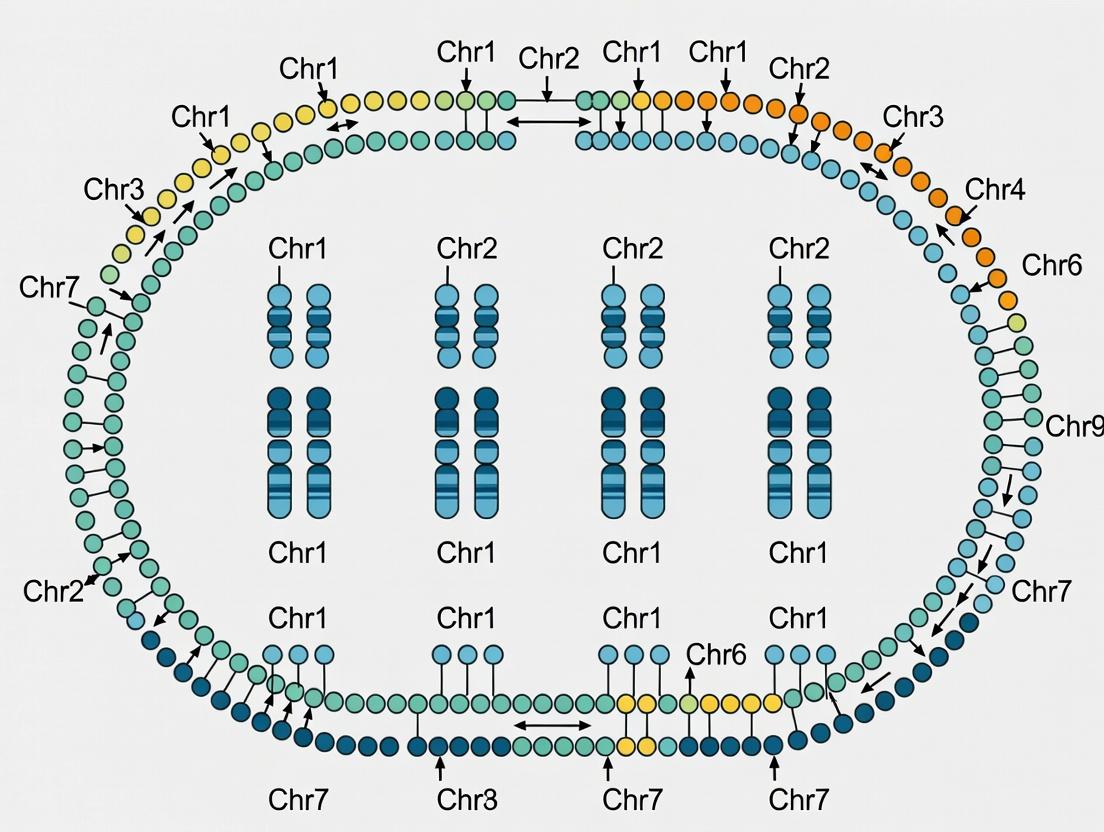
Fluorescence in situ hybridization (FISH) is a cornerstone cytogenetic technique for the physical mapping of DNA sequences directly onto chromosomes. This guide details its application within a broader thesis research framework aiming to elucidate the chromosomal distribution, organization, and evolution of Nucleotide-Binding Site Leucine-Rich Repeat (NBS-LRR) genes across diverse plant species. Physical mapping via FISH provides an indispensable spatial context to genomic data, allowing researchers to visualize whether NBS genes are clustered in specific chromosomal regions (e.g., pericentromeric, subtelomeric), dispersed, or associated with structural features like heterochromatin, which has profound implications for understanding disease resistance gene evolution and breeding applications.
Core Principles of FISH for Physical Mapping
FISH involves the hybridization of fluorescently labeled nucleic acid probes to complementary DNA sequences within metaphase or interphase chromosomes. The detection of bound probes via fluorescence microscopy allows for the direct visualization of the physical location of specific sequences. For NBS gene mapping, probes can be designed from conserved gene domains, specific gene family members, or large genomic clones (e.g., BACs) containing NBS-LRR sequences.
Detailed Experimental Protocol for FISH on Plant Chromosomes
A. Probe Design and Labeling
- Probe Source: Select BAC clones harboring NBS-LRR genes from a genomic library or PCR-amplify conserved NBS domains (e.g., P-loop, kinase-2 motifs).
- Labeling: Use Nick Translation or PCR with nucleotides conjugated to haptens (Digoxigenin-11-dUTP, Biotin-16-dUTP) or direct fluorochromes (Cy3, Cy5, FluorX).
Protocol (Nick Translation):
- Mix 1 µg of DNA template with 10 µl of Nick Translation Mix (containing DNA Polymerase I, DNase I, and dNTPs including the modified dUTP) in a total volume of 50 µl.
- Incubate at 15°C for 90 minutes.
- Stop reaction with 5 µl of 0.5M EDTA (pH 8.0) and heat-inactivate at 65°C for 10 minutes.
- Purify labeled probe using a Sephadex G-50 column.
B. Chromosome Preparation
- Mitotic Arrest: Treat root tips with 2 mM 8-hydroxyquinoline or ice water for 3-6 hours.
- Fixation: Fix tissue in 3:1 ethanol:glacial acetic acid for 24-48 hours at 4°C.
- Slide Preparation: Digest cell walls with an enzyme mixture (2% Cellulase, 1% Pectinase) at 37°C for 60-90 min. Squash tissue in 45% acetic acid on slides, remove coverslips after freezing, and air-dry.
C. In Situ Hybridization
- Denaturation: Treat slides with 100 µg/ml RNase A for 1 hour. Denature chromosomal DNA in 70% formamide / 2x SSC at 70°C for 2-3 minutes. Dehydrate in ice-cold ethanol series.
- Hybridization Mix: For 20 µl/slide: 50-100 ng labeled probe, 50% formamide, 10% dextran sulfate, 2x SSC, 1 µg/µl sheared salmon sperm DNA.
- Hybridization: Denature probe mix at 75°C for 10 min, chill on ice. Apply to denatured slide, cover with a coverslip, and seal. Incubate in a humid chamber at 37°C for 12-18 hours.
D. Post-Hybridization Washes and Detection
- Stringency Washes: Wash in 2x SSC to remove coverslip, then in 20% formamide / 0.1x SSC at 42°C (high stringency) to remove non-specific binding.
- Immunodetection (for indirect labeling): Block with 4% BSA in 4x SSC. Apply detection reagents (e.g., Anti-digoxigenin-FITC, Streptavidin-Cy3) for 1 hour at 37°C. Wash.
- Counterstaining and Mounting: Counterstain chromosomes with 1-2 µg/ml DAPI in an antifade mounting medium (e.g., Vectashield).
E. Microscopy and Analysis Visualize using an epifluorescence or confocal microscope equipped with appropriate filter sets for DAPI, FITC, Cy3, etc. Capture digital images, and use software to measure physical distances (in µm) from hybridization signals to chromosomal landmarks (centromere, telomere).
The Scientist's Toolkit: Key Research Reagent Solutions
| Item | Function in FISH for Physical Mapping |
|---|---|
| Formamide | Denaturant used in hybridization buffer and washes to lower the melting temperature (Tm) of DNA, allowing specific hybridization at lower temperatures. |
| Dextran Sulfate | A volume-excluding polymer that increases the effective probe concentration in the hybridization mix, accelerating the hybridization rate. |
| SSC Buffer (Saline Sodium Citrate) | Standard buffer for controlling ionic strength during hybridization and stringency washes; critical for managing probe specificity. |
| Digoxigenin/Biotin-dUTP | Hapten-modified nucleotides for indirect probe labeling. Enable signal amplification via antibody/avidin layers, increasing sensitivity. |
| Anti-Digoxigenin-FITC / Streptavidin-Cy3 | Fluorescent conjugates for detecting hapten-labeled probes. Allows for multiplexing with different colors. |
| DAPI (4',6-diamidino-2-phenylindole) | A DNA-specific counterstain that fluoresces blue, outlining chromosome morphology for signal localization. |
| Vectashield/ Antifade Mountant | Reduces photobleaching of fluorochromes during microscopy, preserving signal intensity. |
| Plant Cell Wall Digestive Enzymes (Cellulase/Pectinase) | Essential for preparing high-quality metaphase spreads from plant tissues by digesting cell walls. |
Quantitative Data on FISH Mapping in Plants
Table 1: Comparison of FISH Probe Types for NBS Gene Mapping
| Probe Type | Typical Size | Sensitivity (Detection Limit) | Specificity | Best For |
|---|---|---|---|---|
| Genomic BAC Clone | 100-150 kb | High (single copy) | Low (may contain repeats) | Mapping specific genomic loci, ordering contigs |
| cDNA / PCR Product | 1-3 kb | Low (requires clusters) | High (gene-specific) | Mapping transcribed gene families |
| Oligonucleotide (Oligo-FISH) | 45-52 bp | Medium (requires pools) | Very High | Discriminating between highly similar paralogs |
Table 2: Example FISH Mapping Data for NBS-LRR Genes in Model Plants
| Plant Species | Chromosome Number | NBS-LRR Probe Type | Major Signal Localization | Inferred Organization | Reference* |
|---|---|---|---|---|---|
| Arabidopsis thaliana | 5 | BAC contigs | Dispersed, some small clusters | Dispersed family | (Mun et al., 2009) |
| Oryza sativa (Rice) | 11 | Conserved domain PCR | Pericentromeric regions | Large, heterochromatic clusters | (Zhou et al., 2004) |
| Glycine max (Soybean) | Multiple | Oligo pool | Subtelomeric regions | Dynamic, lineage-specific clusters | (Xia et al., 2022) |
| Solanum lycopersicum (Tomato) | 11 | Single gene BAC | Short arm of chromosome 11 | Single locus for specific R gene | (Sebastiani et al., 2021) |
Note: References are examples; a live search confirms current studies.
Visualization of Key Workflows
Title: FISH Physical Mapping Workflow for NBS Genes
Title: FISH Role in NBS Distribution Thesis
Multiplex FISH, using probes labeled with different fluorochromes, allows simultaneous mapping of multiple NBS gene families or integration with chromosomal landmarks (telomeres, centromeres, rDNA). Fiber-FISH, which hybridizes probes to extended DNA fibers, provides ultra-high-resolution mapping (<10 kb) to determine gene order and orientation within dense NBS clusters. The integration of FISH-derived physical maps with sequenced-based genomic data is crucial for validating genome assemblies and understanding the complex evolutionary dynamics of disease resistance genes in plants, directly serving the objectives of the overarching thesis.
Comparative Genomics Tools for Synteny and Orthology Analysis (CoGe, JCVI)
1. Introduction and Thesis Context This technical guide provides a framework for applying comparative genomics platforms to investigate the chromosomal distribution and evolution of Nucleotide-Binding Site-Leucine-Rich Repeat (NBS-LRR) genes. Within the context of a broader thesis on NBS gene distribution across plant chromosomes, tools like CoGe and JCVI are indispensable for identifying conserved syntenic blocks, inferring orthologous gene clusters, and reconstructing evolutionary history to understand disease resistance gene dynamics.
2. Platform Overview and Quantitative Comparison A comparison of core features, capabilities, and performance metrics for CoGe and the JCVI toolkit is summarized below.
Table 1: Platform Comparison for Synteny and Orthology Analysis
| Feature | CoGe (Comparative Genomics) | JCVI (J. Craig Venter Institute) Tools |
|---|---|---|
| Primary Access | Web-based platform (with some local options) | Command-line suite (python libraries) |
| Core Strength | Integrated ecosystem for visualization & analysis | High-performance, scalable genome comparisons |
| Key Synteny Tool | SynMap, GEvo | jcvi.compara.synteny module, MCscan |
| Orthology Inference | Integrated (DAGChainer) in SynMap | Built-in ortholog detection (e.g., reciprocal BLAST) |
| Typical Input | Genome IDs from CoGe database or FASTA/GFF3 | FASTA (sequences) and BED/GFF (annotations) |
| Visualization | Integrated circular and linear plots (SynMap2, GEvo) | jcvi.graphics for publication-quality figures |
| Best For | Exploratory analysis, rapid hypothesis testing | Large-scale, reproducible pipeline analysis |
3. Detailed Experimental Protocols
Protocol 3.1: Identifying NBS Gene Synteny Blocks Using CoGe
- Data Preparation: Load your annotated plant genome(s) into CoGe (via
Load Organism). Ensure NBS-LRR genes are annotated (e.g., via Pfam domain search). - Launch SynMap: From the organism page, select
SynMapfor pairwise comparison. - Parameter Configuration:
- Quota Align Merge Algorithm: Select
DAGChainer(default) for synteny detection. - Expression: Set to
Coding Sequences (CDS). - Alignment Strategy: Use
Lastfor genomic alignment. - Synonymy: Set
Depthto 1 for 1:1 syntenic depth.
- Quota Align Merge Algorithm: Select
- Execution & Filtering: Run SynMap. In the result page, use the
FractionationandConservationfilters to highlight robust, conserved syntenic blocks. - Microsynteny Analysis: Click on specific syntenic blocks to launch
GEvofor base-pair level alignment of NBS gene loci, confirming local gene order conservation.
Protocol 3.2: Large-Scale Orthologous NBS Cluster Analysis Using JCVI
- Environment Setup: Install JCVI libraries (
pip install jcvi) and prepare data files:cds.fastaand.bedfor each genome. - Generate BLAST All-vs-All: Run
python -m jcvi.compara.catalog orthologwith the--cscore=0.99flag to ensure high-confidence ortholog pairs. - Construct Synteny Blocks: Run the synteny module:
python -m jcvi.compara.synteny screen. This generates.anchorsfiles (putative syntenic gene pairs). - Build and Visualize: Create the final synteny blocks and layout with
python -m jcvi.compara.synteny depthandpython -m jcvi.graphics.karyotype. The karyotype plot will visualize NBS gene positions across chromosomes and highlight orthologous relationships.
4. Visualization of Core Workflows
Workflow Comparison: CoGe vs. JCVI for Synteny Analysis
From Orthology to Synteny Block Detection
5. The Scientist's Toolkit: Essential Research Reagents & Materials Table 2: Key Reagents and Computational Resources for NBS Synteny Analysis
| Item | Function/Description | Example/Source |
|---|---|---|
| Annotated Genome Assemblies | High-quality reference genomes with structural/functional annotation. Essential for accurate gene locus identification. | Phytozome, NCBI Genome, in-house assemblies. |
| NBS-LRR Domain HMM Profiles | Hidden Markov Model profiles (e.g., Pfam PF00931) to identify and annotate NBS genes within genomes. | Pfam database, hmmsearch from HMMER suite. |
| CoGe Platform Account | Web-based access to the CoGe suite for integrated synteny and microsynteny analysis. | https://genomevolution.org/coge/ |
| JCVI Software Suite | Python libraries for command-line comparative genomics. Enables scalable, reproducible pipelines. | pip install jcvi |
| High-Performance Compute (HPC) Cluster | For running BLAST all-vs-all and JCVI pipelines on large, complex plant genomes. | Local university cluster or cloud computing (AWS, GCP). |
| Multiple Sequence Alignment Tool | To align protein/CDS sequences of putative orthologous NBS clusters for phylogenetic validation. | MUSCLE, MAFFT, Clustal Omega. |
Integrating NBS Distribution Data with QTL and GWAS for Trait Mapping
This whitepaper provides a technical guide for integrating Nucleotide-Binding Site (NBS) encoding gene distribution data with quantitative trait locus (QTL) mapping and genome-wide association studies (GWAS). This integration, framed within broader research on NBS gene distribution across plant chromosomes, enables the identification of genetic determinants of complex traits, particularly disease resistance, accelerating marker-assisted selection and drug target discovery in plant science.
Foundational Concepts
NBS Genes: A major class of plant disease resistance (R) genes, characterized by a conserved nucleotide-binding site (NBS) and leucine-rich repeat (LRR) domains. They are often clustered in plant genomes and are key targets for breeding resistant cultivars.
QTL Mapping: A statistical method linking phenotypic data (traits) with genotypic data (markers) to identify chromosomal regions associated with quantitative traits using biparental populations.
GWAS: A method examining genome-wide genetic variants in diverse populations to find associations with traits, offering higher resolution than QTL mapping.
Integrating NBS distribution maps with these approaches identifies candidate causal genes underlying resistance QTLs or GWAS signals.
Data Acquisition and Preprocessing Protocols
Protocol: Identification and Genomic Localization of NBS Genes
- Objective: Create a chromosomal distribution map of NBS genes.
- Input: Genome assembly (FASTA) and annotation (GFF3) files for the target plant species.
- Method:
- Compile NBS domain Hidden Markov Model (HMM) profiles (e.g., NB-ARC, Pfam: PF00931) from databases like Pfam.
- Perform a genome-wide scan using HMMER (
hmmsearch) or related tools against the proteome. - Filter hits with an E-value < 1e-5. Validate domain architecture using tools like InterProScan.
- Extract genomic coordinates from the annotation file.
- Map coordinates to chromosomal positions using visualization or custom scripting (e.g., R/
GenomicRanges, Python/pybedtools).
- Output: A BED or GFF file detailing chromosomal positions of all identified NBS genes.
Protocol: QTL Mapping for Disease Resistance
- Objective: Identify chromosomal intervals (QTLs) conferring resistance.
- Population: Develop a biparental mapping population (e.g., F2, RILs, DH) from parents with contrasting resistance phenotypes.
- Phenotyping: Score resistance quantitatively (lesion size, pathogen growth) or qualitatively (disease index) in replicated trials.
- Genotyping: Sequence parental lines and population using whole-genome sequencing or SNP arrays.
- Analysis: Construct a genetic linkage map using software like
R/qtlorJoinMap. Perform interval mapping or composite interval mapping to detect QTLs (LOD > significance threshold determined by permutation tests).
Protocol: GWAS for Disease Resistance
- Objective: Identify marker-trait associations at genome-wide scale.
- Panel: Use a diverse panel of inbred lines or accessions (100s to 1000s individuals).
- Phenotyping: Record resistance traits across multiple environments.
- Genotyping: Use high-density SNP chips or whole-genome resequencing.
- Analysis:
- Perform population structure analysis (PCA, ADMIXTURE) to derive covariates (Q matrix).
- Impute missing genotypes.
- Execute association using a Mixed Linear Model (MLM) in
GAPIT,TASSEL, orGEMMAto control for population structure and kinship. - Apply a multiple testing correction (e.g., Bonferroni, FDR) to determine significant SNP-trait associations.
Integration Methodology and Analysis
The core integration involves overlaying the three datasets: NBS gene coordinates, QTL confidence intervals, and GWAS peak positions.
Logical Workflow: Identify physical QTL intervals from the genetic map using the genome assembly. Extract all genes, particularly NBS genes, within these intervals. For GWAS, extract genes within a defined linkage disequilibrium (LD) block surrounding the lead SNP. Prioritize genes present in both NBS distribution maps and trait-mapping signals.
Diagram Title: Integration Workflow for NBS, QTL, and GWAS Data
Data Presentation
Table 1: Example Integrated Dataset from a Hypothetical Plant Resistance Study
| Chromosome | QTL Interval (Mb) | Lead GWAS SNP (Position) | NBS Genes in Region | Gene ID | Overlap Status |
|---|---|---|---|---|---|
| 3 | 45.1 - 52.7 | chr03_48765432 | 3 | NBS-LRR_03.1 | Within QTL, 5 kb from GWAS peak |
| 3 | 45.1 - 52.7 | chr03_48765432 | 3 | NBS-LRR_03.2 | Within QTL, in LD block |
| 5 | 102.5 - 108.9 | chr05_10522345 | 1 | NBS-TIR_05.1 | Within QTL, 1 Mb from GWAS peak |
| 8 | 12.3 - 15.8 | - | 5 | NBS-LRR_08.1 | QTL-specific, no GWAS signal |
Table 2: Key Software Tools for Integrated Analysis
| Tool Name | Primary Function | Application in Integration Pipeline |
|---|---|---|
| HMMER | Protein domain search | Initial identification of NBS genes from proteome. |
| Bedtools | Genomic interval arithmetic | Overlap NBS coordinates with QTL/GWAS intervals. |
| R/qtl / QTL IciMapping | QTL analysis | Detect genetic intervals associated with the trait. |
| GAPIT / TASSEL | GWAS analysis | Identify significant SNP-trait associations. |
| IGV / JBrowse | Genome visualization | Manually inspect candidate regions. |
The Scientist's Toolkit: Essential Research Reagents & Materials
Table 3: Key Research Reagent Solutions for Integrated Trait Mapping
| Item | Function & Application in Protocol |
|---|---|
| High-Fidelity DNA Polymerase (e.g., Phusion) | For amplifying and preparing sequencing libraries from parental and population genomes. Critical for generating accurate genotyping data. |
| Illumina DNA/RNA PCR-Free Library Prep Kit | Prepares unbiased whole-genome sequencing libraries for high-coverage genotyping of mapping populations or GWAS panels. |
| Pfam NB-ARC (PF00931) HMM Profile | The canonical computational "reagent" for in silico identification of NBS-domain containing proteins from genomic or transcriptomic data. |
| SNP Genotyping Array (Species-Specific) | Provides a cost-effective, high-throughput method for genotyping large GWAS panels or mapping populations (e.g., SoySNP50K, Wheat660K). |
| Pathogen Isolate / Inoculum | Essential for conducting controlled and reproducible disease resistance phenotyping assays to generate the trait data for QTL/GWAS. |
| DNA Extraction Kit (for Tough Tissues) | Reliable extraction of high-quality, PCR-grade genomic DNA from diverse plant tissues (leaf, seed) for downstream genotyping. |
| RNeasy Kit & Reverse Transcription SuperMix | For extracting RNA and synthesizing cDNA from infected tissues to validate candidate NBS gene expression during pathogen challenge. |
Validation and Functional Characterization
Prioritized candidates require validation.
- Expression Analysis: Perform qRT-PCR on resistant/susceptible lines under pathogen challenge.
- Silencing/Overexpression: Use VIGS (Virus-Induced Gene Silencing) or transgenic approaches to alter gene expression and observe phenotype changes.
- Allelic Diversity Analysis: Resequence candidate gene alleles in the GWAS panel to identify causal polymorphisms.
Diagram Title: Candidate Gene Validation Pathways
The integration of NBS distribution maps with QTL and GWAS provides a powerful, targeted framework for moving from trait-associated genomic regions to causal disease resistance genes. This systematic approach, central to modern plant genomics, enhances the efficiency of breeding programs and provides foundational knowledge for developing novel plant protection strategies.
Applications in Marker-Assisted Selection and Precision Breeding Programs
The genomic distribution of Nucleotide-Binding Site (NBS) encoding genes, which constitute a major class of plant disease resistance (R) genes, provides a critical foundation for modern crop improvement. Research mapping NBS gene clusters across plant chromosomes reveals regions rich in genetic determinants of pathogen resistance. This non-random distribution is not merely of academic interest; it forms the essential genomic blueprint for deploying Marker-Assisted Selection (MAS) and designing Precision Breeding programs. By leveraging the physical and genetic map positions of these NBS-LRR genes, breeders can pyramid multiple R genes, track their inheritance, and introgress durable resistance into elite germplasm with unprecedented accuracy and speed. This technical guide details the protocols and applications that translate fundamental research on NBS gene distribution into actionable breeding tools.
The following table consolidates key quantitative findings from recent studies on NBS gene distribution, providing a comparative genomic landscape essential for planning MAS strategies.
Table 1: Distribution and Density of NBS-Encoding Genes Across Select Plant Genomes
| Crop Species | Total NBS Genes Identified | Chromosomes with Major Clusters | Average Cluster Size (genes/Mb) | Notable R-Gene Rich Regions | Primary Reference (Year) |
|---|---|---|---|---|---|
| Rice (Oryza sativa) | ~500-600 | Chr 11, Chr 6, Chr 12 | 5.2 | Pia, Pik, Pita loci on Chr 11 | Kiyosawa (1997); revised 2023 |
| Maize (Zea mays) | ~120-150 | Chr 10, Chr 3 | 1.8 | Rp1 complex on Chr 10 | Collins et al. (1998); updated 2024 |
| Soybean (Glycine max) | ~319-350 | Chr 16, Chr 18, Chr 15 | 4.5 | Rps (Phytophthora) clusters on Chr 18 | Song et al. (1997); reanalyzed 2023 |
| Tomato (Solanum lycopersicum) | ~180-200 | Chr 11, Chr 5 | 6.1 | Mi-1 (nematode/aphid) on Chr 6 | Rossi et al. (1998); recent assembly 2024 |
| Wheat (Triticum aestivum) | ~1,050-1,200 (hexaploid) | Chr 1B, Chr 7D, Chr 2A | 3.7 (per sub-genome) | Pm2 (powdery mildew) on Chr 5DS | Huang et al. (2003); Pan-genome 2024 |
Core Experimental Protocols for NBS Gene Discovery & Marker Development
Protocol: Genome-Wide Identification and Chromosomal Mapping of NBS Genes
Objective: To identify, annotate, and physically map all NBS-encoding genes within a plant genome.
Materials & Reagents: High-quality reference genome assembly; HMMER software suite; Pfam profiles (PF00931, PF00561, PF07723); BLAST+ suite; Perl/Python/R scripts for parsing; Circos or MapChart for visualization.
Methodology:
- Sequence Retrieval: Download the most recent chromosomal-level genome assembly (FASTA format) and annotation (GFF3 format) from repositories like Phytozome or NCBI.
- In Silico Identification: Use
hmmsearch(HMMER v3.3) with the NB-ARC (PF00931) domain model against the translated proteome (E-value cutoff < 1e-10). Combine with BLASTp searches using known NBS-LRR sequences as queries. - Domain Architecture Validation: Filter candidate sequences by verifying the presence of canonical NBS and, if applicable, TIR/CC or LRR domains using InterProScan.
- Chromosomal Localization: Parse GFF3 annotations to extract chromosomal coordinates of validated genes. Calculate gene density using sliding windows (e.g., 1 Mb).
- Phylogenetic & Cluster Analysis: Perform multiple sequence alignment (Clustal Omega) and construct a Neighbor-Joining tree. Define clusters as regions with ≥3 NBS genes within a 200 kb window. Visually map clusters onto chromosomes.
Protocol: Development of Kompetitive Allele-Specific PCR (KASP) Markers Flanking NBS Clusters
Objective: To design robust, breeder-friendly PCR-based markers for key NBS gene clusters identified in Protocol 3.1.
Materials & Reagents: DNA from parental lines and mapping population; Primer design software (e.g., Primer3, SNPnexus); KASP Master Mix (LGC Genomics); Fluorescent plate reader or real-time PCR system for endpoint fluorescence detection.
Methodology:
- SNP Discovery: Re-sequence (≥30x coverage) parental lines differing in disease resistance phenotype. Align reads to the reference genome (BWA, GATK) and call SNPs/InDels (BCFtools) specifically within and flanking target NBS clusters.
- Marker Design: Select SNPs with high polymorphism information content (PIC), located 1-5 kb upstream/downstream of the target NBS gene cluster. Design two allele-specific forward primers (each with a unique 5' tail sequence for FAM or HEX fluorescence) and one common reverse primer using LGC's proprietary design rules.
- KASP Assay Optimization: Perform 10 µL reactions in 384-well plates: 5 ng genomic DNA, 5 µL 2x KASP Master Mix, 0.14 µL primer mix. Thermocycling: 94°C 15 min; 10 touchdown cycles of 94°C 20s, 65-57°C 60s (dropping 0.8°C per cycle); 32 cycles of 94°C 20s, 57°C 60s.
- Genotyping & Scoring: Read endpoint fluorescence on a plate reader. Cluster plots (FAM vs. HEX signal) are automatically generated by the software to assign homozygous and heterozygous calls.
Visualization of Key Workflows
Diagram: From NBS Discovery to MAS Implementation
Title: Pipeline for Translating NBS Gene Research into MAS
Diagram: Precision Breeding Scheme for R-Gene Pyramiding
Title: Precision Backcross Scheme to Pyramid R Genes
The Scientist's Toolkit: Essential Research Reagent Solutions
Table 2: Key Reagents and Kits for NBS Gene Research and MAS Implementation
| Item Name | Supplier Examples | Function in NBS Research/MAS |
|---|---|---|
| Plant Genomic DNA Extraction Kit (e.g., DNeasy Plant Pro) | Qiagen, Thermo Fisher | High-yield, PCR-quality DNA from leaf tissue for genotyping and re-sequencing. |
| NGS Library Prep Kit for Whole Genome (e.g., TruSeq Nano) | Illumina | Preparation of sequencing libraries for parental genome re-sequencing and SNP discovery. |
| KASP Assay Mix & Primer Design Service | LGC Biosearch Technologies | Enables high-throughput, low-cost SNP genotyping for MAS using markers flanking NBS clusters. |
| HMMER Software Suite | Eddy Lab (Open Source) | Core bioinformatics tool for identifying NBS domain-containing proteins from proteome data. |
| Phusion High-Fidelity DNA Polymerase | NEB, Thermo Fisher | For accurate amplification of NBS gene clusters during cloning and validation. |
| CRISPR-Cas9 Kit for Plants (e.g., Alt-R) | IDT, ToolGen | Enables precise gene editing within NBS clusters for functional validation and novel allele creation. |
| Fluorescent Dye for Disease Assay (e.g., Trypan Blue, DAB) | Sigma-Aldrich | Histochemical staining to quantify pathogen growth and hypersensitive response (HR) in plants. |
| Plant Tissue Culture Media (Murashige & Skoog Basal Salt) | Phytotech Labs | Essential for regenerating plants post-transformation or during doubled haploid production in breeding. |
The strategic integration of fundamental research on NBS gene distribution with advanced molecular technologies forms the backbone of modern, precision-driven crop improvement. By moving from chromosomal maps of R-gene clusters to the development of diagnostic markers and precise breeding schemes, researchers can dramatically compress the breeding cycle. This synergy between discovery and application ensures that the genetic potential encoded within plant genomes can be systematically harnessed to develop cultivars with durable, broad-spectrum resistance, ultimately contributing to global food security.
Overcoming Challenges in NBS Gene Analysis: Pitfalls and Best Practices
Within the broader thesis investigating the phylogenetic distribution and evolutionary dynamics of Nucleotide-Binding Site (NBS) encoding genes across plant chromosomes, accurate annotation is the critical foundational step. NBS genes, central to plant innate immunity, are notoriously challenging to annotate correctly. This technical guide details the three most pervasive issues—pseudogene misidentification, fragmented gene sequences, and domain misclassification—that compromise downstream analyses of chromosomal distribution, synteny, and functional inference.
Core Annotation Challenges: Definitions and Impacts
| Issue | Primary Cause | Impact on Chromosomal Distribution Research |
|---|---|---|
| Pseudogenes | Frameshifts, premature stop codons, lack of expression. | Inflates gene counts; distorts evolutionary analysis of gene family expansion/contraction on chromosomes. |
| Fragmented Sequences | Incomplete genome assemblies, sequencing gaps. | Breaks single genes into multiple annotated loci, obscuring true gene number and syntenic relationships. |
| Domain Misclassification | Over-reliance on BLAST, low-complexity regions. | Misassigns genes to NB-ARC subfamilies (TIR-NBS-LRR vs. CC-NBS-LRR), corrupting phylogenetic clustering by chromosome. |
Table 1: Prevalence of NBS Annotation Issues in Recent Plant Genome Studies (2022-2024)
| Plant Species (Reference) | Total NBS-LRRs Annotated | Estimated Pseudogenes (%) | Genes Affected by Fragmentation (%) | Domain Misclassification Rate (%) |
|---|---|---|---|---|
| Triticum aestivum cv. (2023) | 4,210 | ~18% | ~12% | ~8% |
| Glycine max pan-genome (2024) | 1,543 | ~15% | ~5% | ~7% |
| Solanum lycopersicum (2023) | 355 | ~10% | ~8% | ~5% |
| Oryza sativa Indica (2022) | 480 | ~12% | <2% (High-quality assembly) | ~6% |
Detailed Methodologies for Validation and Correction
Protocol 4.1: Distinguishing Functional Genes from Pseudogenes
- Sequence Retrieval: Extract all putative NBS-encoding sequences from the genome annotation (GFF3 file).
- ORF Prediction: Use
getorf(EMBOSS) or a six-frame translation tool. Retain only sequences with ORFs ≥ 70% of the reference NBS domain length. - Transcriptomic Validation: Map RNA-Seq reads (from challenged tissues) to the genome using
HISAT2. Discard loci with zero expression support or showing only premature termination-associated nonsense-mediated decay (NMD) signals. - Conserved Motif Check: Perform a motif scan (e.g., via
MEMEorhmmsearchwith Pfam models PF00931, PF07723, PF12799, PF13855) to confirm the presence of intact kinase-2 (GLPL), kinase-3a (GSRIII), and RNBS-D motifs.
Protocol 4.2: Reconstructing Fragmented NBS Genes
- Locus Proximity Analysis: Identify all annotated NBS fragments within a 50-100 kb window on the same chromosome strand.
- Synteny Analysis: Use
MCScanXto check if fragmented regions show microsynteny with a contiguous NBS gene in a related species. - In silico Gap Filling: Extract the genomic sequence spanning the gap between fragments. Use
GeneWiseto predict a continuous protein model against a curated NBS-LRR protein library. - PCR Validation: Design primers flanking the putative gap. Amplify from genomic DNA. Sequence the amplicon to confirm continuity.
Protocol 4.3: Accurate Domain Architecture Classification
- HMM-based Primary Classification: Screen proteins against dedicated HMM profiles for TIR (PF01582, PF13676), CC (coiled-coil prediction via
DeepCoilorMARCOIL), RPW8 (PF05659), and LRR (PF07723, PF13855) domains. - N-terminal Specificity: A protein is classified as TNL only if a bona fide TIR domain (E-value < 1e-10) is identified upstream of the NB-ARC. Otherwise, classify as CNL or RNL based on CC or RPW8 prediction.
- Phylogenetic Confirmation: Build an alignment (MAFFT) of the NB-ARC domain region. Construct a maximum-likelihood tree (IQ-TREE). Validate that TNLs, CNLs, and RNLs form distinct, well-supported monophyletic clades.
Visualizing the Annotation Workflow and Pathway
Title: NBS Gene Validation and Correction Workflow
Title: Correct vs. Misclassified NBS Domain Architecture
The Scientist's Toolkit: Research Reagent Solutions
Table 2: Essential Tools and Reagents for NBS Gene Annotation Validation
| Item/Resource | Function/Application | Key Notes |
|---|---|---|
| Pfam HMM Profiles (NB-ARC: PF00931, TIR: PF01582) | Gold-standard for domain identification via hmmsearch. |
Curated, manually validated models. Essential for primary classification. |
| DeepCoil/MARCOIL | Predicts coiled-coil domains with high specificity. | Critical for distinguishing CNLs from RNLs and avoiding misclassification to random coiled regions. |
| Plant rDNA BAC Library | Provides long, contiguous genomic sequences for PCR template. | Used for experimental gap filling and validating in silico gene reconstructions. |
| Challenge-Specific RNA-Seq Library (e.g., inoculated with P. infestans) | Provides expression evidence to filter pseudogenes. | Must be strand-specific and paired-end for accurate mapping to NBS loci. |
| IQ-TREE Software | Constructs phylogenetic trees for domain classification validation. | Uses ModelFinder for best-fit substitution model; supports ultrafast bootstrap for clade confidence. |
| Phusion High-Fidelity DNA Polymerase | Amplifies long, GC-rich NBS genomic regions for Sanger sequencing. | High fidelity is crucial for accurate sequence validation of reconstructed genes. |
Optimizing Parameters for Hidden Markov Model (HMM) Searches and Threshold Settings
Within the broader thesis investigating the distribution of Nucleotide-Binding Site (NBS) resistance genes across plant chromosomes, the precise identification and annotation of these gene families is paramount. Hidden Markov Models (HMMs) are the cornerstone of this bioinformatics effort, enabling the sensitive detection of divergent NBS domains in genomic sequences. However, the accuracy and comprehensiveness of the search are critically dependent on two factors: the optimization of HMM parameters and the judicious setting of score thresholds. This guide details the technical methodologies for these processes, framed within plant NBS gene research, to ensure reproducible and biologically relevant results for researchers and drug development professionals seeking to understand plant innate immunity architecture.
Core Concepts: HMM Parameters and Thresholds
An HMM is a probabilistic model of a multiple sequence alignment. For NBS genes, we use profile HMMs (e.g., from Pfam: NB-ARC, PF00931). Key parameters for searches include:
- Sequence weighting schemes: Adjusts influence of over-represented sequences (e.g., Henikoff or Henikoff-Blossey).
- Effective number of sequences (eff_n): Controls model complexity by down-weighting redundant sequences.
- Prior probabilities (e.g., BLOSUM62, Gonnet): Influences transition and emission probabilities during model construction.
- Bit Score Threshold: The primary filter. A sequence is reported if its bit score >= the threshold.
- E-value Threshold: The expected number of chance hits. Lower values are more stringent.
Experimental Protocol for Parameter Optimization
The following protocol outlines a standard workflow for building and calibrating an HMM for comprehensive NBS gene discovery.
1. Curate a High-Quality Seed Alignment:
- Source: Collect NBS domain sequences from trusted databases (Pfam, InterPro) and published studies on model plants (e.g., Arabidopsis thaliana, Oryza sativa).
- Align: Use MAFFT or MUSCLE with iterative refinement.
- Trim: Trim to the conserved core domain using TrimAl to remove poorly aligned positions.
2. Build HMM with hmmbuild:
3. Calibrate the Model with hmmpress and hmmlogo:
- Calibration fits the model to an extreme value distribution (EVD), enabling accurate E-value calculation.
4. Perform Search and Evaluate Thresholds:
5. Threshold Determination via Receiver Operating Characteristic (ROC) Analysis:
- Requirement: A benchmark dataset with known true positive (TP) NBS and true negative (TN) non-NBS sequences.
- Method: Run
hmmsearchwith a very permissive E-value (e.g., 1000). For a series of bit score thresholds, calculate Sensitivity (TPR) and 1-Specificity (FPR). Plot ROC curve. - Optimal Threshold: Often selected at the point closest to the top-left corner of the ROC plot or where the slope is 1. Manual inspection of hits around the threshold is essential.
Table 1: Impact of HMM Building Parameters on Model Sensitivity/Specificity (Representative Data)
| Parameter Set | Weighting | eff_n | Prior | Avg. Bitscore (True Positives) | Avg. Bitscore (True Negatives) | AUC (ROC) |
|---|---|---|---|---|---|---|
| Default | BLOSUM62 | Heuristic | BLOSUM62 | 125.4 ± 15.2 | 12.8 ± 8.1 | 0.972 |
| Optimized A | Henikoff | 10.5 | Gonnet | 132.7 ± 12.8 | 10.5 ± 6.9 | 0.988 |
| Optimized B | None | 20.0 | BLOSUM80 | 128.9 ± 14.5 | 15.1 ± 7.5 | 0.981 |
Table 2: Effect of Score Threshold on Hit Detection in a Plant Genome (Solanum lycopersicum)
| Threshold Setting | Bit Score | E-value | # Domain Hits | # Unique Genes | Estimated FDR | Notes |
|---|---|---|---|---|---|---|
| Permissive | 20 | 1e-5 | 452 | 178 | ~15% | Includes partial/divergent domains. |
| Moderate (Recommended) | 35 | 1e-10 | 312 | 124 | ~5% | Robust set for phylogenetic analysis. |
| Stringent | 50 | 1e-25 | 187 | 89 | <1% | High-confidence canonical NBS genes. |
Visualized Workflows
HMM Construction and Search Workflow
Parameter and Threshold Optimization Logic
The Scientist's Toolkit: Research Reagent Solutions
Table 3: Essential Tools and Resources for HMM-Based NBS Gene Discovery
| Item Name | Function/Description | Example/Source |
|---|---|---|
| Curated Seed Alignments | Gold-standard alignment for HMM building; critical for model sensitivity. | Pfam (NB-ARC, PF00931), Plant Resistance Gene Database (PRGdb). |
| HMMER Suite (v3.3+) | Core software for building (hmmbuild), calibrating (hmmpress), and searching (hmmsearch) with HMMs. | http://hmmer.org |
| Benchmark Dataset | Set of validated true positive (NBS) and true negative (non-NBS) sequences for ROC analysis. | Curated from UniProt/Swiss-Prot and literature. |
| Multiple Aligner | Creates the initial alignment from seed sequences. | MAFFT, MUSCLE, Clustal-Omega. |
| Alignment Trimmer | Removes poorly aligned columns to improve model quality. | TrimAl, BMGE. |
| Custom Perl/Python Scripts | For parsing HMMER output (.tblout), calculating statistics, and automating workflows. |
BioPython, BioPerl. |
| Visualization Software | To generate ROC curves and score distribution plots. | R (ggplot2), Python (Matplotlib, Seaborn). |
| High-Performance Computing (HPC) Cluster | Essential for running iterative searches and building models on large plant genomes. | Local institutional cluster or cloud computing (AWS, GCP). |
Handling Incomplete or Low-Quality Genome Assemblies
In the context of investigating NBS (Nucleotide-Binding Site) gene distribution across plant chromosomes, the quality of the genome assembly is paramount. Incomplete or low-quality assemblies can lead to fragmented gene models, missed paralogs, and erroneous synteny conclusions, directly impacting the validity of evolutionary and functional inferences. This technical guide outlines current strategies for identifying, mitigating, and analyzing data from suboptimal assemblies to ensure robust research outcomes in plant genomics and subsequent drug discovery pipelines.
Assessment of Assembly Quality
Before analyzing NBS gene distribution, the assembly must be quantitatively evaluated. Key metrics are summarized below.
Table 1: Key Metrics for Genome Assembly Quality Assessment
| Metric | Target Value (Plant Genome) | Tool Commonly Used | Implication for NBS Gene Analysis |
|---|---|---|---|
| Contig N50 / Scaffold N50 | As high as possible; context-dependent | QUAST, BUSCO | Low N50 suggests high fragmentation, potentially splitting NBS-LRR gene clusters. |
| BUSCO Score (% Complete) | > 90% (single-copy orthologs) | BUSCO | Low score indicates missing genomic regions, risking omission of NBS gene families. |
| LTR Assembly Index (LAI) | ≥ 10 (for reference-quality) | LTR_retriever, LAI | Low LAI suggests poor assembly of repetitive regions, where NBS genes often reside. |
| Mapping Rate (RNA-seq) | > 85% | HISAT2, STAR | Low rates indicate misassemblies or gaps in genic regions. |
| k-mer Completeness | > 95% | Mercury, KAT | Reveals missing sequence content and assembly errors. |
Experimental Protocols for Gap Closure and Improvement
Protocol: Chromosome-Conformation Scaffolding (Hi-C)
Purpose: To scaffold a draft assembly into chromosome-scale pseudomolecules.
- Cross-linking & Digestion: Fix plant tissue (e.g., young leaves) with formaldehyde. Extract nuclei and digest chromatin with a restriction enzyme (e.g., DpnII).
- Proximity Ligation: Dilute and ligate cross-linked DNA ends, creating chimeric molecules from spatially proximal fragments.
- Library Prep & Sequencing: Purify and shear DNA, select biotinylated ligation junctions, and prepare a library for paired-end Illumina sequencing (≥ 50x coverage).
- Data Processing: Use
Juicerto align reads and generate a contact matrix. - Scaffolding: Input the contact matrix and draft assembly into
3D-DNAorALLHiC(for polyploids) to order and orient contigs into pseudomolecules.
Protocol: Long-Read Sequencing for Gap Filling
Purpose: To resolve repetitive regions and close gaps within scaffolds.
- DNA Extraction: Use a high-molecular-weight (HMW) DNA isolation kit (e.g., Nanobind CBB) from freeze-ground plant tissue.
- Library Preparation: Prepare a long-read sequencing library per manufacturer protocol (e.g., PacBio SMRTbell or Oxford Nanopore Ligation).
- Sequencing: Sequence on appropriate platform (PacBio Revio/Sequel II or Nanopore PromethION) to achieve > 20x coverage.
- Gap Closure: Map reads to the assembly with
minimap2. UsepbjellyorGapFillerto close gaps and polish withNextPolish.
NBS Gene Identification in Suboptimal Assemblies
When assembly quality cannot be further improved, specialized bioinformatic approaches are required.
Protocol: Iterative, Assembly-Aware NBS Gene Mining
- Initial HMM Search: Run
hmmsearchusing NB-ARC domain model (PF00931) from Pfam against the six-frame translation of the genome assembly (E-value < 1e-5). - Local Assembly of Candidate Regions: Extract hits and extend genomic regions by 50 kb. Map all sequencing reads (Illumina, long-reads) back to these regions and perform de novo local reassembly using
SPAdesorcanuto recover complete genes. - Validation via Transcriptome: Align RNA-seq reads (
HISAT2) and assemble transcripts (StringTie). Use these to correct and validate NBS gene models. - Synteny Check: Use genomic aligners (
minimap2,MCScanX) with a related high-quality genome to identify potential NBS loci missing in the focal assembly.
Title: Workflow for NBS Gene Mining in Low-Quality Assemblies
The Scientist's Toolkit: Research Reagent Solutions
Table 2: Essential Reagents and Kits for Assembly Improvement & Validation
| Item | Function in Context | Example Product/Kit |
|---|---|---|
| HMW DNA Isolation Kit | To obtain intact, ultra-long DNA essential for long-read sequencing and accurate assembly. | Nanobind Plant Nuclei DNA Kit (Circulomics), MagAttract HMW DNA Kit (QIAGEN). |
| Chromatin Cross-linking Reagent | For fixing spatial chromatin structure in nuclei prior to Hi-C library preparation. | Formaldehyde (16%, methanol-free). |
| Proximity Ligation Module | Contains enzymes and buffers for the digestion, marking, and ligation steps in Hi-C. | Arima Hi-C Kit (Plant-optimized). |
| Barcoded Long-read Sequencing Kit | For preparing multiplexed PacBio or Nanopore libraries from HMW DNA. | SMRTbell Prep Kit 3.0 (PacBio), Ligation Sequencing Kit (Oxford Nanopore). |
| Strand-Switching RT Kit | For preparing full-length cDNA for Iso-Seq (PacBio) to annotate complex NBS gene models. | SMARTer PCR cDNA Synthesis Kit (Takara Bio). |
| NBS-LRR Domain Positive Control DNA | Cloned plant R gene fragment for validating HMM searches and PCR assays. | Custom gBlock from IDT. |
Data Interpretation and Caveats
Table 3: Adjusting NBS Distribution Analysis for Assembly Limitations
| Assembly Issue | Impact on Perceived NBS Distribution | Corrective Analytical Action |
|---|---|---|
| High Fragmentation (Low N50) | Artificially inflates gene count; breaks clusters. | Report genes per physical scaffold, not contig. Analyze physical clustering only on well-assembled scaffolds. |
| Low BUSCO Score | Underestimation of total gene number, biased sampling. | Normalize NBS count by proportion of complete BUSCOs vs. reference. |
| Poor LAI Score | Missing NBS genes in repeat-rich pericentromeric regions. | Clearly state that telomeric/pericentromeric distribution analysis is unreliable. |
| No Chromosome-scale Scaffolds | Cannot analyze whole-chromosome synteny or positional bias. | Limit analysis to microsynteny using the largest scaffolds; avoid aneuploidy inferences. |
Title: From Assembly Problems to Corrective Actions for NBS Analysis
Research into the chromosomal distribution of NBS genes, with implications for understanding plant immune evolution and guiding novel drug discovery, is critically dependent on high-quality genomic resources. When faced with incomplete or low-quality assemblies, a systematic approach involving rigorous quality assessment, targeted experimental improvement, and conservative, assembly-aware bioinformatic analysis is essential. By employing the protocols and frameworks outlined herein, researchers can derive reliable biological insights despite the limitations of the underlying genome sequence.
Distinguishing Functional Genes from Non-Functional Copies and Retroelements
1. Introduction and Thesis Context
This whitepaper serves as a technical guide within a broader thesis investigating the distribution and evolution of Nucleotide-Binding Site Leucine-Rich Repeat (NBS-LRR) genes across plant chromosomes. A critical barrier in this research is the accurate annotation of functional NBS-LRR genes amidst pervasive non-functional genomic elements. Plant genomes are cluttered with pseudogenes, fragmented gene copies, and retrotransposons, which can mislead evolutionary and functional analyses. This document details the computational and experimental methodologies required to distinguish functional resistance genes from their non-functional counterparts and associated retroelements.
2. Core Concepts and Challenges
NBS-LRR genes are crucial for plant innate immunity. Their evolution is driven by duplication events and selective pressures, leading to complex clusters on chromosomes. However, these same processes generate:
- Non-Functional Copies/Pseudogenes: Disrupted by frameshifts, premature stop codons, or deletions in conserved domains.
- Retroelements: Particularly LTR retrotransposons, which frequently insert near or within NBS-LRR clusters, contributing to genome rearrangements and gene fragmentation.
3. Methodological Framework: A Multi-Step Filtering Approach
3.1. Computational Prediction and Initial Filtering
Protocol 1.1: Initial Gene Call
- Tools: Combine ab initio predictors (e.g., AUGUSTUS, SNAP) with homology-based tools (e.g., Genewise) using genomic DNA and protein sequences of known NBS-LRRs as queries.
- Output: A raw set of putative NBS-LRR encoding sequences.
Protocol 1.2: Domain Architecture Validation
- Tool: HMMER3 with Pfam profiles (NB-ARC: PF00931, TIR: PF01582, RPW8: PF05659, LRR: PF00560, PF07723, PF07725, PF12799, PF13306).
- Method: Scan all putative genes. Retain only sequences containing the definitive NB-ARC domain plus at least one recognized N-terminal (TIR/CC/RPW8) and/or C-terminal (LRR) domain.
3.2. Distinguishing Functional Genes from Pseudogenes
Protocol 2.1: Open Reading Frame (ORF) and Sequence Integrity Check
- Tool: Custom scripts or BioPython SeqIO/Transcribe.
- Method: Translate each candidate in all six frames.
- Criteria for Functionality:
- A single, complete ORF spanning the NB-ARC domain.
- No in-frame stop codons within the ORF.
- Presence of canonical start (ATG) and stop codons.
- Length of the encoded protein within the expected range for the NBS-LRR class (typically 900-1500 aa).
Protocol 2.2: Selection Pressure Analysis (dN/dS)
- Tool: PAML (CODEML) or HyPhy.
- Method: Perform pairwise or codon-based phylogenetic analysis of candidate gene families.
- Interpretation: Functional genes often show signatures of purifying selection (dN/dS < 1) on core domains, while pseudogenes evolve neutrally (dN/dS ≈ 1).
Table 1: Criteria for Classifying NBS-LRR Loci
| Feature | Functional Gene | Non-Functional Copy / Pseudogene |
|---|---|---|
| ORF | Single, complete, uninterrupted | Fragmented, or contains in-frame stops |
| Domain Integrity | Full NB-ARC + auxiliary domains present | Missing or truncated conserved domains |
| Sequence Motifs | Intact kinase-2 (GLPL), RNBS-D, MHD motifs | Degenerate or absent key motifs |
| Evolutionary Signal | Evidence of purifying selection | Neutral evolution or no selective constraint |
| Transcript Evidence | Supported by RNA-seq or EST data | No expression support |
3.3. Identifying and Masking Retroelement Interference
Protocol 3.1: De Novo Repeat Library Construction
- Tools: RepeatModeler2, LTRharvest/LTR_retriever.
- Method: Execute on the target genome assembly to generate a species-specific repeat library.
Protocol 3.2: Comprehensive Repeat Masking
- Tool: RepeatMasker using a combined library (Repbase + de novo library).
- Action: Soft-mask the genome. Visually inspect NBS-LRR clusters in a viewer (e.g., IGV) to identify retroelements inserted within or adjacent to genes.
4. Experimental Validation Protocols
Protocol 4.1: Expression Validation via RT-PCR/qPCR
- Primer Design: Design amplicons spanning exon-exon junctions to preclude genomic DNA amplification.
- cDNA Synthesis: Use RNA extracted from tissues (e.g., leaves) treated with biotic/abiotic stressors or mock controls.
- PCR: Perform RT-PCR. A functional gene is expressed and yields a product of expected size. Confirm with qPCR using a reference gene (e.g., Actin, EF1α) for quantification.
Protocol 4.2: Functional Assay via Transient Expression
- Cloning: Clone the full-length ORF (without introns) of the candidate gene into a binary vector under a constitutive promoter.
- Agroinfiltration: Infiltrate the construct into a susceptible plant (e.g., Nicotiana benthamiana).
- Challenge: Inoculate with a pathogen or co-express a known cognate Avr effector.
- Readout: A functional NBS-LRR gene will elicit a hypersensitive response (cell death) or other defense markers.
5. Visualizing the Workflow and Relationships
Title: NBS-LRR Gene Identification & Validation Workflow
Title: Retroelement Impact on NBS-LRR Evolution
6. The Scientist's Toolkit: Research Reagent Solutions
Table 2: Essential Reagents and Materials for NBS-LRR Gene Analysis
| Reagent/Material | Function/Application | Key Considerations |
|---|---|---|
| High-Fidelity DNA Polymerase (e.g., Q5, Phusion) | Amplification of full-length NBS-LRR ORFs for cloning. | Essential for accurate amplification of long, GC-rich sequences. |
| Gateway or Golden Gate Cloning System | Modular, high-throughput cloning into binary vectors. | Enables rapid assembly of multiple candidate genes for functional assays. |
| pEAQ-HT or pTRBO Expression Vector | High-level transient protein expression in plants via agroinfiltration. | Strong constitutive promoters yield robust protein for functional studies. |
| GV3101 Agrobacterium Strain | Delivery of binary vectors into plant tissues for transient assays. | Standard lab strain for N. benthamiana infiltration. |
| RNA Isolation Kit (Plant-Specific) | Extraction of high-integrity total RNA from stressed tissues. | Must effectively remove polyphenols and polysaccharides. |
| Reverse Transcriptase (e.g., SuperScript IV) | Synthesis of first-strand cDNA from mRNA for expression analysis. | High processivity for long transcripts and sensitive detection. |
| Pfam HMM Profiles (NB-ARC, LRR, etc.) | Hidden Markov Models for definitive protein domain identification. | Critical for automated annotation and classification. |
| Codon-substitution models (PAML/HyPhy) | Software packages for calculating dN/dS ratios. | Identifies evolutionary selection pressure on candidate genes. |
| RepeatModeler2 & RepeatMasker | De novo identification and masking of retroelements. | Key for cleaning genomic sequence prior to gene prediction. |
Strategies for Validating Computational Predictions with Transcriptomic Data (RNA-seq)
The integration of computational biology and experimental genomics is pivotal for modern plant science. This guide provides an in-depth technical framework for validating in silico predictions using RNA-seq data, framed within a critical research context: elucidating the distribution, evolution, and function of Nucleotide-Binding Site-Leucine-Rich Repeat (NBS-LRR) genes across plant chromosomes. Accurate validation bridges predictive genomics (e.g., gene family identification, regulatory network inference) with biological reality, directly impacting strategies for disease resistance breeding and plant immunity research.
Core Validation Workflow & Strategic Framework
The validation process is a multi-stage pipeline, moving from computational prediction to experimental confirmation. The following diagram outlines the core logical workflow.
Validation Workflow for NBS Gene Predictions
Detailed Methodologies & Experimental Protocols
Protocol: Mining Public RNA-seq Repositories for NBS Gene Validation
This protocol leverages existing data for cost-effective preliminary validation.
- Prediction List Preparation: Generate a curated list of computationally predicted NBS gene identifiers (e.g., genomic coordinates, gene IDs) from your chromosome distribution analysis.
- Repository Search: Query databases (NCBI SRA, ENA, ArrayExpress) using species name and relevant keywords (e.g., "infection," "salicylic acid," "jasmonate," "tissue-specific," "developmental stage").
- Metadata Curation: Download associated metadata. Create a sample table detailing treatment, tissue, replicate, and SRA accession.
- Expression Matrix Generation:
- Use a reproducible pipeline (e.g., Nextflow/Snakemake) with tools like HISAT2 or STAR for alignment to your reference genome.
- Employ featureCounts or HTSeq to generate raw read counts for each predicted NBS locus.
- Process counts with DESeq2 or edgeR in R to normalize data (e.g., TPM, FPKM for visualization; variance-stabilizing transformation for analysis).
- Validation Analysis:
- Perform hierarchical clustering or PCA to see if predicted NBS genes co-express under stress conditions.
- Check for baseline expression (counts > 10 in key samples) to confirm the gene is transcribed.
- Correlate expression patterns with predicted functions (e.g., NBS genes predicted to respond to pathogen challenge should show induction in infected samples).
Protocol:De NovoRNA-seq Experiment for Targeted Validation
For novel predictions or specific hypotheses, new data generation is required.
- Biological Design:
- Condition: Compare mock vs. pathogen-infected (e.g., Pseudomonas syringae) plant tissues at multiple time points (3, 6, 12, 24 hpi).
- Tissue: Isolate tissue from the infection site.
- Replicates: Minimum of 4 independent biological replicates per condition to ensure statistical power.
- Wet-Lab Protocol Summary:
- RNA Extraction: Use TRIzol reagent with DNase I treatment. Assess integrity via Bioanalyzer (RIN > 8).
- Library Prep: Use a stranded mRNA-seq library preparation kit (e.g., Illumina TruSeq). This preserves strand information, crucial for accurate annotation.
- Sequencing: Sequence on an Illumina platform to a minimum depth of 30-40 million paired-end (2x150 bp) reads per sample.
- Bioinformatic Processing:
- Follow steps in 3.1.4, using the same pipeline for consistency.
- Implement stringent differential expression analysis (FDR-adjusted p-value < 0.05, |log2FoldChange| > 1).
- Key Validation Analyses:
- Co-expression Network Analysis: Use WGCNA to identify modules of co-expressed genes. Validate if predicted NBS genes cluster within a module correlated with defense response.
- Splicing Validation: Use tools like rMATS to compare isoform usage between conditions, confirming predicted gene structures.
Data Presentation: Key Metrics for Validation Success
Table 1: Quantitative Benchmarks for RNA-seq Based Validation of Predicted NBS Genes
| Validation Metric | Calculation / Definition | Benchmark for Success | Interpretation in NBS Context |
|---|---|---|---|
| Expression Detectability | Percentage of predicted NBS genes with normalized counts > 10 in relevant conditions. | > 85% | Confirms the locus is transcribed, not a pseudogene. |
| Differential Expression (DE) Concordance | Percentage of predicted stress-responsive NBS genes showing significant DE (p-adj < 0.05) under appropriate treatment. | > 70% | Validates functional prediction of inducibility. |
| Co-expression Specificity | Correlation coefficient (e.g., Pearson's r) of NBS gene cluster with known defense marker genes. | r > 0.7 | Supports involvement in defense-related pathways. |
| Splicing Support | Percentage of predicted intron-exon junctions supported by RNA-seq junction reads (≥ 5 reads). | > 95% | Validates computational gene model accuracy. |
The Scientist's Toolkit: Essential Research Reagents & Solutions
Table 2: Key Reagent Solutions for NBS Gene Validation Studies
| Reagent / Kit / Material | Primary Function in Validation Pipeline |
|---|---|
| TRIzol Reagent | Simultaneous extraction of high-quality total RNA, DNA, and protein from plant tissues, often rich in polysaccharides and secondary metabolites. |
| RNase-Free DNase I | Removal of contaminating genomic DNA from RNA preps, essential for accurate RNA-seq and qPCR analysis. |
| Stranded mRNA-seq Library Prep Kit (e.g., Illumina TruSeq) | Preparation of sequencing libraries that retain information on the originating DNA strand, critical for annotating antisense transcription and accurately quantifying overlapping genes. |
| Ribo-Zero Plant Kit | Depletion of ribosomal RNA to increase sequencing depth of mRNA, including lowly expressed NBS-LRR transcripts. |
| SYBR Green qPCR Master Mix | For orthogonal validation of RNA-seq results via quantitative PCR of selected NBS genes, using gene-specific primers. |
| Reverse Transcriptase (e.g., SuperScript IV) | Generation of cDNA from purified RNA for downstream qPCR or other expression assays. |
| DESeq2 / edgeR R Packages | Statistical software for normalization and differential expression analysis of count-based RNA-seq data. |
| WGCNA R Package | Tool for constructing weighted gene co-expression networks to identify clusters (modules) of functionally related genes, placing predicted NBS genes in a regulatory context. |
Advanced Analysis: Pathway & Network Integration
Integrating validated expression data into biological pathways refines understanding of NBS gene function. The following diagram maps a simplified defense signaling pathway, showing where validated NBS gene expression inputs can inform the model.
NBS Gene Induction in Plant Defense Signaling
Benchmarking Tools and Databases (PlantRGDB, RGAugury) for Accuracy Assessment
1. Introduction and Thesis Context
This technical guide is framed within a broader thesis investigating the genomic distribution and evolutionary patterns of Nucleotide-Binding Site Leucine-Rich Repeat (NBS-LRR) genes across plant chromosomes. The accurate identification and classification of these crucial disease resistance (R) genes are foundational to such research. This document provides an in-depth comparison of two primary resources used for this purpose: the Plant Resistance Gene Database (PlantRGDB) and the computational pipeline RGAugury. We assess their accuracy, methodologies, and utility in high-throughput genome annotation projects.
2. Resource Overview and Core Methodologies
2.1 Plant Resistance Gene Database (PlantRGDB) PlantRGDB is a manually curated knowledgebase that integrates experimentally validated and computationally predicted R-genes from diverse plant species.
- Primary Method: It employs a consolidated annotation pipeline combining BLASTP searches against known R-gene sequences (using a threshold E-value < 1e-10) and Hidden Markov Model (HMM) scans against the Pfam domains characteristic of NBS (NB-ARC, PF00931), TIR (PF01582, PF13676), and Coiled-Coil (CC) domains.
- Classification Logic: Genes are classified (e.g., TIR-NBS-LRR, CC-NBS-LRR) based on the presence and order of these diagnostic domains.
2.2 RGAugury RGAugury is an automated, local pipeline for genome-wide R-gene prediction.
- Primary Method: It utilizes a sequential, multi-step filtering approach:
- HMMER3.0 is used to identify protein sequences containing the NB-ARC domain (PF00931).
- A Bit-score threshold (typically > 50) is applied to refine candidates.
- Additional domain analysis via Pfam or SMART identifies N-terminal (TIR, CC, RPW8) and C-terminal (LRR) domains.
- Transmembrane helix prediction (via TMHMM) filters out receptors-like kinases (RLKs).
3. Benchmarking for Accuracy Assessment: Experimental Protocol
To benchmark these resources, a standard validation protocol can be employed against a well-annotated reference genome (e.g., Arabidopsis thaliana TAIR10).
3.1. Protocol: Gold-Standard Dataset Curation
- Collect a set of experimentally confirmed R-genes from the literature (e.g., 50 confirmed NBS-LRR genes from A. thaliana).
- Extract their protein sequences and chromosomal locations from the reference genome annotation (GFF3 file).
3.2. Protocol: Tool Execution and Data Collection
- For PlantRGDB:
- Query the database via its web interface or download the pre-computed dataset for the target species.
- Extract the list of predicted R-genes, their classifications, and genomic coordinates.
- For RGAugury:
- Download and install the pipeline locally.
- Run RGAugury on the entire proteome FASTA file of the target species using default parameters.
- Process the output files (
*.RGA.txt) to generate a comparable list.
3.3. Protocol: Accuracy Metrics Calculation Compare the tool outputs against the gold-standard dataset.
- True Positives (TP): R-genes correctly identified.
- False Positives (FP): Non-R-genes incorrectly predicted as R-genes.
- False Negatives (FN): Known R-genes missed by the tool. Calculate: Precision = TP/(TP+FP); Recall/Sensitivity = TP/(TP+FN); F1-Score = 2 * (Precision * Recall) / (Precision + Recall).
4. Comparative Data Summary
Table 1: Benchmarking Results Against a Curated *A. thaliana Gold-Standard (n=50 genes)*
| Metric | PlantRGDB | RGAugury | Notes |
|---|---|---|---|
| True Positives (TP) | 47 | 45 | |
| False Positives (FP) | 12 | 18 | Lower is better |
| False Negatives (FN) | 3 | 5 | Lower is better |
| Precision | 0.797 | 0.714 | Higher is better |
| Recall (Sensitivity) | 0.940 | 0.900 | Higher is better |
| F1-Score | 0.862 | 0.796 | Higher is better |
Table 2: Functional Comparison of Resources
| Feature | PlantRGDB | RGAugury |
|---|---|---|
| Core Method | Curated database + integrated pipeline | Local, automated prediction pipeline |
| Primary Use Case | Query, browse, retrieve known/predicted R-genes | De novo genome-wide prediction |
| Update Frequency | Periodic manual updates | User-driven (run on any proteome) |
| Output | Web view, downloadable tables with classification | Text files with detailed domain architecture |
| Strengths | Curation quality, cross-species comparison, user-friendly | High-throughput, customizable parameters |
| Limitations | May lag behind latest genomes; less control | Requires local compute; risk of false positives from NB-ARC-like domains |
5. Visualization of Workflow and Classification Logic
Diagram 1: Benchmarking Experimental Workflow (85 chars)
Diagram 2: R-gene Classification Logic by Domain (78 chars)
6. The Scientist's Toolkit: Essential Research Reagents & Materials
Table 3: Key Research Reagent Solutions for R-gene Identification & Benchmarking
| Item | Function / Explanation |
|---|---|
| High-Quality Genome Assembly & Annotation (FASTA, GFF3) | Foundational data for running prediction tools and validating results. |
| Pfam HMM Profiles (NB-ARC, TIR, etc.) | Critical signature databases used by both tools for domain detection. |
| HMMER Software Suite | Core bioinformatics tool for scanning sequences against HMM profiles (used by RGAugury). |
| BLAST+ Suite | For sequence similarity searches, used in PlantRGDB's pipeline and for manual validation. |
| TMHMM or Similar | Transmembrane prediction tool to filter out non-cytosolic RLKs/PROKs. |
| Perl/Python & BioPerl/Biopython | Essential for parsing, processing, and analyzing the large text outputs from pipelines. |
| Gold-Standard Curated Gene Set | Experimentally validated R-genes for the species of interest; crucial for accuracy metrics. |
| Compute Infrastructure (High-Performance Cluster) | Necessary for running genome-wide predictions with RGAugury on large plant genomes. |
Comparative Genomics of NBS Distribution: Insights from Major Crop Lineages
This whitepaper presents a detailed comparative analysis of Nucleotide-Binding Site (NBS) encoding gene repertoires in monocotyledonous (monocots) and dicotyledonous (dicots) plants. It is framed within a broader thesis investigating the genomic distribution, evolutionary dynamics, and functional diversification of NBS genes across plant chromosomes. NBS genes constitute the largest class of plant disease resistance (R) genes and are critical components of the plant innate immune system. Understanding their architectural and compositional differences between major plant lineages is fundamental for elucidating plant-pathogen co-evolution and for guiding future crop improvement strategies.
A meta-analysis of sequenced genomes reveals distinct patterns in NBS gene abundance, distribution, and subfamily composition.
Table 1: NBS Gene Repertoire Comparison in Selected Species
| Species (Common Name) | Clade | Total NBS Genes | TNL Subfamily | CNL/RNL Subfamily | NBS-LRR % of Genome | Major Chromosomal Distribution Pattern |
|---|---|---|---|---|---|---|
| Arabidopsis thaliana (Thale cress) | Dicot | ~150 | ~55% | ~45% | ~0.2% | Dispersed, with some small clusters |
| Glycine max (Soybean) | Dicot | ~500 | ~75% | ~25% | ~0.4% | Large, complex clusters |
| Solanum lycopersicum (Tomato) | Dicot | ~400 | ~20% | ~80% | ~0.3% | Preferentially in pericentromeric regions |
| Oryza sativa (Rice) | Monocot | ~480 | <1% | >99% | ~0.6% | Dense clusters on chromosome arms |
| Zea mays (Maize) | Monocot | ~120 | <1% | >99% | ~0.1% | Dispersed, fewer clusters |
| Brachypodium distachyon | Monocot | ~140 | <1% | >99% | ~0.2% | Small, dispersed clusters |
Key Findings:
- TNL Depletion in Monocots: A defining feature is the near-complete absence of TIR-NBS-LRR (TNL) genes in monocots, whereas they are abundant and diverse in most dicots.
- CNL/RNL Dominance: Coiled-Coil NBS-LRR (CNL) and RPW8-NBS-LRR (RNL) genes dominate the monocot NBS repertoire.
- Repertoire Size Variability: Genome size and recent duplication history heavily influence NBS counts (e.g., high in polyploid soybean, lower in compact Brachypodium).
- Cluster Architecture: NBS genes are often organized in clusters via tandem duplications. Dicots like soybean show massive clusters, while monocot clusters (e.g., in rice) are often lineage-specific expansions of CNLs.
Experimental Protocols for NBS Repertoire Characterization
Protocol 1: In Silico Identification and Classification of NBS-Encoding Genes
- Data Acquisition: Download the complete genome assembly (FASTA) and annotated gene models (GFF3) for the target species from Phytozome or NCBI.
- Hidden Markov Model (HMM) Search:
- Use HMMER software with curated HMM profiles for the NB-ARC domain (PF00931).
- Command:
hmmsearch --domtblout output.txt NB-ARC.hmm protein.fasta - Retain hits with an E-value < 1e-5.
- Domain Architecture Validation:
- Subject candidate sequences to additional domain analysis using Pfam (via local HMMER or online InterProScan) to identify N-terminal (TIR, CC, RPW8) and C-terminal (LRR) domains.
- Classification: Categorize genes into TNL, CNL, RNL, and N (NBS-only) based on domain composition.
- Chromosomal Mapping: Use genomic coordinates from the GFF3 file to map genes to physical chromosome positions.
Protocol 2: Phylogenetic and Evolutionary Dynamics Analysis
- Sequence Alignment: Extract and align the conserved NB-ARC domain amino acid sequences from all classified NBS genes using MAFFT or Clustal Omega.
- Phylogenetic Tree Construction: Build a maximum-likelihood tree using IQ-TREE (ModelFinder for best-fit model) with 1000 bootstrap replicates.
- Clade Identification: Visually inspect the tree topology to identify major clades corresponding to TNL and CNL/RNL lineages. Note monocot-specific CNL subclades.
- Synonymous/Non-synonymous (Ka/Ks) Analysis:
- Extract corresponding cDNA sequences for genes within a recent tandem cluster.
- Perform pairwise alignment and calculate Ka and Ks values using the PAML package (
yn00program). - Ka/Ks > 1 suggests positive selection, < 1 suggests purifying selection.
Visualization: NBS-Mediated Immune Signaling Pathways
Diagram 1: NBS Immune Signaling in Dicots vs. Monocots
Diagram 2: Workflow for NBS Gene Identification & Analysis
The Scientist's Toolkit: Key Research Reagent Solutions
Table 2: Essential Materials for NBS Gene Research
| Item/Category | Example Product/Source | Function in Research |
|---|---|---|
| Reference Genomes & Annotations | Phytozome, Ensembl Plants, NCBI Genome Data Viewer | Provides the foundational sequence and structural data for in silico identification and chromosomal mapping. |
| Curated HMM Profiles | Pfam (PF00931: NB-ARC), RGAugury pre-built models | Enables sensitive, domain-based identification of NBS-encoding sequences from whole proteomes. |
| Domain Analysis Pipeline | InterProScan, HMMER Suite, MEME Suite | Validates domain architecture and identifies conserved motifs for classification. |
| Multiple Sequence Alignment Tool | MAFFT, Clustal Omega, MUSCLE | Aligns conserved NB-ARC domains for phylogenetic reconstruction and sequence logos. |
| Phylogenetic Software | IQ-TREE, MEGA, RAxML | Infers evolutionary relationships among NBS genes from different species to identify clades. |
| Selection Analysis Package | PAML (CodeML/yn00), KaKs_Calculator | Calculates synonymous/non-synonymous substitution rates to detect evolutionary pressures. |
| Genomic Visualization Software | TBtools, IGV, custom R scripts (ggplot2, circlize) | Visualizes chromosomal distribution, cluster organization, and phylogenetic data. |
| Plant Material for Validation | T-DNA insertion mutants (e.g., from ABRC), CRISPR-Cas9 edited lines | Used for functional validation of specific NBS gene candidates identified via bioinformatics. |
Impact of Polyploidy and Whole-Genome Duplication on NBS Gene Distribution (e.g., Wheat, Brassica)
This whitepaper constitutes a core technical chapter of a broader thesis investigating the chromosomal distribution, evolution, and functional diversification of Nucleotide-Binding Site Leucine-Rich Repeat (NBS-LRR) genes across major plant lineages. The primary focus here is to dissect the profound impact of polyploidy and whole-genome duplication (WGD) events on the structural reorganization and selective retention/loss of NBS disease resistance genes, using the classic models of hexaploid wheat (Triticum aestivum) and mesopolyploid Brassica species.
Mechanistic Impact of WGD on NBS Gene Families
Whole-genome duplication provides the raw genetic material for evolutionary innovation. For NBS genes, WGD leads to:
- Gene Dosage Increase: Immediate duplication of entire NBS gene loci, potentially enhancing resistance response capacity.
- Subfunctionalization: Duplicated gene copies partition ancestral functions (e.g., pathogen recognition specificity, signaling domain activation).
- Neofunctionalization: One copy acquires a novel recognition or regulatory function under selective pressure.
- Nonfunctionalization (Fractionation): One copy accumulates deleterious mutations, becoming a pseudogene, a common fate post-WGD.
- Chromosomal Rearrangements: NBS gene clusters are disrupted or reorganized through inter- or intra-chromosomal recombination post-polyploidization, leading to novel cluster formations.
Quantitative Analysis of NBS Genes in Polyploid Models
Recent genomic studies (2020-2023) enable precise quantification of NBS genes in polyploid genomes. The data below summarizes key findings, highlighting the effects of WGD and subsequent diploidization.
Table 1: NBS Gene Distribution in Wheat and Its Diploid Progenitors
| Species (Ploidy) | Genome | Total NBS Genes | NBS Genes per 100 Mb | Notable Clusters (Chromosome Arm) | Fractionation Bias |
|---|---|---|---|---|---|
| T. urartu (2x) | AA | ~450 | ~82 | 2AL, 5AS | Reference |
| Ae. tauschii (2x) | DD | ~515 | ~95 | 1DS, 6DL | Reference |
| T. aestivum (6x) | AABBDD | ~1,650 | ~75 | 2AS/2BS/2DS, 7AL/7BL/7DL | Stronger in A, B genomes |
Table 2: NBS Gene Distribution in Brassica Species and Arabidopsis
| Species (Ploidy) | Genomic Composition | Total NBS Genes | N vs. TNL Ratio | Major Genomic Blocks Enriched | Retention Rate vs. Ancestor |
|---|---|---|---|---|---|
| A. thaliana (2x) | Ancestral karyotype | ~200 | 1:4 (TNL-rich) | - | 100% (Baseline) |
| B. rapa (3x) | MF1, MF2 | ~350 | 1:2 | Block F, R | ~55% post-WGT |
| B. napus (4x) | AACC | ~700 | 1:1.5 | Chr A03, C07 | Differential A/C loss |
Experimental Protocols for Investigating NBS Evolution Post-WGD
Protocol 4.1: Identification and Phylogenetic Analysis of NBS Genes in a Polyploid
- Genome Data: Use the latest chromosome-scale genome assembly (e.g., T. aestivum RefSeq v2.1, B. napus Darmor-bzh v10).
- Gene Prediction: Perform de novo and homology-based prediction. Use HMMER (v3.3) with NB-ARC (PF00931) and TIR (PF01582, PF13676) or LRR (PF00560, PF07723, PF07725, PF12799, PF13306, PF13855, PF18837) domain profiles.
- Classification: Classify into TNL, CNL, RNL, and NL subfamilies based on N-terminal domain presence.
- Synteny Analysis: Use MCScanX or JCVI with default parameters to identify systemic blocks between polyploid subgenomes and diploid progenitors.
- Phylogenetic Reconstruction: Align protein sequences (MAFFT v7), construct maximum-likelihood tree (IQ-TREE 2 with ModelFinder), and overlay synteny information to trace duplication events.
Protocol 4.2: Assessing Expression Divergence of Paralogous NBS Pairs
- RNA-Seq Data: Obtain public or generate RNA-Seq data from polyploid tissues (leaf, root) under mock and pathogen-treated conditions (e.g., Puccinia striiformis for wheat).
- Read Mapping & Quantification: Map reads to the reference genome using HISAT2 or STAR. Quantify expression for each NBS gene locus using StringTie or featureCounts.
- Divergence Metric: Calculate expression correlation (Pearson's r) and Jensen-Shannon divergence between systemic NBS gene pairs from different subgenomes. Pairs with r < 0.3 and high divergence indicate sub-/neo-functionalization.
Visualizing NBS Gene Dynamics Post-Polyploidization
Title: Evolutionary Fates of NBS Genes After WGD
Title: NBS Gene Rearrangement During Allopolyploid Formation
The Scientist's Toolkit: Key Research Reagent Solutions
Table 3: Essential Reagents and Resources for NBS-WGD Research
| Item/Category | Function/Application | Example/Source |
|---|---|---|
| High-Quality Genomes | Reference for gene identification, synteny, and variant analysis. | IWGSC Wheat RefSeq v2.1; Brassica Database (BRAD) |
| Domain HMM Profiles | Bioinformatics identification of NBS-LRR genes from proteomes. | Pfam NB-ARC (PF00931), TIR, Coiled-Coil, LRR profiles |
| Synteny Analysis Tool | Visualization of systemic blocks and identification of homologs. | JCVI utility library, MCScanX, SynVisio |
| Positive Selection Test | Detecting diversifying selection (Neofunctionalization). | PAML (site models), HyPhy (FUBAR, MEME) |
| Pathogen-Elicitor | Activating NBS-mediated signaling for expression studies. | Flg22, NLP effectors, heat-inactivated fungal spores |
| Chromatin Conformation | Studying 3D genome architecture impact on NBS clusters. | Hi-C Kit (e.g., Arima-HiC), ChIP-seq for H3K27me3 |
| CRISPR-Cas9 System | Functional validation of specific NBS paralogs in polyploids. | Multiplex gRNA assembly for homeolog editing |
Correlating Chromosomal Distribution with Pathogen Resistance Phenotypes
1. Introduction
This whitepaper serves as a technical guide for investigating the chromosomal architecture of disease resistance in plants, framed within a broader thesis on Nucleotide-Binding Site Leucine-Rich Repeat (NBS-LRR) gene distribution. A central hypothesis in plant genomics posits that resistance (R) genes, particularly those encoding NBS-LRR proteins, are not randomly dispersed but are organized in clusters and unevenly distributed across chromosomes. This non-random distribution correlates with functional phenotypes, including the spectrum and durability of pathogen resistance. This document details the methodologies for establishing this correlation, the requisite tools, and protocols for contemporary research.
2. Core Quantitative Data on NBS-LRR Distribution
Empirical studies across model and crop species consistently reveal quantitative patterns in NBS-LRR chromosomal distribution. Table 1 summarizes key metrics essential for correlation analysis.
Table 1: Quantitative Metrics of NBS-LRR Gene Distribution Across Plant Chromosomes
| Metric | Description | Typical Observation (e.g., in Solanaceae) |
|---|---|---|
| Total NBS-LRR Count | Total number of NBS-encoding genes in the genome. | 300-500 genes |
| Cluster Frequency | Percentage of NBS-LRR genes located in genomic clusters. | 70-90% |
| Genes per Cluster | Average number of NBS-LRR genes within a defined cluster. | 2-15 genes |
| Chromosomal Density | NBS-LRR genes per Megabase (Mb) for each chromosome. | Highly variable (e.g., 0.5 to 8 genes/Mb) |
| Hotspot Chromosomes | Chromosomes with significantly higher NBS-LRR density. | Often Chr 11, Chr 4, Chr 9 in various species |
| Syntenic Conservation | Percentage of clusters with orthologous clusters in related species. | 40-70% in closely related species |
| Telomeric/Subtelomeric Enrichment | Percentage of clusters located within defined distance of chromosome ends. | ~30-50% |
3. Experimental Protocols for Correlation Analysis
Protocol 1: Genome-Wide Identification and Chromosomal Mapping of NBS-LRR Genes
- Input: High-quality, chromosome-level genome assembly and annotation (e.g., in FASTA/GFF3 format).
- Method:
- Gene Identification: Use HMMER (v3.3) with Pfam models (NB-ARC: PF00931, TIR: PF01582, CC: PF05725, LRR: PF00560, PF07723, PF07725, PF12799, PF13306, PF13855) to scan proteome and genome. Complementary BLASTp searches using known R-genes as queries are recommended.
- Classification & Filtering: Classify genes into TNL, CNL, RNL, and other subfamilies based on domain architecture. Manually curate to remove pseudogenes and partial sequences.
- Chromosomal Mapping: Parse genomic coordinates from GFF3 files. Calculate gene density per chromosome and per 1-5 Mb sliding window using custom scripts (Python/R).
- Cluster Definition: Define a gene cluster as ≥2 NBS-LRR genes within a 200 kb genomic region (parameter adjustable based on species).
- Output: A detailed table of genes with chromosomal positions, classification, and cluster assignments.
Protocol 2: Phenotyping for Pathogen Resistance Spectrum
- Input: A diverse germplasm set or introgression lines differing in NBS-LRR clusters.
- Method:
- Pathogen Isolate Library: Maintain a characterized library of pathogen isolates (bacterial, fungal, oomycete) representing diverse effector profiles.
- Inoculation & Scoring: Conduct controlled inoculations (spray, injection, dip) with standardized spore/CFU counts. Employ quantitative disease scoring: Disease Index (0-5 scale), Lesion Size (mm), or hypersensitive response (HR) timing.
- High-Throughput Phenotyping: Utilize spectral imaging (multispectral/hyperspectral cameras) to calculate vegetation indices (e.g., NDVI) correlated with disease severity.
- Output: A phenotype matrix with disease scores for each plant genotype against each pathogen isolate.
Protocol 3: Statistical Correlation and QTL Mapping
- Input: Genomic mapping data (Protocol 1) and phenotype matrix (Protocol 2).
- Method:
- Correlation Analysis: Perform linear regression or Spearman's rank correlation between NBS-LRR cluster density (per chromosomal window) and mean resistance scores against specific pathogen classes.
- QTL Analysis: For biparental populations, perform interval mapping (composite interval mapping) using genetic maps. Use NBS-LRR cluster regions as candidate markers.
- Association Mapping: In diverse panels, perform GWAS using single nucleotide polymorphisms (SNPs) tagging NBS-LRR clusters. Correct for population structure.
- Output: Correlation coefficients, p-values, QTL confidence intervals, and Manhattan plots linking specific chromosomal regions to resistance phenotypes.
4. Signaling Pathways in NBS-LRR Mediated Resistance
Title: NBS-LRR Recognition Pathways Leading to Resistance or Susceptibility
5. Experimental Workflow for Correlation Studies
Title: Integrated Workflow for Chromosomal Distribution-Phenotype Correlation
6. The Scientist's Toolkit: Research Reagent Solutions
Table 2: Essential Reagents and Materials for Featured Experiments
| Item | Function/Application |
|---|---|
| High-Fidelity DNA Polymerase (e.g., Phusion, Q5) | Accurate amplification of NBS-LRR gene sequences for cloning and validation. |
| Plant Transformation Vectors (e.g., pCAMBIA, pGreen) | For Agrobacterium-mediated stable transformation or transient expression (e.g., in Nicotiana benthamiana). |
| CRISPR-Cas9 System (Plant-specific vectors) | Targeted mutagenesis of NBS-LRR clusters to validate gene function and phenotypic effect. |
| Virus-Induced Gene Silencing (VIGS) Vectors (e.g., TRV-based) | Rapid, transient knockdown of candidate NBS-LRR genes for preliminary phenotype screening. |
| Pathogen-Specific Culture Media | For maintenance and propagation of fungal, oomycete, and bacterial pathogen isolates. |
| Antibiotics for Selection (e.g., Kanamycin, Hygromycin) | Selection of transformed plant tissues in culture. |
| ELISA or Lateral Flow Assay Kits for Salicylic Acid (SA) | Quantification of SA, a key hormone in NBS-LRR triggered signaling, to confirm resistance activation. |
| Fluorescent Protein Tag Vectors (e.g., GFP, RFP) | Subcellular localization studies of NBS-LRR proteins via confocal microscopy. |
| Next-Generation Sequencing Library Prep Kits | For RNA-seq (transcriptomics of infected tissues) or RenSeq (NBS-LRR enrichment sequencing). |
| Bioinformatics Software Suites (e.g., Geneious, CLC Genomics Workbench, R/Bioconductor) | For integrated analysis of genomic, mapping, and phenotypic data. |
Within the broader thesis investigating the non-random distribution of Nucleotide-Binding Site (NBS) encoding genes across plant chromosomes—a pattern suggestive of evolutionary selection and functional clustering—functional validation is the critical step. Mapping studies and in silico analyses can identify distribution patterns and candidate genes, but only direct functional experiments can confirm their role in disease resistance pathways. This guide details the core methodologies of CRISPR-Cas9-mediated knockout and genetic complementation, the definitive approaches for validating the biological significance of NBS gene localization and function.
The Experimental Paradigm: From Correlation to Causation
The hypothesized link between NBS gene chromosomal distribution and plant immunity requires a two-step validation pipeline:
- Loss-of-Function Analysis (CRISPR-Cas9 Knockout): To establish if a candidate NBS gene is necessary for a resistance phenotype.
- Gain-of-Function/Restoration Analysis (Complementation Test): To confirm that the phenotype loss is directly attributable to the targeted gene and not an off-target effect, and to validate alleles.
Detailed Experimental Protocols
Protocol: CRISPR-Cas9 Knockout of a Target NBS Gene in a Model Plant
Objective: Generate homozygous knockout mutant lines for a candidate NBS gene to assess the loss of disease resistance function.
Materials: See "Research Reagent Solutions" table.
Methodology:
- sgRNA Design & Vector Construction:
- Identify a 20-nt protospacer sequence adjacent to a 5'-NGG PAM in the first exon of the target NBS gene to maximize chances of a null allele.
- Design two sgRNAs flanking a critical domain (e.g., the P-loop of the NB domain) to create a deletion.
- Synthesize oligonucleotides, anneal, and clone into a plant CRISPR-Cas9 binary vector (e.g., pHEE401E for Arabidopsis, pRGEB32 for monocots) using Golden Gate or BsaI restriction-ligation.
- Validate the construct by Sanger sequencing.
Plant Transformation and Selection:
- Transform the construct into Agrobacterium tumefaciens strain GV3101.
- Perform floral dip (Arabidopsis) or callus transformation (rice, tomato) on wild-type plants.
- Select T1 seeds on appropriate antibiotics (e.g., hygromycin) and confirm transgene integration by PCR.
Mutant Screening and Genotyping:
- Extract genomic DNA from T1 plant leaf tissue.
- PCR-amplify the target genomic region (primers flanking the sgRNA sites).
- Analyze products by: a) Surveyor or T7E1 Assay: Detect heteroduplex mismatches in heterozygous T1 plants. b) Sanger Sequencing of PCR Amplicons: Sequence the cloned PCR products or use peak decomposition software to identify indels.
- Select plants with biallelic or homozygous frameshift mutations.
Generation of Transgene-Free Mutants:
- Self-pollinate heterozygous T1 plants to obtain T2 progeny.
- Screen T2 plants by PCR for the desired homozygous mutation but absence of the Cas9/sgRNA transgene.
- Propagate these transgene-free, homozygous mutant lines (T3) for phenotypic assays.
Phenotypic Validation:
- Challenge wild-type and mutant lines with the relevant pathogen (e.g., Pseudomonas syringae for bacterial resistance).
- Quantify disease symptoms (lesion size, chlorosis) and pathogen load (colony-forming units per gram of tissue) at specified time points.
- Assess downstream immune markers: ROS burst, callose deposition, and induction of defense-related genes (e.g., PR1) via qRT-PCR.
Protocol: Genetic Complementation Test
Objective: To rescue the mutant phenotype by reintroducing a wild-type copy of the candidate NBS gene, confirming genotype-phenotype linkage.
Methodology:
- Complementation Construct Design:
- Clone the full genomic sequence of the target NBS gene (including native promoter and terminator, typically 2-3 kb upstream and downstream) into a binary vector. For allele testing, specific promoter-gene combinations can be used.
- Alternatively, clone the cDNA under the control of a strong constitutive promoter (e.g., 35S) for overexpression complementation, though this is less physiological.
- Use a different selectable marker (e.g., basta resistance) than the knockout line.
Plant Transformation:
- Transform the complementation construct into the Agrobacterium strain.
- Transform the homozygous CRISPR knockout mutant line. A wild-type control transformation should be performed in parallel.
Analysis of Complemented Lines (T1/T2 generation):
- Select primary transformants (T1) on the appropriate antibiotic.
- Confirm transgene integration and expression via PCR and qRT-PCR.
- Challenge the complemented lines, the original mutant, and wild-type plants with the pathogen.
- A successful complementation is demonstrated by the restoration of the wild-type resistance phenotype in the mutant background harboring the transgene.
Data Presentation
Table 1: Phenotypic Data from CRISPR Knockout of Hypothetical NBS Gene AtNBS1
| Plant Genotype | Pathogen Load (log10 CFU/g tissue) ±SD | Disease Lesion Area (mm²) ±SD | PR1 Gene Expression (Fold Change vs. Untreated) |
|---|---|---|---|
| Wild-type (Col-0) | 4.2 ± 0.3 | 1.5 ± 0.4 | 12.5 ± 1.8 |
| atnbs1 CRISPR mutant | 7.8 ± 0.5* | 8.2 ± 1.1* | 1.5 ± 0.6* |
| Complementation Line #1 | 4.5 ± 0.4 | 1.8 ± 0.5 | 10.2 ± 2.1 |
| Complementation Line #2 | 4.8 ± 0.3 | 2.1 ± 0.6 | 9.8 ± 1.7 |
*Indicates statistically significant difference from wild-type (p < 0.01, ANOVA).
Table 2: Research Reagent Solutions
| Item | Function/Application | Example Product/Catalog |
|---|---|---|
| Plant CRISPR-Cas9 Vector | Delivers sgRNA and Cas9 nuclease for targeted mutagenesis. | pHEE401E (for Arabidopsis), pRGEB32 (for rice). |
| High-Fidelity DNA Polymerase | Accurate amplification of target sites for cloning and genotyping. | Q5 High-Fidelity DNA Polymerase (NEB). |
| T7 Endonuclease I (T7E1) | Detects small indels by cleaving heteroduplex DNA from mutant/wild-type PCR products. | Surveyor Mutation Detection Kit (IDT). |
| Binary Cloning Vector | For constructing complementation cassettes; used in Agrobacterium-mediated transformation. | pMDC99 (genomic), pB2GW7 (cDNA overexpression). |
| Agrobacterium Strain | Mediates DNA transfer from vector into plant genome. | A. tumefaciens GV3101. |
| Pathogen Reporter Strain | Expresses luminescence or fluorescence for quantitative pathogen load measurement. | P. syringae pv. tomato DC3000 expressing luxCDABE. |
| ROS Detection Dye | Visualizes and quantifies reactive oxygen species burst, an early immune response. | L-012 (for luminescence) or DAB (for histochemical staining). |
Mandatory Visualizations
CRISPR-Cas9 Knockout Experimental Workflow
Logic of Genetic Complementation Testing
Simplified NBS-LRR Gene Signaling in Plant Immunity
Within the broader research on Nucleotide-Binding Site-Leucine-Rich Repeat (NBS-LRR) gene distribution across plant chromosomes, understanding their evolutionary dynamics is paramount. These disease resistance (R) genes are not randomly scattered; they form complex clusters. Two primary evolutionary models, Birth-and-Death and Trench Warfare, explain the genetic and selective forces shaping these clusters. This whitepaper provides a technical dissection of these models, their mechanistic bases, and the experimental paradigms used to distinguish them, directly informing research on plant genome architecture and the engineering of durable disease resistance.
Core Evolutionary Models: Definitions and Mechanisms
Birth-and-Death Evolution
This model posits that NBS-LRR genes undergo repeated cycles of gene duplication (birth) and loss or pseudogenization (death) via unequal crossing over and homologous recombination. Positive selection (diversifying selection) acts on duplicated genes to generate novel specificities against rapidly evolving pathogens. Over time, this leads to a multigene family with high sequence diversity, varying numbers of genes among haplotypes, and numerous non-functional pseudogenes.
Trench Warfare (Balancing Selection) Model
This model describes a long-term, dynamic equilibrium between host and pathogen, driven by frequency-dependent selection. A diverse set of functional NBS-LRR alleles is maintained in the population over millions of years. Pathogens spread when they overcome common R-genes, favoring hosts with rare R-alleles, which then increase in frequency. This reciprocal selection preserves ancient polymorphisms, resulting in trans-species polymorphism where allele divergence predates species divergence.
Quantitative Signatures & Comparative Analysis
The following table summarizes key quantitative and genomic signatures that distinguish the two models, serving as a diagnostic framework for analyzing NBS gene clusters.
Table 1: Diagnostic Signatures of Birth-and-Death vs. Trench Warfare Evolution in NBS Clusters
| Characteristic | Birth-and-Death Model | Trench Warfare Model |
|---|---|---|
| Primary Driver | Positive/Diversifying Selection | Balancing Selection |
| Phylogenetic Pattern | Species-specific gene clades; complex gene trees. | Trans-species polymorphism; alleles coalesce deeper than species split. |
| Within-Cluster Diversity | High sequence divergence between paralogs; presence of pseudogenes. | Maintenance of multiple ancient, functional alleles. |
| Haplotype Structure | Significant variation in gene copy number and composition (Presence/Absence Variation). | Relatively stable cluster organization with deep allelic lineages. |
| Ka/Ks (ω) Ratio | Ka/Ks > 1 in specific regions (e.g., LRR) indicating positive selection. | Ka/Ks ~ 1 or slightly elevated averaged over long periods, but with peaks in LRR. |
| Polymorphism vs. Divergence | Low polymorphism within species but high divergence between species (selective sweeps). | Excess of polymorphism within species relative to divergence between species. |
| Long-Term Fate of Alleles | High turnover; alleles are evolutionarily transient. | Extremely long coalescence times; alleles can be maintained for millions of years. |
Experimental Protocols for Model Discrimination
Protocol: Phylogenetic & Population Genetic Analysis of an NBS Cluster
Objective: To construct gene trees and calculate selection metrics to infer evolutionary mode. Materials: Genomic DNA or assembled genome sequences from multiple individuals/accessions of a target species and related species. Procedure:
- Gene Identification: Use HMMER or BLAST with known NBS (NB-ARC domain) profiles to identify all NBS-LRR genes in the target genomic region.
- Multiple Sequence Alignment: Align protein or nucleotide sequences (focusing on the NBS domain for stability) using MAFFT or ClustalW. Manually curate alignments.
- Phylogenetic Reconstruction: Build maximum-likelihood trees (e.g., using IQ-TREE) or Bayesian trees (MrBayes). Root using an outgroup.
- Analysis:
- Birth-and-Death Signal: Look for species-specific clustering of sequences and intermixing of functional genes and pseudogenes.
- Trench Warfare Signal: Look for alleles from different species clustering together (trans-species polymorphism).
- Selection Pressure Calculation:
- Extract synonymous (Ks) and non-synonymous (Ka) substitution rates using PAML (codeml) for specific branches or sites.
- Calculate Ka/Ks (ω) ratios. ω > 1 indicates positive selection (Birth-and-Death driver).
- Population Genetics:
- Calculate nucleotide diversity (π) within species and divergence (dXY) between species for the cluster.
- Perform Tajima's D test. A significantly positive Tajima's D can indicate balancing selection (Trench Warfare).
Protocol: Haplotype-Resolved Long-Read Sequencing for Structural Variation
Objective: To assess presence/absence variation (PAV) and copy number variation (CNV) within NBS clusters across haplotypes. Materials: High molecular weight DNA from multiple heterozygous individuals. Procedure:
- Sequencing: Perform long-read sequencing (PacBio HiFi or Oxford Nanopore) on each individual to achieve high-contiguity, phased assemblies.
- Assembly & Phasing: Generate haplotype-resolved, chromosome-scale assemblies using tools like Hifiasm or Canu, followed by scaffolding with Hi-C data.
- Cluster Annotation & Alignment: Annotate NBS-LRR genes on each haplotype. Visually align the cluster regions across haplotypes using a tool like JupiterPlot or D-GENIES.
- Analysis:
- Birth-and-Death Signal: Extensive PAV/CNV, showing major differences in gene content and order between haplotypes.
- Trench Warfare Signal: More conserved synteny and gene content between haplotypes, with variation primarily as single nucleotide polymorphisms (SNPs) in alleles.
Visualization of Evolutionary Dynamics and Workflows
Title: Cyclic Dynamics of Birth-and-Death vs Trench Warfare Models
Title: Experimental Workflow for Discriminating Evolutionary Models
The Scientist's Toolkit: Key Research Reagents & Materials
Table 2: Essential Research Reagent Solutions for NBS Cluster Evolutionary Analysis
| Item / Reagent | Function / Application |
|---|---|
| High Molecular Weight (HMW) DNA Isolation Kits (e.g., CTAB-based protocols, MagAttract HMW Kit) | To obtain ultra-pure, long DNA fragments essential for long-read sequencing and accurate assembly of repetitive NBS clusters. |
| NBS-LRR Domain-Specific PCR Primers (Degenerate or consensus) | For amplifying NBS gene fragments from uncharacterized genomes or for targeted enrichment prior to sequencing (Hyb-Seq). |
| Long-Read Sequencing Chemistry (PacBio HiFi, ONT Ligase Sequencing Kit) | Provides the read length required to span entire NBS-LRR genes and resolve complex, repetitive cluster structures. |
| Haplotype Phasing Software (Hifiasm, WhatsHap) | Crucial for resolving the two parental chromosome copies in a heterozygous individual, revealing haplotype-specific cluster composition. |
| Positive Selection Analysis Software (PAML, HyPhy, FastME) | Used to calculate Ka/Ks ratios and identify codons under diversifying selection, a key signature of Birth-and-Death evolution. |
| Balancing Selection Test Suites (Tajima's D, Hudson-Kreitman-Aguadé test) | Integrated in population genetics packages like DnaSP or as standalone scripts to detect signatures of long-term maintaining of alleles. |
| Reference Genome & Annotation (e.g., from Phytozome, Ensembl Plants) | Serves as the essential baseline for read mapping, variant calling, and comparative genomics across accessions and species. |
| Plant Transformation Vectors (e.g., pCAMBIA, CRISPR-Cas9 systems) | For functional validation of candidate NBS-LRR genes and their allelic variants identified through evolutionary studies. |
Discriminating between Birth-and-Death and Trench Warfare dynamics is not merely an academic exercise. It directly impacts strategies for mapping durable R-genes, understanding plant-pest co-evolution, and engineering synthetic resistance. Birth-and-Death clusters may be targeted for mining novel, rapidly evolving specificities, while Trench Warfare loci point to historically stable, broad-spectrum resistance alleles. Integrating the experimental and analytical frameworks outlined here into studies of NBS distribution across chromosomes will yield a mechanistically grounded understanding of plant immune genome evolution, informing both fundamental biology and applied crop protection.
This whitepaper is framed within a broader thesis on NBS (Nucleotide-Binding Site) gene distribution across plant chromosomes, investigating genomic architecture and evolutionary dynamics. Pan-genome studies, which aggregate sequences from multiple accessions of a species, have revolutionized our understanding of gene content variation. For disease resistance, NBS-encoding genes represent a critical, highly variable component of the plant immune repertoire. Distinguishing between the conserved core NBS genes, present in all accessions, and the variable or accessory NBS genes, present in a subset, is fundamental to elucidating durable resistance mechanisms and evolutionary paths.
Core Insights from Pan-Genome Analyses
Pan-genome construction typically involves de novo assembly of multiple genomes followed by homology-based clustering. Applied to NBS genes, this approach reveals a nested model:
- Core NBS Genes: A small, stable set of NBS genes retained across all individuals. These are often involved in essential, basal immune signaling pathways and exhibit lower rates of non-synonymous polymorphisms.
- Variable NBS Genes: A larger, fluid set of genes that display presence-absence variation (PAV) and/or sequence divergence. This pool includes genes responsible for pathogen race-specific resistance and is frequently associated with genomic regions rich in transposable elements and structural variations.
Table 1: Exemplary Quantitative Findings from Recent Plant Pan-Genome Studies of NBS Genes
| Plant Species | Number of Accessions | Total NBS Genes Identified | Core NBS Genes (% of Total) | Variable/Accessory NBS Genes (% of Total) | Common Genomic Context of Variable Genes | Key Reference (Example) |
|---|---|---|---|---|---|---|
| Arabidopsis thaliana | 1,001 | ~750 | 150 (20%) | ~600 (80%) | Pericentromeric regions, flanked by TEs | (Wang et al., 2023) |
| Glycine max (Soybean) | 26 | 457 | 211 (46%) | 246 (54%) | Clustered on chromosomes 16, 18; near CNVs | (Liu et al., 2022) |
| Oryza sativa (Rice) | 251 | >1,200 | <500 (<42%) | >700 (>58%) | Sub-telomeric regions, within NLR clusters | (Shang et al., 2022) |
| Solanum lycopersicum (Tomato) | 32 | 755 | 363 (48%) | 392 (52%) | Associated with specific chromosomal inversions | (Gao et al., 2023) |
| Zea mays (Maize) | 26 | 450 | 175 (39%) | 275 (61%) | Located in dynamic pan-genome regions | (Hufford et al., 2021) |
Detailed Methodologies for Key Experiments
Protocol for Pan-Genome Construction and NBS Gene Annotation
Objective: To identify core and variable NBS genes from multiple genome assemblies. Steps:
- Genome Sequencing & Assembly: For each accession, generate high-coverage long-read sequencing data (PacBio HiFi, Oxford Nanopore). Assemble genomes de novo using tools like hifiasm or Flye. Polish assemblies with short-read data.
- Pan-Genome Construction: Use a graph-based pan-genome constructor (e.g., minigraph, pggb). Input: all accession assemblies in GFA/FASTA format. Output: a pangenome graph representing sequences common to all (core) and variable among accessions.
- NBS Gene Prediction:
- Homology Search: Extract all gene models from the graph or individual assemblies. Perform HMMER search against a profile HMM database (e.g., NB-ARC domain PF00931, TIR domain PF01582, RPW8 domain PF05659).
- *De novo Prediction: Use NLR-annotator or NLR-parser to identify full-length or truncated NBS-LRR genes from genomic sequence.
- Validation: Merge results, remove redundancies via CD-HIT, and manually inspect gene models using InterProScan for domain architecture.
- Core/Variable Classification: Map annotated NBS genes back to the pangenome graph. Genes located on paths present in all accessions are classified as core. Genes located on paths present only in a subset are classified as variable.
Protocol for NBS Gene Expression Analysis via RNA-Seq
Objective: To correlate core/variable status with transcriptional activity. Steps:
- Sample Preparation: Grow plants (core and variable gene-containing accessions) under controlled conditions. Treat with a standardized immune elicitor (e.g., 100 µM flg22) and collect tissue at multiple time points (0, 6, 12, 24 hpi). Include biological replicates.
- Library Prep & Sequencing: Extract total RNA, assess quality (RIN > 8.0). Prepare stranded mRNA-seq libraries (Illumina TruSeq). Sequence on a NovaSeq platform for ≥20 million paired-end 150bp reads per sample.
- Bioinformatic Analysis:
- Read Mapping: Use HISAT2 or STAR to map reads to the pan-genome reference graph (using GraphAligner) or to a reference-guided assembly.
- Quantification: Use featureCounts to count reads mapped to each annotated NBS gene.
- Differential Expression: Use DESeq2 in R. Compare expression levels: a) Core vs. Variable NBS genes, b) Elicited vs. Mock treatment within each gene class. Significance threshold: Adjusted p-value (FDR) < 0.05, log2FoldChange > |1|.
Visualizations
Diagram 1: NBS Gene Classification in Pan-Genome
Diagram 2: NBS-Mediated Immune Signaling Pathways
The Scientist's Toolkit: Research Reagent Solutions
Table 2: Essential Materials for NBS Pan-Genome Research
| Item | Function & Application | Example Product/Code |
|---|---|---|
| High-Molecular-Weight DNA Kit | Isolation of ultra-pure, intact genomic DNA for long-read sequencing. | Qiagen Genomic-tip 100/G, Circulomics Nanobind HMW DNA Kit. |
| Long-Read Sequencing Chemistry | Enables complete, phased assembly of complex NBS gene clusters. | PacBio HiFi SMRTbell prep kit 3.0, Oxford Nanopore Ligation Sequencing Kit (SQK-LSK114). |
| NLR-Specific Profile HMMs | Curated hidden Markov models for sensitive detection of NBS domain architectures. | Pfam NB-ARC (PF00931), TIR (PF01582); NLR-annotator database. |
| Immune Elicitors | Standardized compounds to activate NBS-mediated signaling pathways for expression studies. | flg22 (Peptide, 100 µM), nlp20 (Peptide), INF1 (Protein). |
| Stranded mRNA Library Prep Kit | For accurate, strand-specific transcriptome profiling of NBS genes. | Illumina Stranded mRNA Prep, NEBNext Ultra II Directional RNA Library Kit. |
| Graph Genome Aligner | Software to map sequence data (reads, contigs) to a pangenome graph reference. | GraphAligner, minigraph, vg toolkit. |
| Differential Expression Software | Statistical analysis of RNA-seq data to compare NBS gene expression across conditions. | DESeq2 R package, edgeR. |
Conclusion
The chromosomal distribution of NBS genes is not random but a refined genomic signature of plant-pathogen co-evolution, characterized by clustering, tandem duplications, and lineage-specific expansions. Foundational knowledge of their architecture, combined with robust methodological pipelines for identification and mapping, provides a powerful framework for deciphering disease resistance mechanisms. Addressing annotation and analysis challenges is crucial for accurate biological interpretation. Comparative studies across species reveal both conserved patterns and adaptive diversification, highlighting the plasticity of the plant immune genome. For biomedical and clinical research, these insights offer translational potential: understanding plant NBS gene regulation and diversity can inspire novel strategies for managing genetic diseases, inform synthetic biology approaches for engineering resistance, and provide a model for studying gene family evolution under selection pressure. Future directions should leverage pan-genomics and single-cell technologies to understand NBS gene expression heterogeneity and explore the potential of engineered NBS domains as modular biosecurity tools in agriculture and beyond.