CRISPR-Cas9 Functional Validation of VUS: A Comprehensive Guide for Researchers
This article provides a comprehensive guide for researchers, scientists, and drug development professionals on using CRISPR-Cas9 for the functional validation of Variants of Uncertain Significance (VUS).
CRISPR-Cas9 Functional Validation of VUS: A Comprehensive Guide for Researchers
Abstract
This article provides a comprehensive guide for researchers, scientists, and drug development professionals on using CRISPR-Cas9 for the functional validation of Variants of Uncertain Significance (VUS). It explores the foundational challenge VUS pose in genomic medicine, details step-by-step methodological frameworks for designing and executing validation studies, addresses common troubleshooting and optimization strategies for assay robustness, and compares validation approaches to establish clinical relevance. The content synthesizes current best practices to bridge the gap between genetic variant discovery and actionable clinical interpretation.
VUS in Genomic Medicine: The Critical Need for Functional Validation
A Variant of Uncertain Significance (VUS) is a genetic alteration identified through sequencing for which the clinical impact on disease risk or pathogenicity is unknown. The classification follows standardized guidelines (ACMG/AMP), but the absence of functional data is a major contributor to the VUS designation. The proliferation of genetic testing has led to an exponential accumulation of VUSs in databases like ClinVar, creating significant challenges for clinical decision-making, patient counseling, and the development of targeted therapies.
Quantitative Data on the VUS Challenge
Table 1: VUS Prevalence and Classification Challenges
| Metric | Approximate Value / Statistic | Source/Context |
|---|---|---|
| VUS Rate in Clinical Exomes | 20-40% of reported variants | Common in genes like BRCA1, BRCA2, TP53 |
| VUS Entries in ClinVar (2023) | >1,000,000 submissions | Steadily increasing year-over-year |
| Reclassification Rate | ~10-15% over time | Majority reclassified as Benign/Likely Benign |
| Key Barrier to Resolution | Lack of high-throughput functional data | Cited in >80% of unresolved VUSs |
Table 2: Common Evidence Types for VUS Reclassification
| Evidence Type | Description | Typical Data Sources |
|---|---|---|
| Population Data | Allele frequency in gnomAD | Filters common polymorphisms |
| Computational Data | In silico prediction tools (REVEL, SIFT, PolyPhen-2) | Predicts effect on protein function |
| Segregation Data | Co-segregation with disease in families | Often limited for rare variants |
| Functional Data | Direct experimental assessment of variant impact | CRISPR-Cas9 models, biochemical assays |
Application Notes: Integrating CRISPR-Cas9 Functional Validation
The core thesis posits that systematic, high-throughput functional genomics using CRISPR-Cas9 is critical for resolving the VUS backlog. This approach moves beyond bioinformatic predictions to deliver empirical, quantitative data on variant impact in relevant cellular contexts.
Key Application Workflow:
- VUS Prioritization: Use clinical prevalence, gene-disease validity, and in silico scores to select candidates for functional testing.
- Isogenic Cell Line Engineering: Utilize CRISPR-Cas9 to introduce or correct the specific VUS in a controlled cell line background (e.g., hPSCs, immortalized lines).
- Phenotypic Screening: Subject isogenic pairs to assays measuring pathway-specific dysfunction (e.g., DNA damage repair, kinase signaling, transcriptional activity).
- Data Integration & Classification: Correlate functional scores with existing evidence to support pathogenicity or benignity assertions.
Detailed Experimental Protocols
Protocol 1: Generation of Isogenic Cell Lines via CRISPR-Cas9 HDR
Objective: To create a pair of cell lines (Wild-Type vs. VUS) that are genetically identical except for the variant of interest.
Materials:
- Cell Line: Human pluripotent stem cells (hPSCs) or disease-relevant immortalized cell line.
- CRISPR Components:
- Cas9 Nuclease (RNP or plasmid)
- sgRNA targeting the genomic locus
- Single-stranded DNA donor oligo (ssODN) containing the VUS and a silent restriction site for screening.
- Reagents: Lipofectamine CRISPRMAX, Nuclease-Free Duplex Buffer, Opti-MEM, antibiotic selection markers, cloning reagents.
Procedure:
- Design: Design sgRNA with high on-target efficiency (using tools like CRISPOR). Design the ssODN donor template (~100-200 nt) with the VUS flanked by ~60 nt homology arms. Introduce a silent restriction enzyme site or a primer-binding site for PCR-based screening.
- Complex Formation: For RNP delivery, complex purified Cas9 protein (30 pmol) with synthetic sgRNA (30 pmol) in duplex buffer. Incubate 10 min at RT.
- Transfection: Seed cells in a 24-well plate. Transfect with RNP complex + 100-200 pmol ssODN using CRISPRMAX according to manufacturer's protocol.
- Recovery & Cloning: Allow cells to recover for 48-72 hours. For hPSCs, perform single-cell cloning using Accutase and plate in 96-well plates with conditioned medium and Rho kinase inhibitor.
- Screening: Expand clones for 10-14 days. Extract genomic DNA. Perform PCR on the targeted locus and subject amplicons to restriction digest (RFLP) or Sanger sequencing to identify correctly edited clones.
- Validation: Confirm the absence of off-target edits at top-predicted sites by sequencing. Confirm pluripotency markers (if using hPSCs) via flow cytometry.
Protocol 2: Functional Phenotyping via a DNA Damage Repair (DDR) Assay (Example for BRCA1 VUS)
Objective: To quantitatively assess the functional impact of a BRCA1 VUS on homologous recombination (HR) proficiency.
Materials:
- Isogenic Cell Lines: WT and VUS-engineered cell lines (e.g., hPSCs or BRCA1-deficient background).
- Reagents: RAD51 antibody (phospho-Ser4/8, Ser14, Ser371), γH2AX antibody, DAPI, Paraformaldehyde (4%), Triton X-100, DSB Inducer (e.g., 10 Gy ionizing radiation or 1µM Olaparib for 24h).
- Equipment: Immunofluorescence microscope, flow cytometer.
Procedure:
- Induce DSBs: Plate isogenic cells on glass coverslips in 24-well plates. At ~70% confluency, treat cells with 10 Gy IR or vehicle control.
- Fix and Permeabilize: At 4-6 hours post-IR, wash cells with PBS and fix with 4% PFA for 15 min. Permeabilize with 0.5% Triton X-100 for 10 min.
- Immunostaining: Block with 5% BSA for 1 hr. Incubate with primary antibodies (anti-RAD51, anti-γH2AX) overnight at 4°C. Wash and incubate with fluorescent secondary antibodies for 1 hr at RT. Counterstain nuclei with DAPI.
- Imaging & Quantification: Image 5-10 random fields per coverslip at 40x magnification. Quantify the percentage of nuclei with >5 RAD51 foci in γH2AX-positive cells.
- Data Analysis: Compare the mean % of RAD51-positive nuclei between isogenic pairs. A significant reduction in RAD51 foci formation in VUS cells compared to WT indicates HR deficiency, supporting a pathogenic classification.
Visualizations
Title: CRISPR-Cas9 Functional Validation Workflow for VUS
Title: DNA Damage Repair Pathway & VUS Impact Point
The Scientist's Toolkit: Key Research Reagent Solutions
Table 3: Essential Reagents for CRISPR-Based VUS Functional Studies
| Item | Function & Application | Example/Supplier |
|---|---|---|
| High-Efficiency sgRNA | Directs Cas9 to genomic target. Critical for high HDR efficiency. | Synthetic, chemically modified (Alt-R CRISPR-Cas9 sgRNA, IDT) |
| Recombinant Cas9 Protein | For RNP delivery; reduces off-targets and plasmid integration risk. | Alt-R S.p. Cas9 Nuclease V3 (IDT) or TruCut Cas9 Protein (Thermo) |
| HDR Donor Template | Template for precise editing. ssODNs are standard for point mutations. | Ultramer DNA Oligos (IDT) |
| Cloning Reagents | Enables single-cell derivation of isogenic clones. | CloneR (STEMCELL Tech) for hPSCs; Limiting dilution reagents |
| Pathway-Specific Antibodies | Detects functional readouts in phenotypic assays. | Phospho-Histone γH2AX (Ser139), RAD51 (Cell Signaling Tech) |
| Positive Control Inhibitors | Induces pathway-specific defects for assay validation. | Olaparib (PARPi, induces HR deficiency), Bortezomib (proteasome) |
| NGS Off-Target Kit | Validates genomic specificity of engineered clones. | GUIDE-seq or rhAmpSeq Hybridization Capture (IDT) |
Variants of Uncertain Significance (VUS) represent genetic alterations whose clinical and functional impact is unknown. Their interpretation is a central challenge in precision oncology and heritable disease management. In oncology alone, the volume of VUS findings is substantial, as shown in Table 1.
Table 1: Prevalence and Impact of VUS in Clinical Genetic Testing
| Gene | Approx. VUS Rate in Clinical Tests | Clinical Association | Key Challenge |
|---|---|---|---|
| BRCA1 | 5-10% of reported variants | Hereditary Breast & Ovarian Cancer | Distinguishing pathogenic from benign missense changes. |
| TP53 | 3-7% of reported variants | Li-Fraumeni Syndrome, many cancers | Functional impact of missense variants across protein domains. |
| PTEN | ~20% of reported variants | PTEN Hamartoma Tumor Syndrome | Missense variants affecting lipid phosphatase activity. |
| ATM | 15-25% of reported variants | Hereditary cancer predisposition | Interpreting splice region and missense variants. |
| MSH2/MLH1 | 10-15% of reported variants | Lynch Syndrome | Determining effect on mismatch repair complex formation. |
The persistence of VUS creates diagnostic uncertainty, complicates risk assessment for family members, and can preclude access to targeted therapies whose approval is tied to specific pathogenic variants.
CRISPR-Cas9 Functional Validation: A Core Thesis Framework
Resolving VUS requires moving beyond in silico predictions to empirical functional assays. This application note details protocols within a thesis framework utilizing CRISPR-Cas9 genome editing to create isogenic cell models for high-throughput functional characterization of VUS.
Core Experimental Workflow
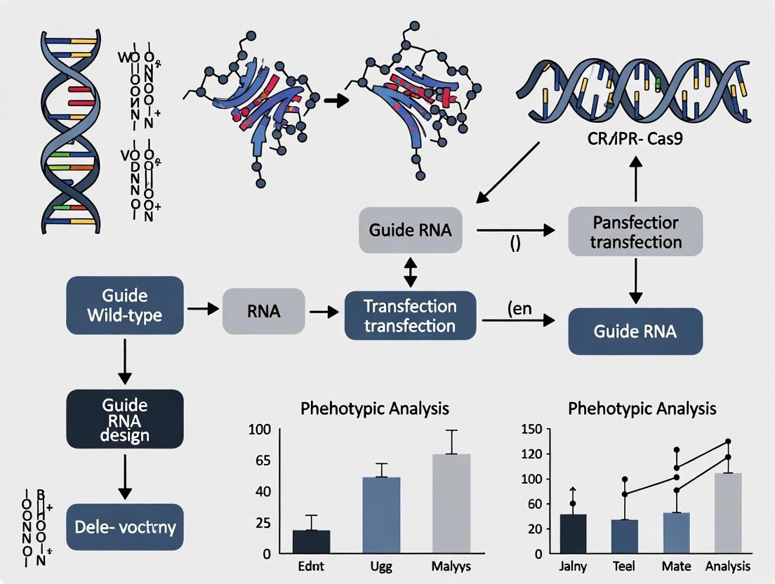
The following diagram outlines the primary workflow for the CRISPR-based functional validation of VUS.
CRISPR-Cas9 VUS Validation Workflow
Key Pathway Analysis for Tumor Suppressor VUS
Many VUS reside in tumor suppressor genes (TSGs) like TP53 and PTEN. Functional validation often hinges on assessing disruption of key signaling pathways. The canonical p53 pathway is a primary assay target.
p53 Pathway Dysfunction by VUS
Detailed Experimental Protocols
Protocol 1: Generation of Isogenic Cell Lines with VUS using CRISPR-Cas9 HDR
Objective: To precisely introduce a specific VUS into a wild-type diploid human cell line (e.g., RPE-1, HAP1) via Homology-Directed Repair (HDR).
Materials: See "Scientist's Toolkit" below.
Procedure:
- Design and Synthesis:
- Design two sgRNAs flanking the target codon (within 10-50 bp) using an online tool (e.g., CHOPCHOP, Benchling). Prioritize on-target efficiency and minimal off-target scores.
- Synthesize a single-stranded oligodeoxynucleotide (ssODN) repair template. Include the VUS nucleotide change(s), flanked by ~60-nt homology arms identical to the non-target strand. Introduce silent restriction site changes or PAM-disrupting mutations in the repair template to prevent re-cutting.
Nucleofection:
- Culture wild-type cells in appropriate medium.
- For one reaction in a 20-µL nucleofection cuvette, complex 1 µg of Cas9 protein (or 1 µg of Cas9 expression plasmid), 150 ng of each sgRNA (as crRNA:tracrRNA duplex or synthetic sgRNA), and 200 ng of ssODN repair template.
- Harvest 1x10⁵ cells, resuspend in nucleofection solution, mix with RNP/ssODN complex, and electroporate using the manufacturer's optimized program.
Clonal Isolation and Genotyping:
- 48 hours post-nucleofection, begin puromycin selection (if using a co-selection marker) for 3-5 days.
- Seed cells at limiting dilution in 96-well plates to obtain single-cell clones. Expand for 2-3 weeks.
- Screen clones by PCR amplification of the target locus and Sanger sequencing. Confirm homozygous or heterozygous introduction of the VUS and the absence of random indel mutations.
Protocol 2: High-Throughput Phenotypic Screening for TSG VUS
Objective: To quantitatively assess the functional impact of TP53 VUS on transcriptional activity and cell growth.
Methods Summary: Isogenic clones are transfected with a p53-responsive luciferase reporter (e.g., PG13-Luc) and treated with a DNA-damaging agent (e.g., 1 µM Nutlin-3a for 24h). Luciferase activity is normalized to a Renilla control.
Table 2: Representative Functional Assay Data for TP53 VUS
| p53 Variant | Transcriptional Activity\n(% of Wild-type) | Growth Suppression in\nSoft Agar Assay | Proposed Classification |
|---|---|---|---|
| Wild-type | 100% ± 8 | Yes (100% inhibition) | Benign (Reference) |
| R175H (Known Pathogenic) | 5% ± 2 | No | Pathogenic |
| VUS: p.R273C | 12% ± 4 | No | Likely Pathogenic |
| VUS: p.P152L | 85% ± 10 | Yes | Likely Benign |
| VUS: p.G245S | 15% ± 5 | No | Likely Pathogenic |
The Scientist's Toolkit: Key Research Reagent Solutions
Table 3: Essential Reagents for CRISPR-Cas9 VUS Validation
| Reagent / Material | Supplier Examples | Function in Protocol |
|---|---|---|
| Recombinant S. pyogenes Cas9 Nuclease | Integrated DNA Technologies (IDT), Thermo Fisher | Catalytic component for inducing double-strand breaks at target genomic locus. |
| Chemically Modified sgRNA (crRNA:tracrRNA) | Synthego, IDT | Guides Cas9 to the specific DNA sequence with high efficiency and stability. |
| Ultramer ssODN Repair Template | IDT, Twist Bioscience | Provides the homology-directed repair template for precise VUS incorporation. |
| Nucleofector System & Kits | Lonza | Enables high-efficiency delivery of RNP complexes into difficult-to-transfect cell lines. |
| p53-Responsive Luciferase Reporter (PG13-Luc) | Addgene (Plasmid #16442) | Measures the transcriptional activity of p53 VUS variants. |
| Isogenic Human Cell Lines (RPE-1, HAP1) | ATCC, Horizon Discovery | Provide a stable, diploid, and genomically characterized background for editing. |
| Next-Generation Sequencing Kit (Illumina MiSeq) | Illumina | For comprehensive off-target analysis and clonal population genotyping. |
Implications for Targeted Therapy
Functional reclassification of VUS directly informs therapeutic eligibility. A VUS reclassified as Likely Pathogenic in BRCA1/2 may qualify a patient for PARP inhibitor therapy. Conversely, a Benign reclassification spares patients from unnecessary prophylactic surgeries. The integration of functional data with clinical databases is critical, as shown in the final logical framework.
From VUS Resolution to Clinical Action
Within the broader thesis on functional validation of Variants of Uncertain Significance (VUS) using CRISPR-Cas9, the "gold standard" for pathogenicity assessment traditionally relies on computational predictors (e.g., CADD, REVEL, PolyPhen-2) and frequency data from population databases (e.g., gnomAD). However, these resources have significant limitations. Computational tools are trained on historical, often biased datasets and may disagree. Population databases, while invaluable, suffer from under-representation of non-European ancestries, leading to misclassification of benign variants unique to certain populations as potentially pathogenic due to their rarity. This necessitates direct functional assays, with CRISPR-Cas9 enabling precise genome editing to model variants in relevant cellular contexts, providing empirical evidence to override or confirm computational predictions.
Quantitative Comparison of Predictor Disagreement Rates
Table 1: Disagreement Rates Among Common Computational Predictors on BRCA1 VUS
| Predictor 1 | Predictor 2 | Concordance Rate (%) | Study/Data Source (Year) |
|---|---|---|---|
| CADD (≥20) | REVEL (≥0.5) | 78% | ClinVar Benchmarking (2023) |
| PolyPhen-2 (Probably Damaging) | SIFT (Deleterious) | 65% | PMC9882345 (2023) |
| MVP (≥0.7) | PrimateAI (Damaging) | 82% | gnomAD v4.0 Analysis (2024) |
| Aggregate Disagreement | (Any two major tools) | ~25-35% | Multiple Cohort Meta-Analysis |
Table 2: Population Allele Frequency Disparities in gnomAD v4.0
| Population Group (gnomAD v4.0) | Mean Exome Sample Count | % of Total Variants Unique to Population | Avg. AF for Unique Variants |
|---|---|---|---|
| European (Non-Finnish) | 72,214 | 12% | 2.1e-05 |
| African/African-American | 24,832 | 41% | 4.8e-05 |
| East Asian | 12,541 | 22% | 3.2e-05 |
| South Asian | 15,820 | 25% | 3.5e-05 |
| All Non-European | ~78,000 | >60% of rare (AF<0.001) variants | 3.8e-05 |
Experimental Protocols for CRISPR-Cas9 Functional Validation
Protocol 3.1: Saturation Genome Editing for High-Throughput VUS Assessment
Objective: To functionally score hundreds to thousands of VUS in a single gene by targeted editing. Materials: Library of sgRNAs covering all possible SNVs in a target exon, HAP1 or RPE1 cells (near-diploid, easy to edit), lentiviral vectors, next-generation sequencing (NGS). Procedure:
- Design & Cloning: Design sgRNA library targeting each nucleotide position in an exon with a "protospacer" sequence. Clone into a lentiviral sgRNA expression vector.
- Viral Production: Produce lentivirus in HEK293T cells using standard packaging plasmids.
- Cell Infection & Selection: Infect HAP1 cells at low MOI (<0.3) and select with puromycin.
- Editing & Outgrowth: Allow 7-14 days for Cas9 editing (via stable expression or delivery) and phenotypic outgrowth.
- NGS & Analysis: Harvest genomic DNA pre- and post-outgrowth. Amplify target region and sequence. Calculate enrichment/depletion scores for each variant based on frequency change. Output: Functional score (e.g., -1 to +1) indicating deleterious, neutral, or beneficial effect.
Protocol 3.2: Isogenic Cell Line Generation via HDR for Specific VUS
Objective: To create and phenotype a single, clinically relevant VUS in a relevant cell model. Materials: CRISPR-Cas9 RNP, ssODN or dsDNA donor template (with VUS and silent restriction site), HEK293 or patient-derived iPSCs, antibiotic selection markers, SURVEYOR or T7E1 assay, flow cytometry antibodies. Procedure:
- Design: Design sgRNA close to the VUS locus. Design a ~100-200nt ssODN donor template containing the VUS, a silent PAM-disrupting mutation, and optionally a restriction site for screening.
- Transfection: Form RNP complexes with Cas9 protein and sgRNA. Co-transfect with donor template into cells via nucleofection.
- Enrichment: Apply antibiotic selection if donor includes a resistance marker.
- Cloning & Screening: Perform limiting dilution to isolate single-cell clones. Screen clones by PCR and restriction digest or Sanger sequencing.
- Functional Assay: Subject validated isogenic clones (VUS vs. WT) to relevant assays (e.g., immunofluorescence for protein localization, Western blot for expression, viability assays for essential genes).
Visualizations
Title: Decision Pathway for VUS Functional Validation
Title: Saturation Genome Editing Workflow
The Scientist's Toolkit: Key Research Reagent Solutions
Table 3: Essential Materials for CRISPR-Cas9 VUS Functional Validation
| Item / Reagent | Supplier Examples | Function in VUS Validation |
|---|---|---|
| High-Fidelity Cas9 | IDT, Thermo Fisher, Synthego | Reduces off-target editing, ensuring isogenic lines differ only at the VUS. |
| Chemically Modified sgRNA | Synthego, IDT (Alt-R) | Enhances stability and editing efficiency, critical for HDR with ssODN donors. |
| Single-Stranded Oligodeoxynucleotide (ssODN) | IDT, Sigma-Aldrich | Serves as the donor template for precise HDR-mediated introduction of the VUS. |
| HAP1 or RPE1 Cells | Horizon Discovery, ATCC | Near-haploid or diploid, genetically stable cell lines ideal for saturation editing screens. |
| Lipofectamine CRISPRMAX | Thermo Fisher | High-efficiency transfection reagent for RNP delivery into various cell types. |
| Nucleofector System & Kits | Lonza | Enables high-efficiency RNP/donor delivery into difficult cells (e.g., iPSCs, primary cells). |
| Surveyor or T7 Endonuclease I | IDT, NEB | Rapid screening tool for identifying mixed indels post-editing before clonal isolation. |
| Next-Generation Sequencing Kit (Illumina) | Illumina, KAPA Biosystems | For deep sequencing of edited pools (saturation editing) or validating clonal lines. |
| CRISPRi/a Inducible Systems | Addgene (Plasmids) | Allows temporal control of gene expression to study dosage-sensitive VUS effects. |
1. Introduction and Application Notes
The interpretation of Variants of Uncertain Significance (VUS) represents a critical bottleneck in genomic medicine and target discovery. Traditional methods, reliant on predictive algorithms or overexpression in non-native cell lines, often fail to capture authentic gene function within a physiologically relevant context. CRISPR-Cas9 technology enables precise genome editing directly in disease-relevant cellular models—such as patient-derived induced pluripotent stem cells (iPSCs) or primary cell lines—allowing for the functional validation of VUS through isogenic comparison. This direct interrogation moves beyond correlation to establish causality, transforming VUS classification and identifying genuine therapeutic targets.
2. Key Quantitative Data Summary
Table 1: Efficiency Metrics for CRISPR-Cas9 Editing in Common Cellular Models for VUS Studies
| Cellular Model | Average Editing Efficiency (Indels) | HDR Efficiency for SNP Knock-in | Clonal Isolation Success Rate | Typical Timeline to Isogenic Line (Weeks) |
|---|---|---|---|---|
| HEK293T | 70-90% | 5-20% | >80% | 3-4 |
| Patient iPSCs | 30-60% | 0.5-5% | 50-70% | 8-12 |
| Primary T-cells | 50-80% | 1-10% | N/A (pooled) | 2-3 (pooled) |
| Cancer Cell Lines | 60-85% | 2-15% | 60-80% | 6-8 |
Table 2: Functional Assay Outcomes from CRISPR-VUS Validation Studies (Representative)
| Gene (VUS) | Cellular Model | Edited Phenotype Assay | Phenotype Result vs. WT | VUS Classification Post-Study |
|---|---|---|---|---|
| BRCA1 (C.150G>T) | iPSC-Derived Mammary Epithelia | RAD51 Foci Formation (DNA Repair) | ~70% Reduction | Pathogenic |
| KCNH2 (G.100A>G) | iPSC-Derived Cardiomyocytes | Action Potential Duration | Prolonged by 25% | Likely Pathogenic |
| MYH7 (E.500C>T) | iPSC-Derived Cardiomyocytes | Contractile Force Measurement | No Significant Change | Benign |
| TP53 (R.175G>A) | Lung Organoid | Apoptosis Post-Chemotherapy | 40% Reduction | Pathogenic |
3. Detailed Experimental Protocols
Protocol 1: CRISPR-Cas9 Mediated Knock-in of a VUS in Human iPSCs to Create an Isogenic Pair
Objective: Introduce a specific single nucleotide VUS into a gene of interest in human iPSCs via Homology-Directed Repair (HDR).
Materials:
- Wild-type human iPSC line.
- Nucleofection system (e.g., Lonza 4D-Nucleofector).
- CRISPR-Cas9 ribonucleoprotein (RNP) complex: Synthetic sgRNA (targeting near VUS locus) and purified Cas9 protein.
- Single-stranded oligodeoxynucleotide (ssODN) HDR template: ~200nt homology arms, incorporating the VUS and silent blocking mutations against re-cutting.
- Clonal isolation reagents: Accutase, 96-well plates, conditioned mTeSR1 with CloneR Supplement.
- Genotyping reagents: Lysis buffer, PCR mix, Sanger sequencing, or T7 Endonuclease I.
Procedure:
- Design & Preparation: Design sgRNA using online tools (e.g., CRISPOR). Order sgRNA and ssODN.
- RNP Complex Formation: Combine 100pmol Cas9 protein and 120pmol sgRNA. Incubate 10min at room temperature.
- iPSC Preparation: Culture iPSCs to ~80% confluency. Dissociate into single cells with Accutase.
- Nucleofection: Pellet 1x10^6 cells. Resuspend in nucleofection solution with RNP complex and 200pmol ssODN. Electroporate using recommended program (e.g., CA-137). Seed into Matrigel-coated wells with recovery medium.
- Clonal Expansion: After 48-72 hours, dissociate and seed at clonal density (500 cells/10cm dish). After 7-10 days, manually pick ~100 colonies into 96-well plates.
- Screening: At near-confluency, split each clone for propagation and lysis. Perform PCR on genomic DNA across the target site. Sequence amplicons to identify correctly edited heterozygous or homozygous VUS clones.
- Validation: Expand positive clones, bank, and confirm pluripotency markers.
Protocol 2: Pooled Functional Screening for VUS Impact on Cell Proliferation
Objective: Rapidly assess the functional impact of multiple VUS in a gene on cellular fitness in a relevant cancer cell line.
Materials:
- Lentiviral sgRNA library targeting exon regions of the gene with protospacer-adjacent motif (PAM) variants to create multiple VUS via NHEJ.
- Target cancer cell line (e.g., HAP1).
- Puromycin.
- Genomic DNA extraction kit.
- PCR primers for NGS library preparation.
- Next-generation sequencer.
Procedure:
- Viral Transduction: Transduce cells at low MOI (0.3) with the lentiviral sgRNA library to ensure single integration. Select with puromycin for 5-7 days.
- Passaging & Harvest: Maintain the pooled population for ~14 population doublings. Harvest genomic DNA at Day 0 (post-selection) and Day 14.
- Amplification & Sequencing: Amplify integrated sgRNA sequences via PCR, adding Illumina adapters and sample indices. Pool and sequence on an NGS platform.
- Analysis: Align reads to the sgRNA library reference. Calculate the depletion or enrichment of each sgRNA from Day 0 to Day 14 using MAGeCK or similar. sgRNAs causing a growth defect (depleted) indicate VUS likely disruptive to essential protein function.
4. Visualizations
Title: CRISPR-Cas9 Isogenic Line Generation for VUS
Title: Functional Assay for a VUS Impact on a Signaling Pathway
5. The Scientist's Toolkit
Table 3: Essential Research Reagent Solutions for CRISPR-Cas9 VUS Studies
| Reagent/Material | Function/Application | Key Consideration |
|---|---|---|
| Recombinant Cas9 Protein | Forms RNP complex for rapid, transient editing. Reduces off-target risk vs. plasmid. | High-purity, endotoxin-free grade is critical for sensitive cells like iPSCs. |
| Chemically Modified sgRNA | Guides Cas9 to target locus. 2'-O-methyl 3' phosphorothioate modifications enhance stability and reduce immune response. | Essential for high efficiency in primary and stem cells. |
| Single-Stranded Oligodeoxynucleotide (ssODN) | Serves as HDR template for precise VUS knock-in. Includes silent mutations to prevent re-cutting. | HPLC-purified; optimal length is 100-200nt total. |
| CloneR Supplement | Enhances survival of single-cell cloned iPSCs post-editing. | Dramatically improves clonal isolation efficiency. |
| Matrigel or Laminin-521 | Defined extracellular matrix for culturing edited iPSCs and derived lineages. | Ensures consistent differentiation into relevant cell models (cardiomyocytes, neurons). |
| T7 Endonuclease I / ICE Analysis Tool | Quickly assesses indel formation efficiency in pooled populations. | Screening tool; does not confirm precise HDR. Sanger sequencing + ICE quantifies mixed outcomes. |
| Validated Antibodies for Disease-Relevant Pathways | Detect functional phenotypes (e.g., phospho-H2AX, cleaved caspase-3, lineage markers). | Enables phenotypic comparison between isogenic wild-type and VUS lines. |
Within the context of a broader thesis on CRISPR-Cas9 functional validation of Variants of Uncertain Significance (VUS), the selection of high-priority candidates for labor-intensive wet-lab studies is a critical bottleneck. This application note outlines a systematic, evidence-based framework for prioritizing VUS by integrating gene function and disease association data, thereby focusing resources on variants most likely to have clinical and biological impact.
Quantitative Prioritization Framework: Scoring Gene & Variant Level Evidence
The following tables summarize key quantitative metrics and data sources for constructing a prioritization pipeline.
Table 1: Gene-Level Prioritization Metrics
| Metric Category | Data Source/Tool | Score Range/Value | Interpretation for Prioritization |
|---|---|---|---|
| Loss-of-Function Intolerance (pLI) | gnomAD | 0 to 1 | pLI ≥ 0.9 indicates high intolerance; variants in such genes are more likely pathogenic. |
| Missense Constraint (Z-score) | gnomAD | ≥ 3.0 | Z-score ≥ 3.0 indicates significant constraint against missense variation. |
| Essential Gene Status | DepMap (CRISPR Screens) | Common Essential (True/False) | Genes essential for cell viability are high-priority targets. |
| Pathway Centrality | STRING DB, KEGG | Degree/Betweenness Centrality | High centrality suggests key regulatory roles; variants may have broad effects. |
| Disease Association Strength | ClinGen, OMIM | Definitive, Strong, Moderate | Genes with definitive/strong disease associations are prioritized. |
Table 2: Variant-Level Evidence Integration
| Evidence Layer | Specific Data | Priority Weight | Example/Threshold |
|---|---|---|---|
| Population Frequency | gnomAD allele frequency | High | AF < 0.00001 (rare) increases priority. |
| Computational Predictors | REVEL, MPC, AlphaMissense | Medium | REVEL score > 0.7 suggests deleteriousness. |
| Conservation | PhyloP, GERP++ | Medium | PhyloP100way > 3.0 indicates high evolutionary conservation. |
| Structural Impact | PredictSNP, FoldX | Medium | Predicted disruption of active site or protein stability. |
| Functional Domain | UniProt, Pfam | High | Variant located in a critical functional domain (e.g., kinase, DNA-binding). |
| Segregation & Case Data | ClinVar submissions | High | Multiple unrelated cases with similar phenotype. |
Experimental Protocol: Tiered CRISPR-Cas9 Validation Workflow
Protocol Title: Functional Assessment of High-Priority VUS using CRISPR-Cas9 Gene Editing in a Disease-Relevant Cell Line.
Objective: To experimentally determine the impact of a prioritized VUS on protein function and cellular phenotype.
Materials: See "The Scientist's Toolkit" below.
Methodology:
Part A: Design and Cloning of Repair Templates
- gRNA Design: Design two CRISPR-Cas9 gRNAs flanking the VUS locus (within 50-100 bp) using an online tool (e.g., CRISPick). Select gRNAs with high on-target and low off-target scores.
- ssODN Repair Template Synthesis: Order a single-stranded oligodeoxynucleotide (ssODN) donor template (~200 nt total). The template must contain:
- Homology arms (≥ 80 nt each) complementary to the genomic sequence flanking the cut sites.
- The desired sequence correction (introducing the VUS or its wild-type counterpart for an isogenic control).
- A silent "blocking" mutation in the PAM sequence of one gRNA to prevent re-cutting.
- Cloning (Optional): For lentiviral delivery, clone the gRNA sequence into a lentiviral CRISPR plasmid (e.g., lentiCRISPRv2).
Part B: Cell Line Engineering
- Cell Culture: Maintain disease-relevant cell line (e.g., HEK293T for ease, or patient-derived iPSCs) under standard conditions.
- Transfection/Nucleofection: Co-deliver the following components:
- RNP Complex: 10 µg Cas9 nuclease protein + 5 µg each of the two synthetic gRNAs (or 2 µg of each plasmid).
- ssODN: 200-500 ng of the purified ssODN repair template. Perform transfection using a high-efficiency method (e.g., nucleofection for primary cells).
- Selection and Clonal Isolation: 48h post-transfection, apply appropriate antibiotic selection (e.g., Puromycin) for 5-7 days if using plasmid systems. For RNP, proceed directly to single-cell sorting via FACS into 96-well plates. Expand clonal populations for 3-4 weeks.
Part C: Genotypic and Phenotypic Validation
- Genotype Screening: Screen clones by genomic PCR of the targeted locus and Sanger sequencing. Identify heterozygous and homozygous edited clones. Confirm the absence of random integration of the ssODN.
- Functional Assays (Tiered Approach):
- Primary Molecular Phenotype: Perform a western blot to assess protein expression level and stability. For kinase variants, conduct an in vitro kinase assay.
- Cellular Phenotype: Based on gene function, execute relevant assays (e.g., cell proliferation assay using Incucyte, apoptosis assay via flow cytometry with Annexin V staining, or immunofluorescence for localization).
- Pathway-Specific Phenotype: If the gene is in a known signaling pathway (see Diagram 1), use a luciferase reporter assay or phospho-specific flow cytometry to measure pathway activity.
Visualizing Key Signaling Pathways & Workflows
Diagram 1 Title: VUS Prioritization Workflow Logic
Diagram 2 Title: VUS Disrupting a Key Signaling Pathway
The Scientist's Toolkit: Research Reagent Solutions
| Reagent/Material | Supplier Examples | Function in Protocol |
|---|---|---|
| Alt-R S.p. Cas9 Nuclease V3 | Integrated DNA Technologies (IDT) | High-purity recombinant Cas9 protein for RNP complex formation; reduces off-target effects. |
| Alt-R CRISPR-Cas9 crRNA & tracrRNA | Integrated DNA Technologies (IDT) | Synthetic gRNA components for RNP assembly; modified for stability and reduced immunogenicity. |
| Neon Transfection System | Thermo Fisher Scientific | Electroporation device for high-efficiency delivery of RNPs into difficult-to-transfect cell lines. |
| CloneSeq Direct Amplicon Sequencing Kit | Swift Biosciences | Enables accurate NGS-based genotyping of edited clones for on-target and off-target analysis. |
| Incucyte Live-Cell Analysis System | Sartorius | Enables real-time, label-free quantification of cellular phenotypes (confluence, death) post-editing. |
| PheNIX (Phenotype-driven NIX) | Broad Institute | Software for computational prioritization of VUS using integrated genomic data. |
| DepMap Portal | Broad Institute | Database for assessing gene essentiality across hundreds of cancer cell lines. |
A Step-by-Step CRISPR-Cas9 Framework for VUS Functional Assays
Application Notes
Within the broader thesis on CRISPR-Cas9 functional validation of Variants of Uncertain Significance (VUS), the generation of isogenic cell lines is a critical step. These lines, which differ only at the specific locus of the VUS, provide the cleanest background for phenotypic comparison. Two primary CRISPR-Cas9 strategies are employed: Non-Homologous End Joining (NHEJ) for knockouts and frameshifts, and Homology-Directed Repair (HDR) for precise nucleotide substitutions or insertions. The choice between HDR and NHEJ depends on the experimental goal—loss-of-function validation (often via NHEJ) versus precise modeling of a patient-specific missense variant (via HDR). This document details the comparative application and protocols for these approaches.
Key Quantitative Comparison of HDR vs. NHEJ Editing Outcomes
Table 1: Comparative Metrics for HDR and NHEJ Editing Strategies in Isogenic Line Creation
| Metric | NHEJ-Based Editing (Knockout) | HDR-Based Editing (Precise VUS) |
|---|---|---|
| Primary Goal | Generate gene knockouts or frameshift mutations. | Introduce precise single-nucleotide variants (SNVs) or small tags. |
| Repair Template Required | No. | Yes (single-stranded oligodeoxynucleotide - ssODN or donor plasmid). |
| Theoretical Efficiency (in dividing cells) | High (can be >80% indel rate). | Low (typically 0.5% - 20% HDR rate). |
| Purity of Desired Edit | Low; heterogeneous mixture of indels. | High; single, defined sequence change. |
| Critical Reagents | Cas9 + sgRNA. | Cas9 + sgRNA + ssODN donor template. |
| Optimal Cell Cycle Phase | All phases, but predominant in G1/S/G2. | Late S and G2 phases. |
| Common Inhibition Strategy | N/A. | Use of NHEJ inhibitors (e.g., Scr7, NU7026) to boost HDR. |
| Primary Screening Method | T7E1 or ICE assay for indels; sequencing for frameshift confirmation. | Allele-specific PCR (AS-PCR) or sequencing of bulk population, then clone screening. |
| Key Challenge | Isolating a clonal line with the desired bi-allelic frameshift. | Overcoming low HDR efficiency and random integration of the donor. |
| Best For Thesis Context | Functional validation of a putative loss-of-function VUS. | Functional comparison of a specific missense VUS against the wild-type allele. |
Protocols
Aim: To model a specific missense VUS in a human diploid cell line (e.g., HEK293T, iPSCs). Materials: See "The Scientist's Toolkit" below. Workflow:
- Design:
- Design sgRNA with Protospacer Adjacent Motif (PAM) site close (<10 bp) to the target VUS locus.
- Design and order a single-stranded oligodeoxynucleotide (ssODN) donor template (~100-200 nt). It should contain the desired VUS flanked by homology arms (50-90 nt each). Incorporate silent mutations in the PAM or seed sequence to prevent re-cutting.
- Nucleofection:
- Culture and harvest 1x10⁶ cells in log growth phase.
- Prepare nucleofection mix: 2 µg Cas9 protein, 1 µg sgRNA (or 2 µg of RNP complex pre-assembled for 10 min at room temp), 2 µl of 100 µM ssODN donor.
- Use appropriate Nucleofector kit and program (e.g., for HEK293T: Kit V, Program B-016).
- Immediately transfer cells to pre-warmed medium.
- Post-Transfection Treatment (Optional):
- To enhance HDR, add NHEJ inhibitor (e.g., 10 µM Scr7) 1 hour post-nucleofection and maintain for 48-72h.
- Screening & Cloning:
- After 72h, harvest a portion for bulk genomic DNA extraction.
- Perform AS-PCR or restriction fragment length polymorphism (RFLP) analysis if a silent marker was introduced.
- Seed the remaining cells at low density (0.5 cells/well) in 96-well plates for clonal expansion.
- After 2-3 weeks, screen individual clones by genomic PCR and Sanger sequencing across the target locus.
- Validation:
- Expand positive clones.
- Validate by full-length gene sequencing to rule off-target edits.
- Bank the final isogenic pair (WT and VUS).
Protocol 2: NHEJ-Mediated Knockout for Loss-of-Function Validation
Aim: To generate a complete gene knockout isogenic line for a putative LoF VUS. Materials: See "The Scientist's Toolkit" below. Workflow:
- Design:
- Design 2-3 sgRNAs targeting early exons of the gene of interest to maximize chances of a frameshift.
- Transfection:
- Transfect cells with plasmid expressing Cas9 and sgRNA(s) (e.g., lentiCRISPRv2) or with pre-assembled RNP complex.
- For RNP, use 2 µg Cas9 protein and 1 µg sgRNA per 1x10⁶ cells.
- Enrichment (Optional):
- If using a plasmid, apply antibiotic selection (e.g., puromycin) 48h post-transfection for 3-5 days.
- Screening & Cloning:
- Harvest bulk cells 72h post-transfection/RNP delivery.
- Extract gDNA and perform T7 Endonuclease I (T7E1) or ICE assay to assess overall indel efficiency.
- Perform limiting dilution cloning as in Protocol 1, step 4.
- Clone Genotyping:
- Screen clonal genomic DNA by PCR and Sanger sequencing.
- Analyze sequencing traces using decomposition tools (e.g., ICE, TIDE) or manually inspect for bi-allelic frameshifts.
- Select a clone with bi-allelic, out-of-frame indels for downstream functional assays.
Diagrams
Title: Decision Workflow for VUS Editing Strategy
Title: CRISPR DSB Repair: NHEJ vs HDR Pathways
The Scientist's Toolkit
Table 2: Essential Reagents for CRISPR Isogenic Line Generation
| Reagent/Material | Function/Description | Example Product/Catalog |
|---|---|---|
| CRISPR-Cas9 Nuclease | Creates a targeted double-strand break (DSB) in the genome. | Recombinant SpCas9 protein (e.g., IDT Alt-R S.p. Cas9 Nuclease V3). |
| sgRNA (crRNA + tracrRNA) | Guides Cas9 to the specific genomic locus via Watson-Crick base pairing. | Synthetic Alt-R crRNA and tracrRNA (IDT); or custom sgRNA plasmid. |
| HDR Donor Template | Provides the homology-directed repair template for precise editing (ssODN for point mutations). | Ultramer DNA Oligo (IDT) or single-stranded donor (ssODN) with homology arms. |
| NHEJ Inhibitor | Small molecule that transiently inhibits the NHEJ pathway to favor HDR. | Scr7 (a DNA Ligase IV inhibitor) or NU7026. |
| Nucleofection System | High-efficiency delivery method for RNP complexes and donor DNA into hard-to-transfect cells. | Lonza Nucleofector 2b/4D with cell line-specific kits. |
| Cloning Medium | Conditioned medium or additive to support single-cell survival and growth during clonal expansion. | CloneR (Stemcell Technologies) or conditioned medium from feeder cells. |
| T7 Endonuclease I | Enzyme used to detect and quantify indel mutations in a mixed population by cleaving mismatched heteroduplex DNA. | NEB T7E1 enzyme (#M0302S). |
| Allele-Specific PCR Primers | Primers designed to selectively amplify the edited allele over the wild-type allele, enabling rapid screening. | Custom primers with the variant at the 3' end. |
| Genomic DNA Extraction Kit | For rapid, high-quality gDNA isolation from bulk or clonal cell populations. | Quick-DNA Miniprep Kit (Zymo Research) or similar. |
Within the broader thesis on CRISPR-Cas9 functional validation of Variants of Uncertain Significance (VUS), precise genome editing is paramount. Introducing a specific VUS into a cellular or animal model requires sgRNAs that drive highly accurate Cas9 cleavage at the intended genomic locus while minimizing off-target activity. This application note details current best practices and protocols for sgRNA design to achieve this critical goal.
Core Principles for On-Target sgRNA Design
The efficacy of a sgRNA is determined by its sequence-specific guide region (typically 20 nucleotides). Key design parameters include:
- GC Content: Optimal between 40-60%.
- Specificity: The 8-12 nucleotides proximal to the PAM (the "seed" region) are critical for specificity.
- PAM Proximity: Design the sgRNA such that the desired edit is within 10-15 base pairs upstream of the PAM site (for SpCas9, PAM = NGG).
- Polymorphism Check: Verify the target sequence is invariant across the genetic background of your model system.
Table 1: Quantitative Parameters for Optimal sgRNA Design
| Parameter | Optimal Range | Rationale |
|---|---|---|
| GC Content | 40% - 60% | Ensures stable DNA-RNA hybridization; extremes reduce efficiency. |
| sgRNA Length | 20 nt (standard) | Standard for SpCas9; truncations (17-18 nt) can increase specificity. |
| Distance from PAM to Edit | < 15 bp | Efficiency of HDR-mediated precise editing drops with distance. |
| Predicted On-Target Score | > 50 (tool-dependent) | Higher scores correlate with increased cleavage activity. |
| MIT Specificity Score | > 50 | Predicts lower off-target potential. |
| Out-of-Frame Score | High (for KO) | Predicts likelihood of frameshift indel for knockout studies. |
Computational Design & Off-Target Prediction Protocol
Objective: To computationally select high-efficiency sgRNAs with minimal predicted off-target sites.
Protocol:
- Input Sequence: Extract 500 bp genomic sequence centered on the VUS locus (GRCh38/hg38). Include the reference and alternate (VUS) allele.
- sgRNA Identification: Use tools like Benchling, CRISPOR, or CHOPCHOP to identify all possible sgRNA sequences with an NGG PAM targeting the region.
- On-Target Scoring: Rank sgRNAs using multiple algorithms (e.g., Doench '16, Moreno-Mateos). Prioritize those with high scores across tools.
- Off-Target Analysis: For the top 5 candidates, run a genome-wide off-target search allowing up to 3-4 mismatches. Tools: Cas-OFFinder, CRISPOR.
- Critical Filter: Discard any sgRNA with a perfect seed match (8-10 bp proximal to PAM) at any other genomic locus.
- Examine off-targets in coding exons, regulatory regions, and known oncogenes/tumor suppressors.
- Final Selection: Choose the sgRNA with the best composite on-target score and the fewest/safest off-target predictions. Design at least two independent sgRNAs per target for validation.
Experimental Validation of Off-Target Effects
Objective: Empirically assess genome-wide off-target cleavage.
Protocol: GUIDE-Seq or CIRCLE-Seq A. GUIDE-Seq Workflow
- Transfection: Co-transfect cells with Cas9:sgRNA RNP complex and the GUIDE-Seq dsODN tag.
- Genomic DNA Extraction: Harvest cells 72 hours post-transfection. Extract high-molecular-weight gDNA.
- Library Preparation:
- Fragment gDNA.
- Repair ends and ligate adaptors with T-overhangs.
- Perform PCR enrichment of tag-integrated fragments.
- Amplify with indexed primers for NGS.
- Sequencing & Analysis: Sequence on an Illumina platform. Align reads to the reference genome and identify GUIDE-Seq tag integration sites as putative off-targets.
Title: GUIDE-Seq Experimental Workflow
B. In Silico vs. Empirical Off-Target Comparison Protocol: Compare computationally predicted off-targets (from Protocol 2) with empirically derived lists from GUIDE-Seq/CIRCLE-Seq.
- Create a Venn diagram of overlapping sites.
- Validate top 5-10 putative off-target sites from each list (computational-only, empirical-only, overlap) by targeted amplicon sequencing (see Protocol 4).
Protocol: Validation of Editing & Specificity
Objective: Quantify on-target editing efficiency and confirm absence of off-target editing at predicted/empirical sites.
Sanger Sequencing & TIDE Analysis (Rapid Screening):
- PCR Amplification: Amplify a 500-800 bp region surrounding the on-target and key off-target loci from transfected cell pool gDNA.
- Sanger Sequencing: Purify PCR products and submit for Sanger sequencing.
- TIDE Analysis: Upload chromatogram files to the TIDE web tool . Deconvolute the complex chromatograms to quantify indel efficiency (%) at the target site.
Targeted Next-Generation Sequencing (Definitive Validation):
- Multiplex PCR: Design amplicons (~250-350 bp) for the on-target and all validated off-target loci. Include sample barcodes.
- Library Prep & Sequencing: Use a high-fidelity polymerase. Pool purified amplicons and sequence on a MiSeq with 2x150 bp reads to achieve >10,000x depth.
- Analysis: Use CRISPResso2 or similar to align reads to reference sequences and quantify the percentage of reads with indels or precise HDR edits.
Title: Targeted NGS Validation Workflow
The Scientist's Toolkit
Table 2: Essential Reagent Solutions for VUS sgRNA Studies
| Item | Function/Application | Example/Note |
|---|---|---|
| High-Fidelity Cas9 Nuclease | Ensures precise DSB formation with minimal off-target strand nicking. | Alt-R S.p. HiFi Cas9 Nuclease V3 |
| Chemically Modified sgRNA | Enhances stability and reduces immunogenicity in cells. | Alt-R CRISPR-Cas9 sgRNA with 2'-O-methyl analogs. |
| HDR Donor Template | Single-stranded oligodeoxynucleotide (ssODN) for precise VUS introduction. | 100-200 nt, homologous arms, phosphorothioate modifications. |
| GUIDE-Seq dsODN Tag | Double-stranded oligo for unbiased, genome-wide off-target profiling. | Tag integration marks DSB sites for NGS detection. |
| CRISPR-Cas9 Transfection Reagent | Efficient delivery of RNP complexes into target cells. | Lipofectamine CRISPRMAX. |
| NGS Library Prep Kit | For targeted amplicon sequencing of on-/off-target loci. | Illumina DNA Prep or NEBNext Ultra II. |
| Genomic DNA Extraction Kit | High-yield, high-purity gDNA for downstream analyses. | DNeasy Blood & Tissue Kit. |
| Off-Target Prediction Tool | Web-based platform for sgRNA design and scoring. | CRISPOR, Benchling. |
Within CRISPR-Cas9 functional validation of Variants of Uncertain Significance (VUS), selecting the appropriate cellular model is a critical determinant of experimental success and biological relevance. This application note provides a comparative analysis of three primary models—induced pluripotent stem cells (iPSCs), immortalized cell lines, and organoids—detailing their specific applications, protocols, and reagent toolkits for VUS research.
Model Comparison & Quantitative Data
Table 1: Comparative Analysis of Model Systems for VUS Functional Validation
| Parameter | Immortalized Cell Lines | Induced Pluripotent Stem Cells (iPSCs) | Organoids |
|---|---|---|---|
| Physiological Relevance | Low; often cancer-derived, genetically aberrant | High; patient-specific, genetically normal background | Very High; recapitulates tissue microanatomy & cell diversity |
| Genetic Manipulation Efficiency | Very High (≥80% editing common) | Moderate to High (30-70% with optimized protocols) | Low to Moderate (10-40%; depends on organoid type) |
| Culture Complexity & Cost | Low; simple, inexpensive media | High; specialized media, growth factors, meticulous culture | Very High; Matrigel, advanced media, growth factor cocktails |
| Throughput / Scalability | Very High (ideal for HTS) | Moderate (improving with automation) | Low (complex, labor-intensive) |
| Multicellular Context | No (mostly monoculture) | No (but can differentiate into many types) | Yes (native tissue-like cell composition) |
| Typical Timeline for CRISPR Experiment (from design to assay) | 3-4 weeks | 8-12 weeks (includes clone isolation, differentiation) | 10-16 weeks (includes generation, editing, expansion) |
| Key Application in VUS Research | Initial high-throughput screening, pathway studies | Disease modeling in relevant cell type, patient-specific effects | Studying variant effects in tissue architecture & cell-cell interactions |
Table 2: Decision Matrix for Model Selection Based on Research Question
| Primary Research Goal | Recommended Primary Model | Key Rationale |
|---|---|---|
| High-throughput drug screening on a genetic variant | Immortalized Line (if a relevant line exists) | Speed, cost, and scalability are paramount. |
| Studying a cardiac ion channel VUS in a patient's background | iPSC-derived cardiomyocytes | Patient-specific genetic context and physiologically relevant cell type. |
| Understanding how a VUS affects intestinal barrier formation | Intestinal Organoids | Complex 3D structure and multiple interacting cell types are essential. |
| Rapid analysis of variant effect on a signaling pathway | Immortalized Line | Allows clean, fast dissection in a controlled, uniform system. |
Detailed Protocols for CRISPR-Cas9 Functional Validation
Protocol 3.1: CRISPR-Cas9 Editing of an Immortalized HEK293T Cell Line for VUS Analysis
Objective: Introduce a single-nucleotide VUS via HDR in HEK293T cells. Materials: See "Research Reagent Solutions" (Section 5). Workflow:
- Design & Synthesis: Design 2-3 sgRNAs targeting near the VUS locus using online tools (e.g., CRISPOR). Order ssODN donor template with the VUS and a silent restriction site for screening.
- Transfection: Seed 2e5 HEK293T cells/well in a 24-well plate. At 70% confluency, co-transfect with 500 ng Cas9 expression plasmid, 250 ng sgRNA plasmid, and 100 pmol of ssODN donor using a standard PEI or lipofectamine protocol.
- Enrichment & Cloning: 48h post-transfection, apply appropriate antibiotic selection (e.g., puromycin for 72h) if the CRISPR plasmid contains a selectable marker. Surviving cells are serially diluted in a 96-well plate to obtain single-cell clones.
- Genotyping: After 2-3 weeks, expand clones. Extract genomic DNA and perform PCR across the target locus. Analyze by Sanger sequencing and/or restriction digest (using the silent site introduced).
- Functional Assay: Culture validated isogenic clones (VUS vs. WT) and perform relevant assays (e.g., Western blot, reporter assay, viability assay).
Diagram Title: CRISPR Workflow for Immortalized Cell Lines
Protocol 3.2: Generation of an Isogenic iPSC Line Harboring a VUS
Objective: Create a genetically matched pair of WT and VUS iPSC lines via CRISPR-Cas9 editing. Materials: See "Research Reagent Solutions" (Section 5). Critical Note: Use integration-deficient Sendai virus or episomal vectors for reprogramming to ensure footprint-free iPSCs. Workflow:
- iPSC Culture: Maintain feeder-free iPSCs in mTeSR Plus on Geltrex-coated plates. Passage as small clumps using EDTA.
- Electroporation: Harvest 1e6 iPSCs as single cells using Accutase. Resuspend in P3 buffer with 5 μg Cas9 protein, 100 pmol synthetic sgRNA, and 200 pmol ssODN donor. Electroporate using a 4D-Nucleofector (program CB-150). Immediately transfer to pre-warmed medium with 10μM ROCK inhibitor.
- Recovery & Single-Cell Sorting: After 72h, dissociate to single cells and sort single, live cells into 96-well plates pre-coated with Geltrex and containing mTeSR Plus with ROCK inhibitor, using a FACS sorter.
- Clone Expansion & Genotyping: Expand clones for 3-4 weeks, with careful medium changes. Screen clones via PCR and sequencing as in Protocol 3.1.
- Pluripotency & Karyotype Check: Confirm pluripotency markers (OCT4, NANOG) via immunofluorescence and perform karyotype analysis on edited clones before banking.
- Directed Differentiation: Differentiate isogenic clones into the relevant disease cell type (e.g., cortical neurons, cardiomyocytes) using established protocols for functional assays.
Diagram Title: Isogenic VUS iPSC Line Generation Workflow
Signaling Pathway Contextualization
A common endpoint in VUS validation is assessing disruption of key pathways. For example, a VUS in a tumor suppressor gene (e.g., PTEN) may hyperactivate the PI3K/AKT pathway.
Diagram Title: PI3K/AKT Pathway Disruption by a PTEN VUS
The Scientist's Toolkit: Research Reagent Solutions
Table 3: Essential Reagents for CRISPR-based VUS Modeling
| Reagent / Material | Function in VUS Research | Example Product/Catalog |
|---|---|---|
| Synthetic sgRNA (Alt-R) | High-fidelity guide RNA for RNP complex formation; reduces off-target effects. | IDT Alt-R CRISPR-Cas9 sgRNA |
| Recombinant Cas9 Protein | For RNP delivery; faster action, lower off-targets than plasmid DNA. | IDT Alt-R S.p. Cas9 Nuclease V3 |
| Single-Stranded Oligo Donor (ssODN) | Template for precise HDR-mediated introduction of the VUS. | IDT Ultramer DNA Oligo |
| CloneR or ROCK Inhibitor (Y-27632) | Enhances survival of single iPSCs post-editing and during cloning. | STEMCELL Technologies CloneR; Tocris Y-27632 |
| Geltrex or Matrigel | Basement membrane matrix for adherent culture of iPSCs and organoids. | Thermo Fisher Geltrex LDEV-Free; Corning Matrigel |
| mTeSR Plus Medium | Feeder-free, defined medium for maintenance of human pluripotent stem cells. | STEMCELL Technologies mTeSR Plus |
| 4D-Nucleofector System & Kit | Electroporation platform for high-efficiency delivery of RNPs into iPSCs. | Lonza 4D-Nucleofector X Kit (P3 Primary Cell Kit) |
| FACS Sorter with 96-well plate deposition | For isolation of single-cell clones into plate format with high viability. | BD FACS Aria III, Sony SH800 |
| KaryoStat+ Assay | High-resolution CNV detection to confirm genomic integrity of edited clones. | Thermo Fisher KaryoStat+ GeneChip |
Within CRISPR-Cas9 functional validation of Variants of Uncertain Significance (VUS), efficient and cell-type-specific delivery of genetic cargo—including Cas9 nucleases, guide RNAs, and donor templates—is the critical bottleneck. The choice of delivery method directly impacts editing efficiency, cytotoxicity, and experimental outcome reliability. This Application Note provides optimized protocols and comparative data for three core delivery modalities, framed specifically for functional genomics workflows in VUS research.
The optimal method is contingent upon cell type (primary, immortalized, stem cell), desired outcome (knockout, knock-in), and experimental scale. The table below synthesizes current performance data for common mammalian cell models in VUS studies.
Table 1: Performance Metrics of Delivery Methods for Common Cell Types in CRISPR Validation
| Cell Type | Recommended Method | Average Efficiency (Editing%) | Key Advantage | Primary Limitation | Optimal Use Case in VUS Research |
|---|---|---|---|---|---|
| HEK293T | Lipofection (LNP) | 85-95% | High efficiency, ease of use | Cytotoxicity at high dose | Rapid gRNA validation, Cas9/sgRNA co-delivery |
| Jurkat T-Cells | Electroporation (Neon) | 70-85% | High efficiency in "hard-to-transfect" cells | High cell mortality | Immune gene variant studies |
| HAP1 | Viral Transduction (Lentivirus) | >90% (stable) | Near-complete population transduction | Lentiviral size constraints | Creating isogenic cell lines for functional assays |
| iPSCs | Electroporation (4D-Nucleofector) | 60-75% | Maintains pluripotency post-editing | Low cell survival | Creating disease models with patient-derived variants |
| Primary Fibroblasts | Viral Transduction (AAV6) | 40-60% (HDR) | Low immunogenicity, high HDR rate | Cargo size limit (~4.7 kb) | Precise knock-in of VUS for allele correction |
| HepG2 | Lipofection (Polymer-based) | 50-70% | Low serum sensitivity | Variable batch-to-batch | Metabolic pathway variant analysis |
Detailed Experimental Protocols
Protocol 3.1: Lipid Nanoparticle (LNP) Transfection for Adherent Cells (e.g., HEK293T)
Objective: Co-deliver Cas9 mRNA and sgRNA for rapid knockout generation.
Research Reagent Solutions:
| Reagent/Material | Function/Explanation |
|---|---|
| Lipofectamine CRISPRMAX Cas9 Transfection Reagent | Lipid formulation optimized for ribonucleoprotein (RNP) or mRNA/gRNA delivery. |
| Cas9 mRNA (truncated poly-A) | High-stability mRNA for robust Cas9 expression with lower immunogenicity than plasmid DNA. |
| Chemically modified sgRNA | Modified bases enhance stability and reduce off-target effects. |
| Opti-MEM I Reduced Serum Medium | Serum-free medium for complex formation, minimizing lipid-serum interactions. |
Procedure:
- Day 0: Seed HEK293T cells in a 24-well plate at 1.5 x 10^5 cells/well in complete growth medium. Incubate to reach ~80% confluency at time of transfection.
- Day 1 – Complex Formation: For each well, prepare two tubes:
- Tube A (Diluted RNA): Dilute 500 ng Cas9 mRNA and 250 ng sgRNA in 25 µL Opti-MEM.
- Tube B (Diluted Lipid): Dilute 1.5 µL CRISPRMAX reagent in 25 µL Opti-MEM. Incubate for 5 minutes at RT.
- Combine the contents of Tube A and Tube B. Mix gently and incubate for 15-20 minutes at RT to form RNA-lipid complexes.
- Transfection: Add the 50 µL complex dropwise to the cell well. Gently rock the plate.
- Post-Transfection: Replace medium with fresh complete medium 6-8 hours post-transfection.
- Analysis: Harvest cells 48-72 hours post-transfection for genomic DNA extraction and T7E1 or NGS analysis of editing efficiency.
Protocol 3.2: Nucleofector Electroporation for Suspension Cells (e.g., Jurkat)
Objective: Deliver pre-assembled Cas9 RNP for high-efficiency editing with minimal off-target effects.
Research Reagent Solutions:
| Reagent/Material | Function/Explanation |
|---|---|
| Neon Transfection System 10µL Kit (Invitrogen) | Electroporation tips and buffers optimized for high viability in sensitive cells. |
| Alt-R S.p. Cas9 Nuclease V3 | High-fidelity Cas9 protein for RNP formation. |
| Alt-R CRISPR-Cas9 sgRNA (chemically modified) | Synthetic sgRNA, ready for complexing with Cas9 protein. |
| Recombinant Albumin | Added to recovery medium to improve post-electroporation cell health. |
Procedure:
- RNP Complex Assembly: For one reaction, combine 3 µg (6 pmol) Alt-R Cas9 protein and 1.5 µg (12 pmol) sgRNA in a sterile microcentrifuge tube. Add Resuspension Buffer R to bring total volume to 10 µL. Incubate at room temperature for 20 minutes.
- Cell Preparation: Harvest 2 x 10^5 Jurkat cells in log growth phase. Centrifuge at 300 x g for 5 minutes. Aspirate supernatant completely.
- Electroporation Setup: Resuspend cell pellet in 10 µL Resuspension Buffer R. Combine with the 10 µL pre-assembled RNP complex. Mix gently. Draw the entire 20 µL mixture into a Neon 10 µL Tip.
- Electroporation: Electroporate using the pre-optimized program for Jurkat cells: 1400V, 10ms, 3 pulses. Immediately transfer the electroporated cells into 500 µL pre-warmed complete medium supplemented with 0.1% recombinant albumin in a 24-well plate.
- Recovery & Analysis: Incubate cells at 37°C, 5% CO2. Expand cells as needed. Analyze editing efficiency by flow cytometry (if co-transfected with a fluorescent marker) or genomic cleavage assay 72-96 hours post-electroporation.
Protocol 3.3: Lentiviral Transduction for Stable Cell Line Generation (e.g., HAP1)
Objective: Generate a polyclonal or monoclonal cell population stably expressing Cas9 for sequential VUS targeting.
Research Reagent Solutions:
| Reagent/Material | Function/Explanation |
|---|---|
| LentiCas9-Blast (or -GFP) plasmid (Addgene) | Second-generation lentiviral vector for constitutive Cas9 expression. |
| psPAX2 (packaging plasmid) | Provides gag, pol, rev viral proteins. |
| pMD2.G (envelope plasmid) | Provides VSV-G glycoprotein for broad tropism. |
| Polybrene (Hexadimethrine bromide) | Cationic polymer that enhances viral attachment to target cells. |
| Lenti-X Concentrator (Takara) | PEG-based solution for gentle, high-efficiency viral precipitation. |
Procedure:
- Day 0 – Virus Production: Seed HEK293T cells in a 10 cm dish to reach 70-80% confluency the next day in DMEM + 10% FBS (no antibiotics).
- Day 1 – Transfection: Co-transfect cells with 10 µg LentiCas9-Blast, 7.5 µg psPAX2, and 2.5 µg pMD2.G using a calcium phosphate or PEI-based method.
- Day 2 & 3 – Harvest: Replace medium 8 hours post-transfection with 10 mL fresh complete medium. Collect viral supernatant at 48 and 72 hours post-transfection. Pool supernatants, filter through a 0.45 µm PES filter.
- Concentration (Optional): Mix filtered supernatant with Lenti-X Concentrator (3:1 ratio). Incubate overnight at 4°C. Centrifuge at 1500 x g for 45 minutes at 4°C. Resuspend pellet in 1/100th original volume in cold PBS.
- Day 4 – Transduction: Seed 2 x 10^5 HAP1 cells per well in a 12-well plate. Add concentrated virus (MOI ~5) and 8 µg/mL Polybrene. Spinoculate by centrifuging the plate at 800 x g for 30 minutes at 32°C, then return to incubator.
- Day 5 – Selection: Replace medium 24 hours post-transduction. Begin selection with 5-10 µg/mL Blasticidin (or sort for GFP+) 48 hours post-transduction. Maintain selection for 7 days to generate a stable polyclonal Cas9-expressing cell line.
Visualizing the Decision Workflow and Mechanism
Diagram 1: CRISPR Delivery Method Selection Workflow
Diagram 2: Key Intracellular Barriers to Genetic Cargo Delivery
Application Notes
Within the functional validation of Variants of Uncertain Significance (VUS) using CRISPR-Cas9, establishing robust phenotypic readouts is paramount. The following notes detail the application of key assays to elucidate the functional consequence of a VUS, linking genetic perturbation to observable cellular behavior.
Reporter Assays for Signaling Pathway Activity
CRISPR-engineered isogenic cell lines (wild-type vs. VUS) are used to probe specific pathway activities. Luciferase-based reporters (e.g., NF-κB, p53, Wnt/β-catenin) provide a quantitative, high-throughput measure of transcriptional output changes induced by the VUS. This is critical for VUS in signaling hubs or transcription factors.
Cell Viability and Proliferation Profiling
Determining if a VUS confers a growth advantage or induces cytotoxicity is a fundamental phenotypic readout. Long-term proliferation assays and real-time cell analysis can reveal subtle fitness differences. For suspected oncogenic VUS, sensitivity to targeted therapeutics can be co-assayed.
Cell Migration and Invasion Assays
For VUS in genes implicated in metastasis or cell adhesion, functional validation requires assessment of motile phenotypes. Transwell and wound healing assays quantify migration and invasion potential in isogenic backgrounds, connecting genotype to metastatic propensity.
Molecular Profiling for Deep Phenotyping
Bulk or single-cell RNA sequencing, proteomic analyses, and phospho-flow cytometry provide multi-parametric signatures of VUS impact. This unbiased approach can uncover novel affected pathways and generate hypotheses for more focused assays.
Table 1: Summary of Key Phenotypic Readouts for VUS Validation
| Assay Category | Example Assays | Key Measured Parameters | Typical Timeline | Throughput |
|---|---|---|---|---|
| Reporter Assay | Dual-Luciferase, SEAP | Luminescence (RLU), Fluorescence | 24-72 hours | High (96/384-well) |
| Cell Viability | CTG, MTS, Real-time Cell Analysis | IC50, Doubling Time, Confluence | 72-144 hours | Medium-High |
| Migration | Transwell (Boyden Chamber), Wound Healing | Migrated Cell Count, Wound Closure % | 6-48 hours | Medium |
| Molecular Profiling | RNA-seq, LC-MS/MS Proteomics | Differential Expression, Pathway Enrichment | Days-Weeks | Low |
Detailed Protocols
Protocol 1: Dual-Luciferase Reporter Assay for Pathway Activity
Objective: Quantify the impact of a specific VUS on a defined signaling pathway (e.g., NF-κB) in CRISPR-corrected vs. uncorrected cells.
Materials:
- Isogenic cell pairs (VUS and CRISPR-corrected control).
- Pathway-specific reporter plasmid (Firefly luciferase under responsive element).
- Renilla luciferase control plasmid (pRL-TK or similar) for normalization.
- Transfection reagent (e.g., lipofectamine 3000).
- Dual-Luciferase Reporter Assay System.
- White, flat-bottom 96-well plates.
- Luminometer with injectors.
Procedure:
- Day 1: Seed 2.5 x 10⁴ cells per well in 100 µL complete medium.
- Day 2: Co-transfect cells with 100 ng pathway reporter plasmid and 10 ng Renilla control plasmid per well using manufacturer's protocol.
- Day 3: Apply pathway-specific agonist/inhibitor or vehicle control for 6-24 hours as optimized.
- Lysis & Measurement: Aspirate medium. Add 30 µL 1X Passive Lysis Buffer, shake 15 min. Program luminometer to inject 50 µL Luciferase Assay Reagent II, measure Firefly luminescence (2-10 sec), then inject 50 µL Stop & Glo Reagent, and measure Renilla luminescence.
- Analysis: Calculate the ratio of Firefly/Renilla luminescence for each well. Normalize the mean ratio of treated VUS cells to treated control cells to determine fold-change in pathway activity.
Protocol 2: Real-Time Cell Proliferation & Viability Assay (Incucyte or Equivalent)
Objective: Continuously monitor growth kinetics and viability of isogenic cell lines to detect fitness defects.
Materials:
- Isogenic cell pairs.
- Real-time cell analysis instrument.
- Specialized 96-well tissue culture plates.
- Labeling dye (e.g., Cytolight Green for nuclei).
Procedure:
- Day 1: Seed cells at optimized density (e.g., 2-5 x 10³ cells/well) in 100 µL medium. Include triplicates for each genotype/condition.
- Setup: Add 25 µL of 5x Cytolight Green reagent directly to each well. Gently swirl plate.
- Acquisition: Place plate in imaging system inside incubator. Program to scan every 2-6 hours. Acquire phase and green fluorescence (ex/em ~450/525 nm) images from 4-9 sites per well.
- Analysis: Use integrated software to segment and count labeled nuclei per image over time. Generate growth curves (cell count vs. time). Calculate population doubling times from the exponential growth phase.
Protocol 3: Transwell Migration Assay
Objective: Assess the migratory capacity of cells harboring a VUS compared to CRISPR-corrected controls.
Materials:
- Isogenic cell pairs, serum-starved.
- 24-well Transwell inserts with 8.0 µm pore polycarbonate membrane.
- Serum-free medium and medium with chemoattractant (e.g., 10% FBS).
- Crystal violet stain (0.5% w/v in 25% methanol) or Calcein-AM.
- 4% Paraformaldehyde (PFA).
- Cotton swabs.
Procedure:
- Preparation: Add 500-750 µL of complete medium (chemoattractant) to the lower chamber of a 24-well plate.
- Seeding: Gently place the Transwell insert. Seed 5-10 x 10⁴ serum-starved cells in 200-300 µL serum-free medium into the upper chamber. Triplicate per genotype.
- Incubation: Incubate at 37°C, 5% CO₂ for 6-24 hours (optimize to avoid saturation).
- Fixation & Staining: Remove insert, carefully swab the interior (top) membrane with a cotton bud to remove non-migratory cells. Rinse with PBS. Fix cells on bottom side in 4% PFA for 10 min. Stain with 0.5% crystal violet for 15 min. Rinse thoroughly in water. Air dry.
- Quantification: Image membranes under a brightfield microscope (10x objective, 5 random fields/membrane). Count migrated cells manually or using ImageJ software. Normalize mean VUS cell count to control.
Protocol 4: Molecular Profiling via Bulk RNA-Sequencing
Objective: Obtain transcriptome-wide differential expression profile induced by the VUS.
Materials:
- Isogenic cell pairs, biological triplicates.
- TRIzol or equivalent RNA isolation reagent.
- DNase I, RNase-free.
- Stranded mRNA library prep kit.
- Bioanalyzer/TapeStation.
- Next-generation sequencing platform.
Procedure:
- RNA Isolation: Harvest cells directly in TRIzol. Extract total RNA following manufacturer's protocol. Include DNase I treatment. Assess RNA integrity (RIN > 8.5) and quantity.
- Library Preparation: Using 500 ng-1 µg total RNA, perform poly-A selection, fragmentation, cDNA synthesis, adapter ligation, and PCR amplification using a stranded mRNA kit.
- QC & Pooling: Validate library size (~350 bp insert) and concentration via Bioanalyzer. Pool libraries equimolarly.
- Sequencing: Sequence on an Illumina NovaSeq 6000 (or equivalent) to a minimum depth of 30 million 150 bp paired-end reads per sample.
- Bioinformatic Analysis: Align reads to reference genome (STAR). Generate count matrices (featureCounts). Perform differential expression analysis (DESeq2). Conduct pathway enrichment analysis (GSEA, Enrichr).
The Scientist's Toolkit: Research Reagent Solutions
Table 2: Essential Materials for Phenotypic Screening in VUS Validation
| Reagent/Material | Supplier Examples | Primary Function in VUS Research |
|---|---|---|
| CRISPR-Cas9 Nucleofection Kit | Lonza (4D-Nucleofector), Thermo Fisher (Neon) | Efficient delivery of RNP complexes for precise genome editing to create isogenic pairs. |
| Dual-Luciferase Reporter Assay System | Promega | Quantifies transcriptional activity from specific pathway reporters with internal normalization. |
| Real-Time Cell Analysis Instrument | Sartorius (Incucyte), ACEA (xCELLigence) | Enables label-free, kinetic monitoring of cell proliferation, viability, and confluence. |
| Matrigel Basement Membrane Matrix | Corning | Used to coat Transwell inserts for invasion assays, modeling extracellular matrix penetration. |
| Cell Viability Assay (CTG) | Promega (CellTiter-Glo) | Luminescent ATP quantitation for high-throughput cytotoxicity and proliferation screens. |
| Phosflow Antibodies & Fixation Buffer | BD Biosciences | Enables detection of phosphorylation-dependent signaling events via flow cytometry. |
| Stranded mRNA Library Prep Kit | Illumina (TruSeq), NEB (NEBNext) | Prepares high-quality RNA-seq libraries for transcriptomic profiling of VUS impact. |
| Single-Cell RNA-seq Kit | 10x Genomics (Chromium) | Enables deconvolution of heterogeneous cellular responses to a VUS at single-cell resolution. |
Diagrams
Diagram Title: Workflow for CRISPR VUS Validation with Phenotypic Readouts
Diagram Title: Mechanism of a Pathway Reporter Assay for VUS Impact
Diagram Title: Transwell Migration Assay Protocol Steps
Application Notes
Within CRISPR-Cas9 functional validation of Variants of Uncertain Significance (VUS), the implementation of essential controls is the cornerstone of rigorous experimental design and data interpretation. These controls establish the baseline and dynamic range necessary to assign functional consequence to a VUS. The wild-type control defines normal gene function, the null/knockout control establishes the complete loss-of-function phenotype, and the known pathogenic variant control provides a benchmark for a clinically relevant dysfunctional state. Together, they enable the calibration of assays—from cellular proliferation and viability to downstream signaling output and gene expression profiles—allowing researchers to confidently categorize a VUS as benign, loss-of-function, gain-of-function, or dominant-negative by direct comparison.
Protocols
Protocol 1: Generation of Isogenic Cell Line Controls via CRISPR-Cas9
Objective: To create a matched set of wild-type, null (knockout), and known pathogenic variant cell lines from a parental cell line.
- Design gRNAs: Design two gRNAs for each target: one for the wild-type allele (to introduce a synonymous or benign edit via HDR) and one to create a frameshift indel via NHEJ (for null). For the pathogenic variant, design a gRNA near the known mutation site and a single-stranded DNA donor template (ssODN) containing the variant.
- Nucleofection: Co-transfect the parental cell line (e.g., HEK293T, HAP1) with the Cas9 expression plasmid (or RNP complex) and the respective gRNA(s) and donor DNA using an appropriate nucleofection system.
- Clonal Isolation: 48-72 hours post-transfection, single cells are sorted by FACS into 96-well plates. Allow clonal outgrowth for 2-3 weeks.
- Genotype Validation: Extract genomic DNA from clones. Perform PCR amplification of the target locus and validate by Sanger sequencing (for precise edits) or T7 Endonuclease I assay/TIDE analysis (for indels). Confirm the absence of off-target edits at top-predicted sites.
- Expand and Bank: Expand validated clonal lines, cryopreserve aliquots, and perform mycoplasma testing.
Protocol 2: Functional Validation via Cell Viability/Proliferation Assay
Objective: To quantitatively compare the growth phenotype of VUS lines against essential controls.
- Seed Cells: Seed triplicate wells of a 96-well plate with 1,000-2,000 cells per well for each cell line: Parental, Wild-Type Edited, Null (KO), Known Pathogenic, and VUS.
- Incubate & Measure: Incubate plates at 37°C, 5% CO₂. At 0, 24, 48, 72, and 96 hours, add a luminescent (e.g., CellTiter-Glo) or colorimetric (e.g., MTT) viability reagent according to the manufacturer's instructions.
- Data Analysis: Plot relative luminescence/absorbance vs. time. Normalize all readings to the 0-hour time point for each line. Calculate area under the curve (AUC) for growth. Statistical significance is determined via one-way ANOVA with post-hoc tests comparing VUS to each control.
Protocol 3: Signaling Pathway Output Assay (e.g., Phospho-protein Western Blot)
Objective: To assess the impact of a VUS on a key downstream signaling pathway.
- Stimulate and Lyse: Serum-starve all isogenic cell lines for 12-16 hours. Stimulate with the appropriate ligand (e.g., EGF for EGFR pathways) for a defined time course (e.g., 0, 5, 15, 60 min). Immediately lyse cells in RIPA buffer with protease and phosphatase inhibitors.
- Western Blot: Resolve equal protein amounts by SDS-PAGE. Transfer to PVDF membrane. Probe with primary antibodies against: the total protein of interest, the phosphorylated/active form of the protein, and a loading control (e.g., GAPDH, β-Actin).
- Quantification: Image using a chemiluminescent imager. Quantify band intensities. For each time point, calculate the ratio of phospho-protein/total protein, then normalize this ratio to the unstimulated wild-type control (set to 1.0).
Data Presentation
Table 1: Quantitative Summary of Functional Assay Results for Isogenic TP53 VUS Models
| Cell Line Genotype | Proliferation AUC (72h) (Mean ± SD) | p53 Protein Level (Relative to WT) | p21 Transcript Induction (Fold over Baseline) | Cisplatin IC₅₀ (μM) |
|---|---|---|---|---|
| Wild-Type (Edited) | 1.00 ± 0.05 | 1.00 | 10.2 ± 1.5 | 2.1 ± 0.3 |
| Null (Knockout) | 1.52 ± 0.08* | 0.05* | 1.1 ± 0.3* | 12.5 ± 1.8* |
| Known Pathogenic (R175H) | 1.48 ± 0.07* | 1.95* | 2.3 ± 0.6* | 11.8 ± 2.1* |
| VUS A (R267Q) | 1.04 ± 0.06 | 0.92 | 9.8 ± 2.0 | 2.4 ± 0.4 |
| VUS B (P152L) | 1.41 ± 0.09* | 1.12 | 3.1 ± 0.8* | 8.9 ± 1.2* |
*p < 0.001 vs. Wild-Type control (One-way ANOVA with Dunnett's test).*
Visualizations
Title: CRISPR-Cas9 Workflow for Isogenic Control & VUS Cell Line Generation
Title: Logical Framework for Control-Based VUS Interpretation
The Scientist's Toolkit
Table 2: Essential Research Reagent Solutions for CRISPR Validation
| Item | Function & Application |
|---|---|
| RNP Complex (Cas9 + gRNA) | The direct delivery of pre-formed ribonucleoprotein complexes increases editing efficiency and reduces off-target effects and plasmid persistence compared to plasmid-based delivery. |
| HDR Donor Template (ssODN) | A single-stranded oligodeoxynucleotide template used to guide homology-directed repair for precise introduction of point mutations (pathogenic or VUS) or synonymous edits (wild-type control). |
| Clonal Isolation Matrix | A low-attachment, cell culture-ready hydrogel or specialized media that supports the survival and outgrowth of single cells following FACS sorting, critical for generating pure isogenic lines. |
| T7 Endonuclease I / ICE Analysis | Enzymatic and in silico tools for detecting and quantifying the presence of insertion/deletion (indel) mutations at the target site, essential for validating null/knockout clones. |
| Cell Viability Assay (Luminescent) | A homogeneous, ATP-quantifying assay (e.g., CellTiter-Glo) providing a sensitive and high-throughput readout of metabolically active cells for proliferation and drug response studies. |
| Phospho-Specific Antibodies | Antibodies that specifically recognize the phosphorylated (activated) state of signaling proteins, enabling the measurement of pathway activity downstream of the gene of interest. |
Optimizing CRISPR-Cas9 VUS Validation: Troubleshooting Common Pitfalls
Within the functional validation of Variants of Uncertain Significance (VUS) using CRISPR-Cas9, precise genome editing via Homology-Directed Repair (HDR) is paramount. This process enables the introduction of specific VUS into model cell lines for phenotypic characterization. However, the inherently low efficiency of HDR relative to the error-prone Non-Homologous End Joining (NHEJ) pathway remains a critical bottleneck, leading to excessive screening workload and delayed timelines. These application notes provide current, evidence-based protocols and reagents to enhance HDR efficiency and streamline the isolation of correctly edited monoclonal cell lines.
Core Strategies to Modulate DNA Repair Pathways
The primary strategy to improve HDR outcomes involves the temporal or pharmacological modulation of key DNA repair pathway components to favor HDR over NHEJ.
Signaling Pathway: Key Nodes for HDR Enhancement
The following diagram illustrates the CRISPR-Cas9-induced double-strand break (DSB) repair pathway decision node and points of intervention.
Title: CRISPR DSB Repair Pathway and Intervention Points
Quantitative Comparison of HDR-Enhancing Agents
Recent studies validate small molecules and synchronization strategies for improving HDR outcomes, summarized in the table below.
Table 1: Efficacy of HDR-Enhancing Treatments
| Intervention | Proposed Mechanism | Reported HDR Increase (vs. Control) | Key Considerations |
|---|---|---|---|
| SCR7 | Inhibits DNA Ligase IV (NHEJ) | 2- to 9-fold | Cytotoxicity observed at higher doses; specificity debated. |
| NU7026 | Inhibits DNA-PKcs (NHEJ) | 3- to 5-fold | Potent NHEJ block; can increase off-target effects. |
| RS-1 | Stabilizes RAD51 filaments (HDR) | 2- to 4-fold | Can be cell-type specific; may require optimization. |
| L755507 | β3-AR agonist, enhances HDR | ~3-fold | Novel pathway; less validated across diverse cell lines. |
| Cell Cycle Synchronization (S/G2 phase) | Restricts editing to HDR-competent phases | 3- to 8-fold | Requires reversible arrest agents (e.g., thymidine, nocodazole). |
Detailed Experimental Protocols
Protocol 1: Combined Pharmacological and Cell Cycle Synchronization for HDR
This protocol maximizes HDR efficiency by treating cells in the S/G2 phase with a cocktail of NHEJ inhibitor and HDR enhancer.
Materials: See "The Scientist's Toolkit" below. Procedure:
- Seed and Synchronize: Plate HEK293T or target cells to reach 40-50% confluency in 24 hours. Treat with 2 mM thymidine for 18 hours to arrest at G1/S boundary.
- Release and Transfer: Wash cells 3x with pre-warmed PBS and release into fresh, pre-warmed complete medium. Incubate for 4-5 hours to allow cells to progress into S/G2 phase.
- Transfection & Drug Treatment: At the time of release, perform CRISPR-Cas9 RNP + ssODN HDR template transfection using your preferred method (e.g., lipofection, nucleofection). Concurrently, add the drug cocktail:
- 10 µM SCR7 (or 5 µM NU7026)
- 5 µM RS-1
- Post-Transfection: Replace medium with fresh, complete medium 12-16 hours post-transfection.
- Recovery and Analysis: Allow cells to recover for 72 hours before assessing editing efficiency via next-generation sequencing (NGS) or enrichment PCR.
Protocol 2: High-Throughput Pre-Screening for Clone Isolation
This workflow enables rapid identification of plates containing correctly edited clones prior to single-cell sorting, drastically reducing workload.
Procedure:
- Pooled Transfection: Perform HDR editing (using Protocol 1) on a large cell population (e.g., a 10cm dish).
- Limited Dilution Plating: At 48-72 hours post-transfection, detach and count cells. Plate cells at a very low density (e.g., 0.5 cells/well) into multiple 96-well plates. In parallel, plate the remaining bulk population into several 96-well plates at a high density (~500 cells/well) for "plate pools."
- Genomic DNA (gDNA) Harvest from Plate Pools: After 7-10 days, when plate pools are ~80% confluent, harvest gDNA directly from each well using a quick lysis buffer (e.g., 25µL of 25mM NaOH/0.2mM EDTA, 95°C for 30 min, then neutralize with 25µL of 40mM Tris-HCl).
- Primary PCR Screening: Perform a first-round PCR on plate pool lysates using primers flanking the edit site. Use a touchdown PCR program to ensure specificity.
- Secondary PCR (or RFLP) Screening: Use 1 µL of the primary PCR product as template for a second, nested PCR. For known edits that create/disrupt a restriction site, perform Restriction Fragment Length Polymorphism (RFLP) analysis on the secondary product.
- Targeted Single-Cell Clone Expansion: Only proceed to expand the single-cell clones from the 96-well plates that correspond to the plate pools which screened positive in Step 5. This avoids expanding hundreds of negative clones.
Title: Pre-Screening Workflow for Efficient Clone Isolation
The Scientist's Toolkit: Research Reagent Solutions
Table 2: Essential Materials for High-Efficiency HDR Workflows
| Reagent/Material | Function & Rationale | Example Product/Catalog |
|---|---|---|
| Chemically Synthesized ssODN | HDR template. Use ultramer-length, phosphorothioate-modified ends to resist exonuclease degradation. | IDT Ultramer DNA Oligo |
| Alt-R S.p. Cas9 Nuclease V3 | High-activity, high-fidelity Cas9 protein for RNP complex formation. | Integrated DNA Technologies (IDT) |
| Alt-R CRISPR-Cas9 sgRNA | Synthetic, chemically modified sgRNA for enhanced stability and RNP compatibility. | Integrated DNA Technologies (IDT) |
| SCR7 (or NU7026) | Small molecule NHEJ inhibitor to suppress the dominant repair pathway. | Sigma-Aldrich (SML1545) / Tocris (3712) |
| RS-1 (RAD51 stimulant-1) | Small molecule HDR enhancer that stabilizes RAD51 nucleoprotein filaments. | Sigma-Aldrich (SML1599) |
| Cell Cycle Synchronization Agents | To enrich for HDR-competent S/G2 phase cells (e.g., Thymidine, Nocodazole). | Sigma-Aldrich (T9250, M1404) |
| Lipofectamine CRISPRMAX | Lipid-based transfection reagent optimized for Cas9 RNP delivery. | Thermo Fisher Scientific |
| QuickExtract DNA Solution | Rapid, single-tube gDNA extraction solution for high-throughput lysate preparation from 96-well plates. | Lucigen (QE09050) |
| NGS-based HDR Analysis Service | For accurate, quantitative assessment of precise editing efficiency and purity. | Illumina MiSeq / Amplicon-EZ (Genewiz) |
Within the broader thesis on the functional validation of Variants of Uncertain Significance (VUS) using CRISPR-Cas9, establishing the specificity of genetic perturbations is paramount. Off-target editing can confound phenotypic readouts, leading to incorrect interpretations of a VUS's pathogenicity. This Application Note details integrated experimental and computational protocols to rigorously identify and quantify CRISPR-Cas9 off-target effects, ensuring high-confidence validation in VUS research.
Core Experimental Protocol: CIRCLE-Seq for Comprehensive Off-Target Identification
Principle: CIRCLE-Seq (Circularization for In Vitro Reporting of Cleavage Effects by Sequencing) is an in vitro, cell-free method that uses circularized, purified genomic DNA to achieve highly sensitive, unbiased identification of Cas9 nuclease cleavage sites.
Detailed Protocol:
A. Genomic DNA Preparation and Circularization
- Isolate Genomic DNA: Extract high-molecular-weight genomic DNA (>40 kb) from target cell lines (e.g., HEK293T, patient-derived iPSCs) using a phenol-chloroform protocol or a commercial kit.
- Fragment DNA: Sheer or enzymatically digest 5 µg of gDNA to an average size of 300-500 bp.
- End-Repair & A-tailing: Use a DNA End Repair and A-tailing Module (e.g., NEBnext Ultra II) to create 5’-phosphorylated, 3’-dA-tailed ends.
- Circularize: Incubate A-tailed DNA with a single-stranded DNA splint oligo complementary to the ends and T4 DNA ligase. This circularizes fragments lacking free ends.
- Digest Linear DNA: Treat with a double-strand-specific exonuclease (e.g., Plasmid-Safe ATP-Dependent DNase) to degrade all remaining linear DNA, enriching for successfully circularized molecules. Purify circular DNA.
B. In Vitro Cas9 Cleavage
- Complex Formation: Assemble a cleavage reaction containing:
- 100 ng of circularized genomic DNA.
- 100 nM purified recombinant SpCas9 nuclease.
- 200 nM target-specific sgRNA (chemically synthesized, 2’-O-methyl modified).
- 1X Cas9 reaction buffer. Incubate at 37°C for 2 hours.
- Re-Linearization: Post-cleavage, the Cas9-induced double-strand break linearizes the circular DNA. Purify the DNA.
C. Library Preparation & Sequencing
- End Repair & Adapter Ligation: Perform a second end-repair and ligate sequencing adapters with unique dual indices.
- PCR Enrichment: Amplify libraries with 12-15 PCR cycles using a high-fidelity polymerase. Only fragments that were cleaved by Cas9 (now linear) will amplify.
- Sequencing: Pool libraries and sequence on an Illumina platform (minimum 5 million 150bp paired-end reads per sample).
D. Data Analysis
- Alignment: Map reads to the reference genome (e.g., GRCh38) using BWA-MEM or Bowtie2.
- Junction Site Calling: Identify read pairs where one read maps in the forward direction and its mate maps in the reverse direction, indicating a precise junction formed by Cas9 cleavage and re-ligation.
- Off-Target Scoring: Rank sites by read depth at junctions. Compare to an untreated control sample to filter background. Validate top candidate sites in cellulo.
Table 1: Comparison of Major Off-Target Detection Methods
| Method | Principle | Sensitivity | Throughput | Key Limitation |
|---|---|---|---|---|
| CIRCLE-Seq | In vitro cleavage of circularized gDNA | Very High (detects rare events) | High | In vitro context may not reflect chromatin state |
| GUIDE-Seq | Integration of a dsODN tag at cleavage sites in vivo | High | Medium | Requires transfection of dsODN; efficiency varies by cell type |
| Digenome-Seq | In vitro cleavage of genomic DNA, whole-genome sequencing | High | High | High sequencing depth/cost; high background noise |
| SITE-Seq | In vitro cleavage of biotinylated gDNA, streptavidin capture | High | Medium | Complex protocol; requires large gDNA input |
| Targeted Amplicon-Seq | Deep sequencing of predicted off-target loci | Low (biased) | High (multiplexed) | Relies on prediction algorithms, misses novel sites |
Table 2: Typical CIRCLE-Seq Output for a Model Gene (e.g., MYH7) VUS Targeting Experiment
| Target Site (VUS locus) | Total Reads (Millions) | Candidate Off-Target Loci Identified | Reads at Top Off-Target Locus | Mismatches in Top Off-Target sgRNA Seed Region |
|---|---|---|---|---|
| MYH7 c.2161C>T (p.Arg721Trp) | 7.2 | 18 | 45,892 | 2 (positions 8 & 12) |
| Non-Targeting Control sgRNA | 6.8 | 0 | N/A | N/A |
Computational Tools for Prediction and Analysis
Protocol: Integrated Computational Pipeline
- Primary Prediction: Input sgRNA sequence into multiple algorithms.
- Cas-OFFinder: Searches for genomic sites with up to 6 mismatches, bulges, and considers different PAMs.
- CRISPRseek: A comprehensive R/Bioconductor package for design and off-target identification.
- Ranking & Prioritization: Compile results and rank sites using a consensus score. Prioritize sites in:
- Coding exons or regulatory regions.
- Genes with known function in the pathway of interest (e.g., sarcomere function for MYH7).
- Validation Design: Design PCR primers for amplicon sequencing of the top 10-15 predicted + top 5 CIRCLE-Seq identified loci from cellular DNA.
The Scientist's Toolkit: Research Reagent Solutions
Table 3: Essential Materials for Off-Target Validation Workflow
| Item | Function/Description | Example Product/Cat. No. |
|---|---|---|
| High-Fidelity DNA Polymerase | Accurate amplification for NGS library prep and amplicon validation. | NEB Q5 High-Fidelity, Thermo Fisher Platinum SuperFi II |
| T4 DNA Ligase | Critical for DNA circularization in CIRCLE-Seq. | NEB T4 DNA Ligase (M0202) |
| Plasmid-Safe ATP-Dependent DNase | Degrades linear DNA, enriching circularized fragments. | Lucigen Plasmid-Safe DNase (E3101K) |
| Recombinant SpCas9 Nuclease | For in vitro cleavage assays. | IDT Alt-R S.p. Cas9 Nuclease V3 |
| Chemically Modified sgRNA | Enhances stability and reduces immune response for in cellulo work. | Synthego sgRNA, IDT Alt-R CRISPR-Cas9 sgRNA |
| dsODN for GUIDE-Seq | Double-stranded oligodeoxynucleotide tag for in vivo off-target integration. | IDT GUIDE-Seq Duplex (Custom) |
| Illumina-Compatible UDI Adapters | For multiplexed NGS library preparation. | Illumina Unique Dual Index Sets |
| Target Enrichment/PCR Kit | For targeted deep sequencing of candidate off-target loci from cellular gDNA. | Takara SeqCap, IDT xGen Amplicon Panels |
Visualizations
Diagram 1: Off-target validation workflow for VUS studies.
Diagram 2: CIRCLE-Seq experimental principle.
Within CRISPR-Cas9 functional validation of Variants of Uncertain Significance (VUS) research, robust phenotypic assays are critical. They are the primary readout linking a genetic perturbation to a biological function. Assay failure or variability directly compromises the validation of a VUS. These Application Notes address common pitfalls in phenotypic assays—specificity, sensitivity, and reproducibility—and provide actionable protocols for troubleshooting within the context of isogenic cell line models.
Specificity: Is the Measured Phenotype Linked to the Target?
Specificity ensures the observed phenotype is due to the CRISPR-mediated edit of the VUS and not off-target effects or confounding variables.
Common Challenges & Solutions:
- Challenge: Off-target CRISPR edits causing parallel phenotypes.
Solution: Utilize multiple sgRNAs targeting the same VUS. Concordant phenotypes across guides increase confidence. Employ control cell lines (e.g., non-targeting sgRNA, wild-type rescue). Perform targeted NGS on top predicted off-target sites.
Challenge: Assay measures a correlated, but not direct, biological outcome.
- Solution: Choose orthogonal assays. For a proliferative VUS, complement a viability assay (e.g., ATP-based) with direct cell counting and cell cycle analysis.
Key Protocol: Off-Target Assessment via Targeted NGS
Methodology:
- Design: Use in silico tools (e.g., Cas-OFFinder) to identify top 10-20 potential off-target sites for each sgRNA, prioritizing sites with ≤3 mismatches in the seed region.
- PCR Amplification: Design primers flanking each predicted off-site. Amplify genomic DNA from edited and parental cell pools (minimum n=3 independent cultures).
- Library Prep & Sequencing: Use a high-fidelity polymerase for amplicon generation. Barcode samples for multiplexing. Sequence on a platform with sufficient depth (≥10,000x).
- Analysis: Align sequences to reference genome. Use tools like CRISPResso2 to quantify indel frequencies at each locus. Compare edited to parental sample frequencies.
Table 1: Specificity Validation Data Table
| sgRNA ID | On-Target Indel Efficiency (%) | Top 3 Predicted Off-Target Loci | Off-Target Indel Frequency in Edited Pools (%) | Phenotype Score (e.g., % Growth Inhibition) |
|---|---|---|---|---|
| VUSGuideA | 85 | Chr4:55210123 | 0.15 | 72 ± 5 |
| Chr12:88345219 | 0.08 | |||
| Chr19:41156782 | 0.02 | |||
| VUSGuideB | 78 | Chr4:55210123 | 0.22 | 68 ± 7 |
| Chr2:101245633 | 0.11 | |||
| Chr9:43217890 | 0.05 | |||
| NTCtrlGuide | N/A | N/A | N/A | 3 ± 2 |
Diagram: Specificity Confirmation Workflow
Title: Specificity Confirmation Workflow for CRISPR VUS Models
Sensitivity: Can the Assay Detect Meaningful Biological Differences?
Sensitivity determines the assay's ability to distinguish the (often subtle) phenotypic impact of a VUS from wild-type.
Common Challenges & Solutions:
- Challenge: High assay background noise obscures subtle effects.
Solution: Optimize signal-to-background (S/B) and Z'-factor. Increase replicate number (biological, not technical). Use assay windows with robust positive/negative controls (e.g., known pathogenic variant as positive control).
Challenge: Phenotype is context-dependent (e.g., requires specific media, co-culture, or stressor).
- Solution: Perform assay under a panel of physiologically relevant conditions (e.g., nutrient stress, drug treatment).
Key Protocol: Z'-Factor Calculation for Assay Quality Control
Methodology:
- Plate Design: On a 96-well plate, include minimum 12 wells each for a strong positive control (e.g., cells with a known pathogenic knockout) and a negative control (wild-type/isogenic control).
- Assay Execution: Run the phenotypic assay (e.g., fluorescence-based viability). Record raw signal for each well.
- Calculation: Compute the Z'-factor for each assay plate:
Z' = 1 - [ (3σ_positive + 3σ_negative) / |μ_positive - μ_negative| ]where σ = standard deviation, μ = mean signal. - Interpretation: Z' ≥ 0.5 indicates an excellent assay suitable for subtle VUS detection. Z' between 0 and 0.5 is marginal. Z' < 0 indicates no separation between controls; the assay requires optimization.
Table 2: Sensitivity Metrics for a Cell Viability Assay
| Assay Plate | Positive Control Mean (RFU) ± SD | Negative Control Mean (RFU) ± SD | Signal-to-Background | Z'-Factor | Pass/Fail (Z'≥0.5) |
|---|---|---|---|---|---|
| 20240912_A | 15500 ± 1200 | 42000 ± 1800 | 2.71 | 0.62 | Pass |
| 20240912_B | 16800 ± 3500 | 41000 ± 2200 | 2.44 | 0.38 | Fail (High Variance) |
| 20240915_A | 16200 ± 950 | 43500 ± 1400 | 2.69 | 0.68 | Pass |
Reproducibility: Can the Result be Replicated Over Time and Across Hands?
Reproducibility is the cornerstone of validation. Irreproducible phenotypes cannot inform VUS classification.
Common Challenges & Solutions:
- Challenge: Cell passage number and culture conditions drift.
Solution: Use low-passage, authenticated, mycoplasma-free cell banks. Standardize culture protocols (seeding density, media change schedule, confluency at assay). Record and track passage number for every experiment.
Challenge: Assay reagent lot-to-lot variability.
Solution: Where possible, qualify a critical reagent lot for the duration of a project. Include control samples that benchmark assay performance across lots.
Challenge: Data analysis inconsistencies.
- Solution: Use automated, scripted analysis pipelines (e.g., in R or Python) instead of manual gating/calculations. Document all parameters.
Key Protocol: Longitudinal Reproducibility Tracking
Methodology:
- Reference Standard: Create a frozen vial of "assay control cells" (e.g., a mix of known responsive and non-responsive cell lines).
- Scheduled Testing: Thaw a vial of the reference standard every 4-6 weeks or with each new critical reagent lot. Run the core phenotypic assay alongside current experimental samples.
- Quality Control Chart: Plot the historical mean phenotype value (e.g., normalized viability %) for the reference standard on a control chart with upper and lower control limits (e.g., ±3 standard deviations from the grand mean).
- Action: Any result outside control limits triggers an investigation into reagents, equipment, or technique before experimental data is accepted.
Diagram: Key Factors Influencing Assay Reproducibility
Title: Key Factors Influencing Assay Reproducibility
The Scientist's Toolkit: Research Reagent Solutions
Table 3: Essential Materials for Phenotypic Assay Troubleshooting
| Item | Function & Relevance to VUS Validation | Example (Brand) |
|---|---|---|
| CRISPR-Cas9 Components | Generate isogenic cell lines for functional comparison. Essential for specific phenotype attribution. | TrueCut Cas9 Protein v2 (Thermo), Synthego sgRNA kits. |
| High-Fidelity DNA Polymerase | Accurate amplification for on-/off-target analysis via NGS. Critical for specificity confirmation. | Q5 High-Fidelity DNA Pol (NEB). |
| Cell Line Authentication Service | Confirms cell line identity. Prevents misidentification from invalidating longitudinal reproducibility. | STR Profiling (ATCC). |
| Mycoplasma Detection Kit | Regular screening prevents phenotypic artifacts caused by contamination. | MycoAlert PLUS (Lonza). |
| Validated Phenotypic Assay Kits | Robust, optimized reagents for viability, apoptosis, etc. Improves sensitivity and reproducibility. | CellTiter-Glo (Promega), Incucyte Caspase-3/7 Dye (Sartorius). |
| Reference Control Cell Lines | Provide consistent positive/negative controls for Z'-factor and longitudinal tracking. | Engineered lines with known pathogenic/benign variants. |
| Automated Data Analysis Software/Scripts | Removes operator-dependent variability from data processing, enhancing reproducibility. | CRISPResso2 (Broad), Custom Python/R scripts. |
Within CRISPR-Cas9 functional validation of Variants of Uncertain Significance (VUS) research, a critical challenge is distinguishing genuine functional impacts from background phenotypic noise introduced by clonal variation. This variation arises from random genomic integration of editing constructs, off-target effects, and inherent cellular heterogeneity. Effective management requires parallel strategies for analyzing isolated monoclonal lines and complex pooled populations, each offering distinct advantages in throughput, statistical power, and biological insight for VUS characterization.
Table 1: Comparative Analysis of Clonal and Pooled Population Strategies
| Parameter | Monoclonal Line Analysis | Pooled Population Analysis |
|---|---|---|
| Primary Application | Deep phenotypic characterization, mechanism of action studies, low-throughput validation. | High-throughput screening, genetic interaction mapping, fitness/death assays. |
| Throughput (Genotypes) | Low (typically 3-10 clones/VUS) | High (100s-1000s of gRNAs/variants per experiment) |
| Data Output | Rich, multi-parametric data per clone (e.g., morphology, omics, sub-cellular localization). | Primarily single, scalable readout (e.g., sequencing count, viability, fluorescence intensity). |
| Key Advantage | Controls for clonal variation; reveals effect size distribution and penetrance. | Identifies strong, consistent phenotypes; accounts for positional effects statistically. |
| Major Limitation | Labor-intensive; may miss phenotypes masked by clonal compensation. | Can miss subtle or context-dependent phenotypes; complex deconvolution. |
| Statistical Power | Achieved through biological replicates of each clone. | Achieved through high replicate numbers within the pool. |
| Typical Readouts | Western blot, imaging, detailed functional assays, RNA-seq. | NGS (amplicon-seq), bulk cell viability, FACS-based sorting & sequencing. |
| Cost per Data Point | High | Low |
Table 2: Recommended Clone Numbers and Analysis Depth for VUS Validation
| Validation Stage | Recommended # Clones/VUS | Recommended # Control Clones (WT/Null) | Essential Analyses |
|---|---|---|---|
| Initial Phenotyping | 3-5 (isogenic) | 3-5 each (WT & knockout) | Targeted functional assay, basic viability/proliferation. |
| Mechanistic Follow-up | 2-3 (from initial set) | 2-3 each | Omics (RNA-seq, proteomics), subcellular localization, pathway-specific assays. |
| Rescue Validation | 3 (engineered with rescue construct) | N/A | Reversion of phenotype via cDNA complementation or alternative editing. |
Experimental Protocols
Protocol 1: Generation and Isolation of Monoclonal CRISPR-Edited Lines for VUS Analysis
Objective: To create and isolate single-cell derived clonal populations harboring a specific VUS for controlled functional studies.
Materials: See "The Scientist's Toolkit" below.
Method:
- Design & Delivery: Design CRISPR-Cas9 reagents (RNP or plasmid) to introduce the VUS alongside a silent restriction site or sequencing tag for screening. Transfect or electroporate target cells.
- Dilution Cloning: 48-72 hours post-editing, trypsinize and count cells. Serially dilute to 0.5-1 cell per 100 µL in pre-conditioned growth media. Seed 100 µL/well into five 96-well plates. Alternatively, use FACS to deposit single cells into 96- or 384-well plates.
- Clonal Expansion: Culture plates for 2-3 weeks, monitoring weekly for single-colony wells. Re-feed carefully with 50% media exchange weekly.
- Genotyping Screening: a. At ~50% confluence, split each clone: 2/3 for expansion, 1/3 for lysis (using direct PCR lysis buffer). b. Perform first-pass PCR of the target locus. c. Analyze products via T7 Endonuclease I or Surveyor assay for heteroduplex detection. Sequence PCR products from potential heterozygous or homozygous edited clones. d. Identify clones with the desired VUS and correct silent marker. Discard clones with indels or incorrect edits.
- Expansion & Banking: Expand 3-5 validated monoclonal lines per VUS. Create master and working cell banks, and record population doubling times.
Protocol 2: Competitive Proliferation Assay Using Pooled Populations
Objective: To assess the fitness impact of multiple VUS variants in a pooled, high-throughput format.
Method:
- Pooled Library Design: Design a lentiviral sgRNA library targeting VUS loci and control (non-targeting, essential gene, and safe-harbor targeting gRNAs). Include a minimum of 3-5 sgRNAs per VUS target.
- Library Production & Transduction: Generate high-complexity lentiviral library. Transduce target cells at a low MOI (~0.3) to ensure most cells receive one sgRNA. Include a puromycin selection marker; select cells for 5-7 days.
- Passaging & Sampling: Maintain the pooled population at a minimum representation of 500 cells per sgRNA. Passage cells every 3-4 days, harvesting a sample of at least 5 million cells at each passage (T0, T7, T14, T21).
- Genomic DNA Extraction & NGS Prep: Isolate gDNA (Qiagen Maxi Prep). Perform a two-step PCR: (1) Amplify the integrated sgRNA cassette from gDNA. (2) Add Illumina adaptors and sample barcodes.
- Sequencing & Analysis: Sequence on an Illumina platform to a depth of >500 reads per sgRNA. Align reads to the sgRNA library. Calculate fold-change depletion/enrichment for each sgRNA (e.g., using MAGeCK or PINTA algorithms) relative to T0. Hit VUS are those whose targeting sgRNAs are significantly depleted (FDR < 0.05) across multiple guides.
Visualizations
Title: Monoclonal Line Generation & Analysis Workflow
Title: Pooled CRISPR Fitness Screen Workflow
Title: Separating VUS Signal from Clonal Noise
The Scientist's Toolkit
Table 3: Essential Research Reagents and Solutions
| Item | Function in Clonal/Pooled Analysis | Example Product/Brand |
|---|---|---|
| CRISPR-Cas9 RNP Complex | Enables precise, transient editing for monoclonal line generation with reduced off-target risk. | Alt-R S.p. Cas9 Nuclease V3 (IDT) |
| Lentiviral sgRNA Library | Delivers a pooled guide library for high-throughput, barcoded fitness screens. | Custom or pre-designed (e.g., Brunello, GeCKO) libraries from VectorBuilder or Addgene. |
| CloneSelect Imager | Automates identification and monitoring of single-cell derived colonies in multi-well plates. | Sartorius CloneSelect Imager |
| Direct PCR Lysis Buffer | Allows rapid genotyping from minimal clonal cell samples without DNA extraction. | QuickExtract DNA Extraction Solution (Lucigen) |
| Next-Generation Sequencing Kit | For amplicon sequencing of sgRNA barcodes from pooled screens or clonal validation. | Illumina MiSeq Reagent Kit v3 |
| Cell Counting Beads | Absolute quantification of cell number in pooled screens for accurate normalization. | CountBright Absolute Counting Beads (Thermo Fisher) |
| Analysis Software (MAGeCK) | Statistical toolkit for identifying significantly enriched/depleted sgRNAs/VUS in pooled screens. | MAGeCK (open-source) |
| Fluorescence-Activated Cell Sorter (FACS) | For single-cell deposition into plates and sorting pooled populations based on reporter signals. | BD FACSAria III |
Thesis Context: Within CRISPR-Cas9 functional validation of Variants of Uncertain Significance (VUS) research, confirming the precise introduction of a specific edit without unintended on- or off-target modifications is paramount. This application note details a multi-modal validation strategy.
Validating a precise CRISPR-Cas9 edit requires a hierarchical approach, from initial screening to deep characterization. Sanger sequencing, Droplet Digital PCR (ddPCR), and Next-Generation Sequencing (NGS) offer complementary strengths in specificity, sensitivity, and throughput.
Quantitative Comparison of Validation Methods
The following table summarizes the core attributes of each validation technique.
Table 1: Comparison of Key Validation Methodologies
| Method | Key Metric | Typical Sensitivity | Throughput | Primary Application in Validation | Cost per Sample |
|---|---|---|---|---|---|
| Sanger Sequencing | Sequence Chromatogram | ~15-20% variant allele frequency | Low to Medium | Initial clone screening, confirming homozygous edits | Low |
| ddPCR | Absolute Quantification | ≤0.1% variant allele frequency | Medium | Quantifying editing efficiency, detecting low-frequency indels | Medium |
| Targeted NGS | Read Depth & Variant Calls | ~0.1-1% variant allele frequency (depends on depth) | High | Comprehensive on-target analysis, detecting heterogeneity, off-target screening | High |
Detailed Experimental Protocols
Protocol: Sanger Sequencing for Initial Clone Validation
Objective: Confirm the intended DNA sequence at the target locus in isolated single-cell clones. Materials: Clonal genomic DNA, locus-specific PCR primers, PCR reagents, sequencing purification kit. Procedure:
- PCR Amplification: Design primers ~300-500bp flanking the edited region. Perform standard PCR (35 cycles) using clonal gDNA.
- PCR Clean-up: Purify amplicons using a spin column or enzymatic clean-up kit.
- Sequencing Reaction: Set up a Sanger sequencing reaction with one forward or reverse primer (5-10 ng/µL template, 3.2 pmol primer). Use a standard cycle sequencing protocol.
- Purification & Run: Remove unincorporated dyes via precipitation or column purification. Run on a capillary sequencer.
- Analysis: Align chromatograms to the reference sequence using tools like SnapGene or CRISPResso2 for direct visualization of base changes.
Protocol: ddPCR for Editing Efficiency Quantification
Objective: Absolutely quantify the percentage of alleles containing a specific edit in a bulk edited population. Materials: ddPCR Supermix for Probes (No dUTP), two primer/probe assays (FAM for edit, HEX for reference locus), Droplet Generator, QX200 Droplet Reader. Procedure:
- Assay Design: Design two TaqMan assays: a FAM-labeled probe specific to the novel sequence created by the edit, and a HEX-labeled probe targeting a conserved reference sequence within the amplicon.
- PCR Setup: Prepare a 20 µL reaction mix containing 1x ddPCR Supermix, each primer at 900 nM, each probe at 250 nM, and ~10-50 ng of bulk edited population gDNA.
- Droplet Generation: Transfer 20 µL of the mix to a DG8 cartridge. Add 70 µL of Droplet Generation Oil. Generate droplets in the QX200 Droplet Generator.
- PCR Amplification: Carefully transfer the emulsified droplets to a 96-well PCR plate. Seal and run PCR: 95°C for 10 min, then 40 cycles of 94°C for 30 sec and 58-60°C (assay-specific) for 1 min, then 98°C for 10 min (ramp rate: 2°C/sec).
- Droplet Reading & Analysis: Read the plate in the QX200 Droplet Reader. Use QuantaSoft software to set thresholds and calculate the concentration (copies/µL) of edited (FAM+) and reference (HEX+) alleles. Editing efficiency = [FAM]/[HEX] x 100%.
Protocol: Targeted Amplicon NGS for Comprehensive Analysis
Objective: Perform deep sequencing of the on-target region to assess editing precision, heterogeneity, and potential low-frequency indels. Materials: High-fidelity PCR master mix, Illumina platform-specific adapter primers, bead-based clean-up kit, NGS instrument (e.g., MiSeq). Procedure:
- Amplicon Library Preparation: Perform two-step PCR. Primary PCR: Amplify the target locus from gDNA (bulk or clonal) using locus-specific primers with overhangs. Use high-fidelity polymerase (8-15 cycles). Clean up amplicons.
- Indexing PCR: Add unique dual indices (i5 and i7) and full Illumina adapter sequences via a second, limited-cycle PCR (6-10 cycles).
- Library Clean-up & Quantification: Pool libraries and clean using SPRI beads. Quantify via qPCR (library quantification kit).
- Sequencing: Dilute and denature the library according to Illumina protocols. Load on a MiSeq or iSeq system with a paired-end 2x300 or 2x150 cycle kit to ensure sufficient overlap.
- Bioinformatic Analysis: Use a pipeline (e.g., CRISPResso2, BWA-GATK) to align reads to the reference genome, quantify the percentage of reads with perfect HDR, indels, or other unintended modifications, and assess sample-level heterogeneity.
Diagrams
Diagram Title: Hierarchical Validation Workflow for CRISPR Editing
Diagram Title: Logical Flow from Editing to Functional Assays
The Scientist's Toolkit: Research Reagent Solutions
Table 2: Essential Reagents and Materials for Validation
| Item | Function / Application | Example Provider/Catalog |
|---|---|---|
| High-Fidelity DNA Polymerase | Reduces PCR errors during amplicon generation for Sanger and NGS library prep. | NEB Q5, Thermo Fisher Platinum SuperFi II |
| ddPCR Supermix for Probes (No dUTP) | Optimized reagent mix for droplet digital PCR with probe-based assays. | Bio-Rad #186-3024 |
| TaqMan SNP Genotyping Assay | Custom or pre-designed probes for allele-specific quantification in ddPCR. | Thermo Fisher (Custom) |
| Illumina-Compatible Adapter Primers | Adds sequencing adapters and dual indices during amplicon library construction. | IDT for Illumina DNA/RNA UD Indexes |
| SPRIselect Beads | Size selection and clean-up of NGS amplicon libraries. | Beckman Coulter B23318 |
| Sanger Sequencing Purification Kit | Removes unincorporated primers/dNTPs for clean sequencing chromatograms. | Thermo Fisher ExoSAP-IT |
| CRISPResso2 Software | Key bioinformatics tool for quantifying editing outcomes from NGS and Sanger data. | Open Source (GitHub) |
| Genomic DNA Isolation Kit | High-quality, high-molecular-weight gDNA extraction from edited cells. | Qiagen DNeasy Blood & Tissue Kit |
1. Introduction Within the broader thesis on CRISPR-Cas9 functional validation of Variants of Uncertain Significance (VUS), establishing robust phenotypic assays is critical. The pathophysiological impact of a VUS is often revealed through subtle changes in cellular phenotypes, such as viability, proliferation, migration, or reporter gene expression. A poorly optimized assay, with arbitrary timepoints and dosages, can obscure these subtle effects, leading to false negatives or misinterpretation of variant pathogenicity. This Application Note details a systematic, two-phase framework for determining optimal timepoints and dosages for phenotypic readouts, specifically within the context of isogenic cell lines (wild-type vs. VUS-corrected/knocked-in) generated via CRISPR-Cas9.
2. Foundational Principles & Experimental Design The optimization process is driven by two core principles: (1) Dynamic Range Maximization: Identify conditions that maximize the detectable difference between positive and negative controls. (2) Signal-to-Noise Optimization: Define conditions where the phenotypic signal (from genetic perturbation) is strongest relative to background assay variability. A sequential, two-phase approach is recommended:
- Phase 1: Dosage-Response at a Fixed, Middle Timepoint. Establishes the dynamic range of the assay and identifies sub-toxic, effective dosages of any stimuli or compounds used.
- Phase 2: Kinetic Timecourse at Selected Dosages. Defines the optimal window for phenotypic observation post-stimulation or post-genetic perturbation.
3. Phase 1 Protocol: Dosage-Response Curve Establishment
- Objective: To determine the half-maximal effective concentration (EC50) or inhibitory concentration (IC50) of a stimulus (e.g., a DNA-damaging agent, a pathway agonist) and select sub-toxic, physiologically relevant dosages for timecourse analysis.
- Materials: Isogenic cell pairs (WT and VUS), stimulus compound, DMSO/vehicle control, cell culture plates, relevant assay reagents (e.g., CellTiter-Glo for viability, Incucyte Caspase-3/7 dye for apoptosis).
- Method:
- Seed isogenic cells in 96-well plates at a density determined by pilot growth curves (e.g., 20-30% confluency).
- Allow cells to adhere overnight.
- Prepare a serial dilution of the test stimulus (e.g., 8 concentrations across a 3-4 log range). Include a vehicle-only control (0 concentration).
- Treat cells in triplicate or quadruplicate for each genotype and dosage.
- Incubate for a predetermined, intermediate timepoint (e.g., 48 hours) based on literature or pilot data.
- Perform the phenotypic readout (e.g., luminescence-based viability, fluorescence-based apoptosis).
- Normalize data: (Signalsample / Signalvehicle control) * 100%.
- Data Analysis: Fit normalized data to a 4-parameter logistic (4PL) curve using software (GraphPad Prism, R) to calculate EC50/IC50. Select 2-3 dosages for Phase 2: one near the EC50/IC50, one lower, and one higher, ensuring the highest dosage does not exceed 80-90% cytotoxicity in control cells.
Table 1: Example Dosage-Response Data for Olaparib in Isogenic BRCA1 VUS Cell Lines
| Cell Line (Genotype) | Olaparib IC50 (µM) | 95% Confidence Interval | R² of Curve Fit | Max Inhibition (%) at 10 µM |
|---|---|---|---|---|
| WT (BRCA1 Wild-Type) | 4.2 | 3.8 - 4.7 | 0.99 | 95 |
| VUS1 (BRCA1 p.R1699Q) | 1.5 | 1.2 - 1.9 | 0.98 | 98 |
| VUS2 (BRCA1 p.S1715A) | 3.8 | 3.3 - 4.4 | 0.97 | 92 |
4. Phase 2 Protocol: Kinetic Timecourse Analysis
- Objective: To identify the timepoint where the phenotypic difference between isogenic genotypes is maximal for a given dosage.
- Materials: Isogenic cell pairs, selected dosages from Phase 1, real-time capable assay instrumentation (e.g., Incucyte, live-cell imager, plate reader with environmental control).
- Method:
- Seed cells as in Phase 1. Include sufficient technical replicates for each timepoint or use real-time monitoring.
- Treat cells with selected dosages (including vehicle).
- Initiate phenotypic measurement at a defined interval post-treatment (e.g., 4-6 hours).
- For endpoint assays: harvest replicate plates at multiple timepoints (e.g., 0, 24, 48, 72, 96h). For real-time assays: take measurements automatically at defined intervals.
- Perform readout and normalize data to time-zero or vehicle control at each respective timepoint.
- Data Analysis: Plot phenotype (e.g., normalized cell confluence, fluorescence intensity) vs. time for each genotype and dosage. Calculate the difference (Δ) between genotypes (e.g., WT - VUS) at each timepoint. The optimal timepoint is where |Δ| is maximized and the signal is stable.
Table 2: Phenotypic Difference (Δ Viability: WT - VUS1) Over Time at 2 µM Olaparib
| Timepoint (hours) | WT Viability (% of Ctrl) | VUS1 Viability (% of Ctrl) | Δ (WT - VUS1) |
|---|---|---|---|
| 24 | 92 ± 5 | 85 ± 4 | 7 |
| 48 | 65 ± 6 | 42 ± 5 | 23 |
| 72 | 40 ± 7 | 18 ± 4 | 22 |
| 96 | 22 ± 8 | 10 ± 3 | 12 |
5. Integrated Workflow Diagram
Workflow for Assay Optimization in VUS Validation
6. The Scientist's Toolkit: Key Research Reagent Solutions
Table 3: Essential Materials for Phenotypic Assay Optimization
| Item | Function & Relevance to VUS Studies |
|---|---|
| CRISPR-Cas9 Ribonucleoprotein (RNP) Kits (e.g., IDT Alt-R, Synthego) | For precise generation of isogenic cell lines. Essential for creating the clean genetic background needed to attribute phenotypes directly to the VUS. |
| Real-Time Cell Analysis (RTCA) or Live-Cell Imaging Systems (e.g., Incucyte, xCELLigence) | Enables continuous, label-free or fluorescent monitoring of phenotypes (proliferation, death, morphology) across the entire kinetic timecourse without disturbing cells. |
| Luminescent Cell Viability/Cytotoxicity Assays (e.g., CellTiter-Glo, RealTime-Glo) | Homogeneous, sensitive "add-mix-read" assays to quantify cell health and proliferation at endpoint timepoints. Critical for dosage-response curves. |
| Pathway-Specific Reporter Cell Lines (e.g., NF-κB, p53, Wnt reporters) | Engineered cell lines with luciferase or GFP under control of specific response elements. Allow direct functional readout of pathway activity perturbed by a VUS. |
| DNA Damage Response (DDR) Assay Kits (e.g., γH2AX, p53, p21 detection) | Immunofluorescence or luminescence-based kits to quantify specific DDR markers, highly relevant for VUS in genes involved in genome maintenance (e.g., BRCA1, ATM). |
| High-Content Screening (HCS) Reagents & Instrumentation | Multiplexed fluorescent dyes and automated imagers to capture complex phenotypes (e.g., nuclear morphology, cytoskeletal organization, biomarker colocalization) in a single assay. |
7. Protocol for an Optimized DNA Damage Sensitivity Assay This integrated protocol applies the optimized conditions from Phases 1 & 2.
- Title: Functional Validation of a CHEK2 VUS via Clonogenic Survival Post-Irradiation.
- Optimized Conditions: Based on prior optimization: 4 Gy ionizing radiation (IR), 10-day incubation, readout by colony formation.
- Procedure:
- Seed: Plate 500-1000 cells (isogenic WT and CHEK2 VUS) per well in 6-well plates. Plate in triplicate for each condition (Untreated, 2 Gy, 4 Gy, 6 Gy).
- Treat: 24h post-seeding, irradiate plates using a calibrated X-ray or γ-irradiator. Return cells to incubator.
- Culture: Incubate for 10 days, refreshing medium every 3-4 days.
- Fix & Stain: Aspirate medium. Fix cells with 4% formaldehyde for 20 min. Stain with 0.5% crystal violet (in 25% methanol) for 30 min. Rinse gently with water and air dry.
- Quantify: Image plates. Manually count colonies (>50 cells) or dissolve stain in 10% acetic acid and measure absorbance at 590 nm.
- Analyze: Normalize absorbance of treated wells to untreated controls. Plot survival fraction vs. radiation dose. Compare survival curves between isogenic lines; a significant difference indicates functional impact of the VUS.
Benchmarking Success: Validating and Interpreting CRISPR-Cas9 VUS Data
Application Notes
Within CRISPR-Cas9 functional validation studies of Variants of Uncertain Significance (VUS), interpreting experimental data to classify variants is the critical endpoint. This framework provides a standardized approach for translating quantitative and qualitative functional data into a ternary classification: Pathogenic, Benign, or Ambiguous. This classification directly informs the potential for drug targeting and patient stratification.
The core of the framework rests on integrating orthogonal data types from engineered cellular models. Key assays measure: 1) Gene/Protein Function (e.g., enzymatic activity, reporter assays), 2) Cellular Phenotype (e.g., proliferation, apoptosis, morphology in 2D/3D cultures), and 3) Molecular Phenotype (e.g., transcriptomics, proteomics, downstream pathway modulation). No single assay is definitive; confidence is built through concordance.
Quantitative Data Integration Table
Table 1: Thresholds for Classifying Variant Functional Impact Based on Normalized Assay Data. Data is normalized to wild-type (WT) control = 100% and Null/KO control = 0% function.
| Assay Category | Specific Assay Example | Interpretive Thresholds |
|---|---|---|
| Gene/Protein Function | Luciferase-based transcriptional activity assay | Pathogenic: ≤ 30% of WT activityBenign: ≥ 70% of WT activityAmbiguous: 31-69% of WT activity |
| Cellular Phenotype | Cell viability (ATP-based) at 96h in isogenic lines | Pathogenic: ≥ 150% or ≤ 50% of WTBenign: 80-120% of WTAmbiguous: 51-79% or 121-149% of WT |
| Molecular Phenotype | qPCR of known downstream target gene | Pathogenic: Expression ≤ 40% or ≥ 200% of WTBenign: Expression 80-125% of WTAmbiguous: Expression 41-79% or 126-199% of WT |
| High-Throughput Fitness | Pooled CRISPR screen essentiality score (χ-score) | Pathogenic: χ-score ≤ -1.0 (loss-of-essentiality) or ≥ +1.0 (gain-of-essentiality)Benign: χ-score -0.5 to +0.5Ambiguous: χ-score -0.99 to -0.51 or +0.51 to +0.99 |
Final Classification Logic: A variant is classified as Pathogenic if ≥2 orthogonal assays meet pathogenic thresholds, with none meeting benign thresholds. A Benign classification requires ≥2 assays meeting benign thresholds, with none pathogenic. Results falling into conflicting categories or meeting thresholds for "Ambiguous" in ≥2 key assays result in an Ambiguous classification, necessitating further study.
Protocols
Protocol 1: CRISPR-Cas9 Mediated Generation of Isogenic Cell Lines for VUS Analysis
Objective: To create homozygous VUS and wild-type control lines from a parental cell line.
Materials:
- Parental diploid cell line (e.g., HAP1, RPE1, or relevant cancer line).
- pSpCas9(BB)-2A-Puro (PX459) V2.1 plasmid.
- Oligonucleotides for sgRNA design (targeting variant locus).
- Single-stranded oligodeoxynucleotide (ssODN) homology-directed repair (HDR) template containing the VUS.
- Lipofectamine 3000 transfection reagent.
- Puromycin.
- Cloning discs or FACS for single-cell cloning.
- PCR primers for genomic validation.
- Sanger sequencing reagents.
Procedure:
- Design: Design a sgRNA with the PAM site proximal to the VUS. Design an ssODN HDR template (~100-200 nt) centered on the cut site, incorporating the VUS and silent blocking mutations to prevent Cas9 re-cutting.
- Cloning: Clone the sgRNA oligo duplex into the BbsI site of PX459. Validate by sequencing.
- Transfection: Plate 2e5 cells/well in a 6-well plate. Co-transfect 1 µg of PX459-sgRNA plasmid and 100 pmol of ssODN using Lipofectamine 3000 per manufacturer's instructions.
- Selection: At 48h post-transfection, apply puromycin (1-3 µg/mL, cell line-dependent) for 48h to select for transfected cells.
- Cloning: Replate cells at low density (~0.5 cells/well) in 96-well plates via limiting dilution or using cloning discs from a 10cm dish. Allow colonies to form for 10-14 days.
- Screening: Expand clones and isolate genomic DNA. Perform PCR amplification of the target locus and sequence by Sanger sequencing. Identify homozygous VUS and WT clones.
- Validation: Freeze multiple validated clones to account for clonal variation.
Protocol 2: High-Throughput Cellular Fitness Assay via Pooled Competition
Objective: Quantitatively compare the relative fitness of isogenic VUS and WT lines in a co-culture.
Materials:
- Isogenic WT and VUS cell lines, each stably expressing a unique, heritable fluorescent marker (e.g., GFP vs. RFP) or barcode.
- Flow cytometer or fluorescence plate reader.
- Cell culture medium.
Procedure:
- Initiation: Mix the two isogenic lines (WT and VUS) in a 1:1 ratio (e.g., 1e5 cells each) and seed into a 6-well plate. This is Day 0.
- Passaging: Culture cells, passaging them every 3-4 days at a fixed total seeding density (e.g., 1e5 cells/well) to maintain exponential growth. Maintain the culture for 15-20 population doublings.
- Sampling: At each passage (e.g., Days 0, 3, 7, 10, 14), sample an aliquot of cells (~10,000 cells).
- Analysis: For fluorescently tagged lines, analyze the proportion of GFP+ (WT) vs. RFP+ (VUS) cells by flow cytometry. For barcoded lines, extract genomic DNA and quantify barcode abundance by qPCR.
- Calculation: Calculate the log2(fold change) of the VUS:WT ratio over time relative to Day 0. A linear regression slope of this log2 ratio over time yields a fitness coefficient (χ). A negative χ indicates a growth defect (potential pathogenicity); a positive χ indicates a growth advantage.
Visualizations
Title: VUS Functional Validation and Classification Workflow
Title: Signaling Consequence of Benign vs. Pathogenic Variants
The Scientist's Toolkit: Research Reagent Solutions
Table 2: Essential Reagents for CRISPR-Cas9 VUS Functional Studies
| Reagent/Material | Function in VUS Validation | Example Product/Catalog |
|---|---|---|
| Nuclease-Competent Cas9 Vector | Enables targeted DNA cleavage at the VUS locus for knock-in or knock-out. | Addgene #62988 (pSpCas9(BB)-2A-Puro V2.1) |
| High-Fidelity DNA Polymerase | Accurate amplification of genomic target loci for sequencing validation of edits. | Q5 High-Fidelity DNA Polymerase (NEB) |
| Single-Stranded Oligodeoxynucleotide (ssODN) | Serves as the precision repair template for introducing the specific VUS via HDR. | Ultramer DNA Oligos (Integrated DNA Technologies) |
| Fluorescent Protein Expressing Lentiviruses | Tags isogenic lines for pooled competition assays (Protocol 2). | pLV-EF1a-GFP and pLV-EF1a-RFP (Vector Builder) |
| Cell Viability/Proliferation Assay Kit | Quantifies cellular fitness phenotype in monolayer culture. | CellTiter-Glo Luminescent Viability Assay (Promega) |
| Next-Generation Sequencing Library Prep Kit | For deep sequencing of edited pools or transcriptomic analysis of variant effects. | KAPA HyperPrep Kit (Roche) |
| Genomic DNA Purification Kit | Rapid, high-quality DNA extraction for PCR screening and barcode analysis. | DNeasy Blood & Tissue Kit (Qiagen) |
| Lipid-Based Transfection Reagent | Efficient delivery of CRISPR plasmids and ssODNs into hard-to-transfect lines. | Lipofectamine 3000 (Invitrogen) |
The functional validation of Variants of Uncertain Significance (VUS) is a critical bottleneck in translational genomics. Within a broader thesis on CRISPR-Cas9 functional validation, this Application Note details a multi-omics integration framework. The core strategy involves using CRISPR-Cas9 to engineer isogenic cell lines harboring a VUS, followed by high-content phenotyping (e.g., proliferation, apoptosis, drug sensitivity). To move beyond the phenotype and elucidate mechanistic drivers, these CRISPR-generated phenotypes are systematically correlated with deep molecular profiling via transcriptomics (bulk or single-cell RNA-seq) and proteomics (mass spectrometry). This integrated approach transforms a descriptive phenotype into a network of causal molecular events, providing functional evidence for VUS pathogenicity and identifying potential therapeutic targets.
Experimental Workflow & Protocols
Core Experimental Workflow
Title: Multi-Omics CRISPR Validation Workflow
Detailed Protocols
Protocol 2.2.1: Generation of Isogenic Cell Lines via CRISPR-Cas9 HDR
- Objective: Introduce a specific VUS into a wild-type cell line.
- Materials: See "Scientist's Toolkit" (Table 1).
- Method:
- Design & Synthesis: Design single-guide RNA (sgRNA) targeting the genomic locus and a single-stranded oligodeoxynucleotide (ssODN) donor template containing the VUS and silent restriction site for screening.
- RNP Complex Formation: Complex 20 pmol of purified S.p. Cas9 protein with 20 pmol of sgRNA (synthetic or in vitro transcribed) for 15 min at 25°C.
- Nucleofection: Mix RNP complex with 100 pmol of ssODN donor. Electroporate 2e5 HEK293T or relevant parental cells (e.g., hTERT-immortalized lines) using a 4D-Nucleofector (Lonza) with program CM-130.
- Clonal Isolation: 48h post-nucleofection, seed cells at limiting dilution in 96-well plates. Expand clones for 2-3 weeks.
- Genotype Validation: Isolate genomic DNA. Perform PCR amplification of the target region and validate via Sanger sequencing and restriction fragment length polymorphism (RFLP) analysis (if silent cutter site was introduced).
Protocol 2.2.2: High-Content Phenotypic Screening
- Objective: Quantify differential phenotypes between isogenic pairs.
- Method:
- Assay Setup: Seed wild-type (WT) and VUS (MUT) isogenic cells in 96-well imaging plates at 2000 cells/well in triplicate.
- Perturbation (Optional): Treat cells with a relevant stressor (e.g., DNA damage agent, targeted inhibitor) for 24-72 hours.
- Staining & Imaging: Fix cells, stain for nuclei (Hoechst 33342), cytoskeleton (Phalloidin), and apoptosis (Cleaved Caspase-3). Image using a high-content imager (e.g., ImageXpress Micro).
- Analysis: Quantify parameters: cell count (proliferation), integrated Phalloidin intensity (cell size/morphology), and Caspase-3 positivity (apoptosis) using MetaXpress or CellProfiler software.
Protocol 2.2.3: Parallel Multi-Omics Profiling
- A. RNA-seq Sample Preparation:
- Harvest WT and MUT cells at 70% confluency in biological quadruplicate.
- Extract total RNA using a silica-membrane kit with on-column DNase digestion.
- Assess RNA Integrity (RIN > 9.5) via Bioanalyzer.
- Prepare libraries using a stranded mRNA poly-A selection kit. Sequence on an Illumina NovaSeq platform for >30M paired-end 150bp reads per sample.
- B. Proteomics Sample Preparation (Label-Free Quantification):
- Lyse parallel cell pellets in RIPA buffer with protease inhibitors.
- Digest proteins using the S-Trap micro column protocol: reduce with DTT, alkylate with iodoacetamide, digest with trypsin (1:50) for 1h at 47°C.
- Desalt peptides using C18 StageTips. Dry and resuspend in 0.1% formic acid.
- Analyze via LC-MS/MS on a timsTOF Pro (Bruker) coupled to an Evosep One LC, using a 21min gradient. Acquire data in DIA-PASEF mode.
Data Integration & Analysis Pathway
Title: Multi-Omics Data Integration & Analysis Pathway
Example Data Output & Interpretation
Table 1: Representative Multi-Omics Correlation Data for a Hypothetical Tumor Suppressor VUS
| Gene / Protein | RNA-seq Log2FC (MUT/WT) | Proteomics Log2FC (MUT/WT) | Phenotype Correlation (r) with Proliferation | Key Pathway Association (GO/KEGG) |
|---|---|---|---|---|
| MYC | +2.15 | +1.78 | +0.92 | Cell Cycle, p53 signaling |
| CDKN1A (p21) | -1.87 | -1.45 | -0.89 | p53-mediated senescence |
| BCL2 | +1.23 | +0.95 | +0.76 | Apoptosis inhibition |
| PARP1 | +0.68 | +1.12 | +0.81 | DNA Damage Repair |
| SLC2A1 (GLUT1) | +1.45 | +1.33 | +0.84 | Glycolysis / Metabolic Reprogramming |
FC: Fold Change; r: Pearson correlation coefficient across all assayed phenotypes.
Table 2: The Scientist's Toolkit: Essential Research Reagents & Platforms
| Item | Function & Role in Workflow | Example Product / Platform |
|---|---|---|
| S.p. Cas9 Nuclease | CRISPR-mediated DNA cleavage for precise genome editing. | Alt-R S.p. Cas9 Nuclease V3 (IDT) |
| Chemically Modified sgRNA | Increases stability and reduces immune responses in cells. | Alt-R CRISPR-Cas9 sgRNA (IDT) |
| ssODN Donor Template | Homology-directed repair (HDR) template for VUS introduction. | Ultramer DNA Oligo (IDT) |
| 4D-Nucleofector System | High-efficiency delivery of RNP complexes into difficult cell lines. | Lonza 4D-Nucleofector X Unit |
| High-Content Imager | Automated acquisition of quantitative phenotypic data. | ImageXpress Micro Confocal (Molecular Devices) |
| Stranded mRNA Library Prep Kit | Preparation of sequencing libraries for transcriptomics. | NEBNext Ultra II Directional RNA (NEB) |
| S-Trap Micro Columns | Efficient, detergent-insensitive digestion for proteomics. | S-Trap micro spin columns (ProtiFi) |
| LC-MS/MS with DIA-PASEF | High-sensitivity, high-throughput label-free proteomics. | timsTOF Pro / Evosep One (Bruker/Evosep) |
| Bioinformatics Pipeline | Integrated analysis of RNA-seq, proteomics, and phenotypic data. | Nextflow nf-core/rnaseq, MaxQuant/DIA-NN, R Statistical Environment |
Mechanistic Pathway Elucidation
Based on integrated data (e.g., Table 1), a perturbed signaling pathway can be modeled.
Title: Example Signaling Pathway from Integrated VUS Data
Introduction Within the broader thesis on CRISPR-Cas9 functional validation of Variants of Uncertain Significance (VUS), this analysis examines the synergistic relationship between computational/population-based guidelines and direct experimental evidence. The 2015 American College of Medical Genetics and Genomics and Association for Molecular Pathology (ACMG/AMP) guidelines provide a structured framework for variant classification. CRISPR-Cas9 functional assays deliver the direct, mechanistic evidence required to satisfy specific evidence criteria within this framework, moving VUS into definitive pathogenic or benign categories.
Application Notes: Bridging the Evidence Gap The ACMG/AMP framework uses criteria codes (e.g., PS3/BS3 for functional evidence). A significant bottleneck is the lack of well-validated functional studies for most genes. CRISPR-based validation directly addresses this.
Table 1: ACMG/AMP Evidence Criteria Satisfied by CRISPR Validation
| ACMG/AMP Code | Evidence Strength | Description | How CRISPR Validation Provides Evidence |
|---|---|---|---|
| PS3 | Pathogenic, Strong | Well-established functional studies show a deleterious effect. | Isogenic cell models show significant impact on gene expression, protein function, or pathway activity. |
| BS3 | Benign, Strong | Well-established functional studies show no deleterious effect. | Isogenic cell models demonstrate no significant difference from wild-type controls. |
| PVS1 | Pathogenic, Very Strong | Null variant in a gene where LOF is a known mechanism of disease. | CRISPR-engineered knockouts confirm loss-of-function phenotype, supporting the variant's null effect. |
CRISPR validation is particularly crucial for PP3/BP4 (Computational/Predictive Evidence). These criteria have moderate weight and often leave variants as VUS. A CRISPR functional assay can transform a PP3+BP4 VUS into a PS3+PM2+PP3 definitive classification.
Protocols: Key Methodologies for CRISPR-Based Functional Validation Protocol 1: Generation of Isogenic Cell Lines for Missense VUS Objective: To create a precise single-nucleotide edit representing a VUS and its wild-type control in a relevant cell line.
- Design: Design two sgRNAs flanking the target codon and a single-stranded oligodeoxynucleotide (ssODN) donor template containing the VUS. Design a separate ssODN for the wild-type sequence.
- Delivery: Co-transfect RNP complexes (Cas9 protein + sgRNA) and the ssODN donor into the target cell line (e.g., iPSC-derived cardiomyocytes for a cardiac gene) via nucleofection.
- Cloning & Screening: Single-cell clone the transfected population. Screen clones by Sanger sequencing across the target locus.
- Validation: Confirm the absence of off-target edits at top-predicted sites via targeted NGS. Expand validated homozygous edited clones.
Protocol 2: Multiplexed Growth Advantage/Disadvantage Assay for Tumor Suppressor VUS Objective: Quantitatively assess the functional impact of VUS on cell proliferation.
- Pooled Library Construction: Create a lentiviral sgRNA library targeting VUS and known pathogenic/benign controls in the gene of interest, with non-targeting controls.
- Infection & Selection: Infect a diploid cell line at low MOI to ensure one integration per cell. Select with puromycin.
- Time-Course Passaging: Passage cells for 14-21 days, harvesting genomic DNA at multiple time points (T0, T7, T14, T21).
- NGS & Analysis: Amplify integrated sgRNA sequences by PCR and sequence. Calculate the fold-depletion or enrichment of each sgRNA relative to T0 using MAGeCK or similar software. VUS that phenocopy known pathogenic sgRNA depletion profiles provide strong PS3 evidence.
Visualizations
CRISPR Validation Integrates with ACMG Framework
Isogenic Cell Line Generation Workflow
The Scientist's Toolkit: Key Research Reagent Solutions
Table 2: Essential Materials for CRISPR VUS Validation
| Item | Function | Example/Notes |
|---|---|---|
| Recombinant Cas9 Protein | Catalyzes DNA cleavage. High-purity, RNase-free protein for RNP complex formation. | Alt-R S.p. Cas9 Nuclease V3. |
| Synthetic sgRNAs | Guides Cas9 to the target genomic locus. Chemically modified for stability. | Alt-R CRISPR-Cas9 sgRNA, with 2'-O-methyl analogs. |
| ssODN Donor Template | Provides homology-directed repair template for precise editing. HPLC-purified. | 100-200 nt Ultramer DNA Oligos. |
| Nucleofector System | High-efficiency delivery of RNPs and donors into hard-to-transfect cells. | Lonza 4D-Nucleofector. |
| CloneSEQ Technology | Allows isolation and genotyping of single cells in one workflow. | Berkeley Lights Beacon Optofluidic System. |
| Pathogenicity Reporter Assays | Quantifies impact on splicing or protein function in a high-throughput format. | Saturation Genome Editing (SGE) pipelines or dual-luciferase splice assays. |
Within the broader thesis on CRISPR-Cas9 functional validation of Variants of Uncertain Significance (VUS) research, this application note details successful VUS reclassification case studies. The strategic reclassification of VUS into pathogenic or benign categories is critical for precision medicine, enabling correct diagnosis, risk assessment, and therapeutic decisions. Recent advancements, particularly leveraging functional assays and genome editing, have accelerated this process. This document provides protocols and analyses for key studies in hereditary cancer (BRCA1/2) and Mendelian disorders.
Table 1: Summary of Key VUS Reclassification Studies
| Study / Consortium (Year) | Gene(s) | Number of VUS Initially | Number Successfully Reclassified | Primary Method(s) Used | Key Outcome/Impact |
|---|---|---|---|---|---|
| BRCA1 Functional Study (Findlay et al., 2018) | BRCA1 | 96 | 34 (20 Benign, 14 Pathogenic) | Saturation Genome Editing (CRISPR-Cas9 + HAP1 cell viability) | Defined functional scores for all SNVs in 13 exons; high concordance with ClinVar. |
| BRCA2 Exon 27 Study (Fayer et al., 2021) | BRCA2 | 3,893 variants tested in silico | 251 predicted functional | Deep mutational scanning (CRISPR-Cas9 + proliferation) | Assayed all possible SNVs in exon 27; provided likelihood ratios for pathogenicity. |
| ClinGen ENIGMA BRCA1/2 Expert Panel (2020) | BRCA1, BRCA2 | >500 reviewed | Continuous updates | Multifactorial: Computational, pedigree, tumor histopathology, functional data. | Established a curated list of clinically actionable variants; integrated functional evidence. |
| Arrhythmogenic Right Ventricular Cardiomyopathy (ARVC) Study (Jünemann et al., 2022) | PKP2 | 23 | 11 Pathogenic, 5 Benign | CRISPR-Cas9 knockout in hiPSC-derived cardiomyocytes + electrophysiology | Linked specific VUS to loss-of-function cellular phenotypes, validating pathogenicity. |
| CFTR2 Project (Ongoing) | CFTR | >300 variants curated | >200 with phenotypic consequence | Clinical/Functional: Sweat chloride, pancreatic function, electrophysiology. | Publicly available database correlating CFTR genotypes with clear phenotypes. |
Detailed Experimental Protocols
Protocol 1: Saturation Genome Editing for BRCA1 VUS Functional Assessment
Adapted from Findlay et al., Nature 2018.
Objective: To functionally assay the effect of all possible single nucleotide substitutions across critical exons of BRCA1.
Key Research Reagent Solutions:
- HAP1 Cells: Near-haploid human cell line allowing clear phenotype from single gene edits.
- CRISPR-Cas9 Plasmid Library: Delivers SpCas9 and a complex library of single-guide RNAs (sgRNAs) targeting the exon of interest and containing all possible variant templates.
- Homology-Directed Repair (HDR) Template Library: Oligo pool encoding every possible single-nucleotide change in the targeted exon(s), plus synonymous barcodes.
- Illumina Sequencing Reagents: For deep sequencing of barcodes pre- and post-selection.
- Puromycin: For selection of transfected cells.
- CellTiter-Glo Luminescent Cell Viability Assay: To measure cell proliferation.
Methodology:
- Library Design & Cloning: Design an sgRNA library targeting the exon(s) of interest. Synthesize an oligo pool containing HDR templates for every possible SNV in the exon, each linked to a unique 15-nt barcode. Clone into a lentiviral vector.
- Cell Line Engineering & Transfection: Generate a parental HAP1 cell line with stable, inducible expression of Cas9. Transduce cells with the lentiviral template library at low MOI to ensure single-variant integration.
- Editing & Selection: Induce Cas9 expression to cut the genomic target. The cell repairs the break using the provided HDR template, introducing the variant. Apply puromycin to select for successfully transfected cells.
- Phenotypic Selection (Viability Assay): Culture the pool of variant-containing cells for ~14 population doublings. Variants that impair BRCA1 function (pathogenic) will drop out of the population over time.
- Sequencing & Analysis: Extract genomic DNA at Day 0 (reference) and Day 14 (post-selection). Amplify and sequence the variant barcodes. The functional score for each variant is calculated as log2(relative abundance Day 14 / Day 0). Low/negative scores indicate pathogenicity.
Protocol 2: hiPSC-Cardiomyocyte Model for PKP2 VUS Validation
Adapted from Jünemann et al., Stem Cell Reports 2022.
Objective: To determine the functional impact of PKP2 VUS associated with ARVC using an isogenic hiPSC-derived cardiomyocyte (hiPSC-CM) model.
Key Research Reagent Solutions:
- Control hiPSC Line: Well-characterized, disease-free line (e.g., from HIPSCI project).
- CRISPR-Cas9 RNP Complexes: Ribonucleoprotein complexes of SpCas9 protein and specific sgRNAs for precise editing.
- Single-Stranded Oligodeoxynucleotides (ssODNs): Homology templates for introducing specific VUS.
- Cardiomyocyte Differentiation Kit (e.g., Gibco PSC Cardiomyocyte Differentiation Kit): For consistent generation of hiPSC-CMs.
- Patch Clamp Electrophysiology Setup: For action potential duration (APD) measurement.
- Immunofluorescence Antibodies: For desmosomal proteins (e.g., PKP2, Plakoglobin) and gap junctions (Connexin 43).
Methodology:
- CRISPR-Cas9 Genome Editing: Electroporate control hiPSCs with RNP complexes and ssODNs encoding the VUS. Single-cell clone and expand. Validate edits via Sanger sequencing and off-target analysis.
- Generation of Isogenic hiPSC-CMs: Differentiate the parental and VUS-containing hiPSC clones into cardiomyocytes using a directed, small-molecule protocol. Harvest cells at day 15-30 for analysis.
- Functional Phenotyping (Electrophysiology): Perform whole-cell patch clamp on single hiPSC-CMs. Measure action potential parameters, particularly APD at 50% and 90% repolarization (APD50, APD90). Pathogenic PKP2 variants typically cause prolonged APD.
- Structural Phenotyping (Immunofluorescence): Fix hiPSC-CMs and stain for desmosomal proteins (PKP2, plakoglobin) and Connexin 43. Image using confocal microscopy. Quantify signal intensity and localization. Pathogenic variants often cause abnormal protein aggregation or reduced gap junction formation.
- Data Integration: Correlate electrophysiological dysfunction with structural defects. Variants causing significant APD prolongation and desmosomal disarray are classified as pathogenic.
Visualizations
Title: CRISPR-Cas9 Saturation Genome Editing Workflow for VUS
Title: VUS Reclassification Through Integrated Evidence
The Scientist's Toolkit: Key Research Reagent Solutions
Table 2: Essential Reagents for CRISPR-Cas9 Functional Validation of VUS
| Item / Solution | Primary Function in VUS Validation | Example Use Case / Rationale |
|---|---|---|
| Cas9-Expressing Cell Lines | Provides the nuclease enzyme constitutively or inducibly for genome editing. | HAP1-Cas9, HEK293T-Cas9, or hiPSC-Cas9 lines offer standardized, efficient editing platforms. |
| sgRNA Libraries & HDR Template Pools | Targets the genomic locus and provides the donor DNA for introducing specific variants. | Custom oligo pools from vendors like Twist Bioscience enable saturation genome editing studies. |
| High-Fidelity Cas9 Variants (e.g., SpCas9-HF1) | Reduces off-target editing while maintaining high on-target activity. | Critical for creating clean isogenic models in hiPSCs to avoid confounding phenotypes. |
| Ribonucleoprotein (RNP) Complexes | Pre-formed complexes of Cas9 protein and sgRNA for direct delivery. | Increases editing efficiency and reduces off-targets/delivery time in primary or hiPSCs. |
| Next-Generation Sequencing (NGS) Kits | For deep sequencing of barcodes, amplicons, and whole-genome off-target analysis. | Essential for quantifying variant abundance in pooled screens and validating edited clones. |
| Cell Viability/Proliferation Assays | Measures the growth effect of gene disruption (e.g., for tumor suppressors). | Assays like CellTiter-Glo quantify the fitness defect caused by pathogenic VUS in haploid screens. |
| Differentiation Kits for hiPSCs | Generates relevant cell types (cardiomyocytes, neurons, hepatocytes) for phenotyping. | Enables functional assessment of VUS in disease-relevant tissues (e.g., ARVC in cardiomyocytes). |
| Phenotype-Specific Detection Reagents | Measures disease-relevant cellular outputs (e.g., calcium dyes, IF antibodies, metabolic probes). | Links the genetic variant to a quantifiable pathological cellular phenotype. |
Within the broader thesis on CRISPR-Cas9 functional validation of Variants of Uncertain Significance (VUS), this document outlines structured application notes and protocols for translating functional assay data into clinically actionable resources. The primary pathways are submission to ClinVar and integration into clinical practice guidelines. This process is critical for bridging the gap between experimental research and clinical decision-making.
Pathways for Data Submission and Integration
Submission of Functional Data to ClinVar
ClinVar is a central, public archive for interpreting the clinical significance of genomic variants. Submitting functional evidence is crucial for reclassifying VUS.
Key Steps:
- Obtain a Submission Account: Register as a submitter via the ClinVar Submission Portal.
- Assay Validation: Ensure functional assays meet minimum guidelines for clinical validity (e.g., CLIA-validation or established research benchmarks).
- Data Curation: Categorize findings based on the variant's effect on protein function (e.g., loss-of-function, gain-of-function, neutral).
- Evidence Categorization: Assign evidence codes per ClinVar specifications (e.g., PS3: Well-established functional studies supportive of a damaging effect).
- Submit Interpretation: Provide the variant identifier, functional assay description, interpretation (Pathogenic, Benign, etc.), and supporting evidence.
Table 1: ClinVar Functional Evidence Codes Relevant to CRISPR-Cas9 Studies
| Evidence Code | Description | Application to CRISPR-Cas9 Functional Data |
|---|---|---|
| PS3 | Well-established in vitro/vivo functional studies supportive of damaging effect. | Data from robust, replicated CRISPR-based assays showing significant loss/gain-of-function. |
| BS3 | Well-established in vitro/vivo functional studies show no damaging effect. | CRISPR assays demonstrating wild-type or neutral functional activity. |
| PP3 | Computational evidence supportive of damaging effect. | Used in conjunction with PS3; bioinformatic predictions corroborated by CRISPR data. |
| BP4 | Computational evidence suggests no impact. | Used with BS3; benign predictions supported by functional assays. |
Integration into Clinical Practice Guidelines
Functional data can inform updates to gene-specific guidelines (e.g., ACMG/AMP) or drug development companion diagnostic criteria.
Key Pathways:
- Publication in Peer-Reviewed Journals: Data published in high-impact journals is more likely to be reviewed by guideline committees.
- Submission to Professional Societies: Direct submission of evidence to groups like the ACMG or disease-specific professional organizations for consideration in guideline updates.
- Regulatory Submission: For drug development, functional data supporting a biomarker's role can be submitted to regulatory bodies (e.g., FDA) as part of a Pre-Submission or Biomarker Qualification package.
Table 2: Comparison of Data Submission Pathways
| Pathway | Primary Audience | Data Format Required | Turnaround for Impact | Key Challenge |
|---|---|---|---|---|
| ClinVar Submission | Clinicians, Labs, Researchers | Structured submission (VCF, tabular) | Moderate (weeks-months) | Standardizing assay description and clinical significance. |
| Guideline Committee Review | Clinical Practitioners | Published manuscript or evidence dossier | Slow (months-years) | Achieving consensus on evidence strength and clinical utility. |
| Regulatory Submission | Drug Developers, Regulators | Highly structured reports (e.g., eCTD) | Very Slow (years) | Demonstrating analytical and clinical validity for actionability. |
Detailed Experimental Protocol: CRISPR-Cas9 Saturation Genome Editing Functional Assay
This protocol is for functional validation of missense VUS via saturation genome editing and high-throughput phenotyping, generating data suitable for clinical submission.
Objective: To assess the functional impact of all possible single-nucleotide variants in a critical protein domain.
Materials & Reagents:
- Cell Line: HAP1 or RPE1 (haploid or near-haploid, diploid) cells.
- CRISPR-Cas9 Components: SpCas9 protein, sgRNA targeting the genomic locus of interest.
- Repair Template: A synthesized oligo pool containing all possible nucleotide substitutions for the targeted region, flanked by homology arms (~60-90 bp each).
- Culture Media: Appropriate complete growth medium and antibiotic selection medium.
- Flow Cytometry Antibodies: For surface protein detection or intracellular staining relevant to the gene's function.
- Next-Generation Sequencing (NGS) Library Prep Kit.
Procedure:
- Design & Synthesis: Design sgRNA to cut near the target domain. Synthesize oligo pool encoding all possible variants in the defined window.
- Cell Transfection: Electroporate the Cas9 ribonucleoprotein (RNP) complex and the repair template oligo pool into 10^7 cells. Include a non-transfected control.
- Recovery & Selection: Allow cells to recover for 48-72 hours, then apply appropriate selection (e.g., puromycin if a resistance marker is co-introduced) for 7-10 days to enrich for edited cells.
- Phenotyping by FACS: Harvest cells and stain for the relevant functional readout (e.g., cell surface expression, phosphorylation status). Sort cells into discrete bins based on phenotypic severity (e.g., "Wild-type," "Intermediate," "Null").
- NGS Library Preparation & Sequencing: Isolate genomic DNA from each sorted population and the pre-sort input. Prepare amplicon sequencing libraries for the edited region. Sequence on an Illumina platform to sufficient depth (>500x).
- Data Analysis:
- Map sequencing reads and count variant frequencies in input and each phenotypic bin.
- Calculate an enrichment score for each variant:
log2((freq_bin_null / freq_bin_wt) / (freq_input_null / freq_input_wt)). - Classify variants as functional (enrichment score > threshold), non-functional (enrichment score < threshold), or intermediate.
Deliverable: A quantitative functional score for each tested variant, suitable for interpretation using ACMG/AMP rules (e.g., applying PS3/BS3).
The Scientist's Toolkit: Key Research Reagent Solutions
Table 3: Essential Materials for CRISPR-Cas9 Functional Validation Studies
| Item | Function | Example/Supplier |
|---|---|---|
| CLD Cas9 Nuclease | Creates targeted double-strand breaks in DNA. Essential for genome editing. | Integrated DNA Technologies (IDT) Alt-R S.p. Cas9 Nuclease. |
| Synthetic sgRNA | Guides Cas9 to the specific genomic target locus. | Synthesized as crRNA+tracrRNA or as a single guide RNA (sgRNA). |
| Pooled Repair Oligos | Contains all possible variant sequences to introduce via homology-directed repair (HDR). | Twist Bioscience or Agilent SureSelect oligo pools. |
| Electroporation System | Enables efficient delivery of RNP complexes and repair templates into cells. | Lonza 4D-Nucleofector. |
| Flow Cytometry Sorter | Allows high-throughput separation of cells based on functional phenotypes. | BD FACSAria III. |
| NGS Library Prep Kit | For preparing sequencing libraries from amplified genomic regions. | Illumina TruSeq DNA PCR-Free Kit. |
| Haploid Cell Line | Simplifies functional analysis by eliminating the second allele. | HAP1 cells (Horizon Discovery). |
Visualizations
Title: Pathways from Functional Data to Clinical Action
Title: CRISPR Saturation Genome Editing Workflow
Conclusion
CRISPR-Cas9 functional validation represents a transformative approach for resolving the clinical ambiguity of VUS, directly linking genetic variation to biological consequence. By integrating a robust foundational understanding, precise methodological execution, rigorous troubleshooting, and systematic data interpretation, researchers can generate high-evidence data to reclassify VUS. This not only advances fundamental biological knowledge but also paves the way for more precise diagnosis and personalized therapeutic strategies. Future directions will involve scaling these assays via high-throughput screening platforms, standardizing validation protocols across laboratories, and further integrating functional data with AI-driven models to accelerate variant interpretation on a genomic scale, ultimately fulfilling the promise of precision medicine.