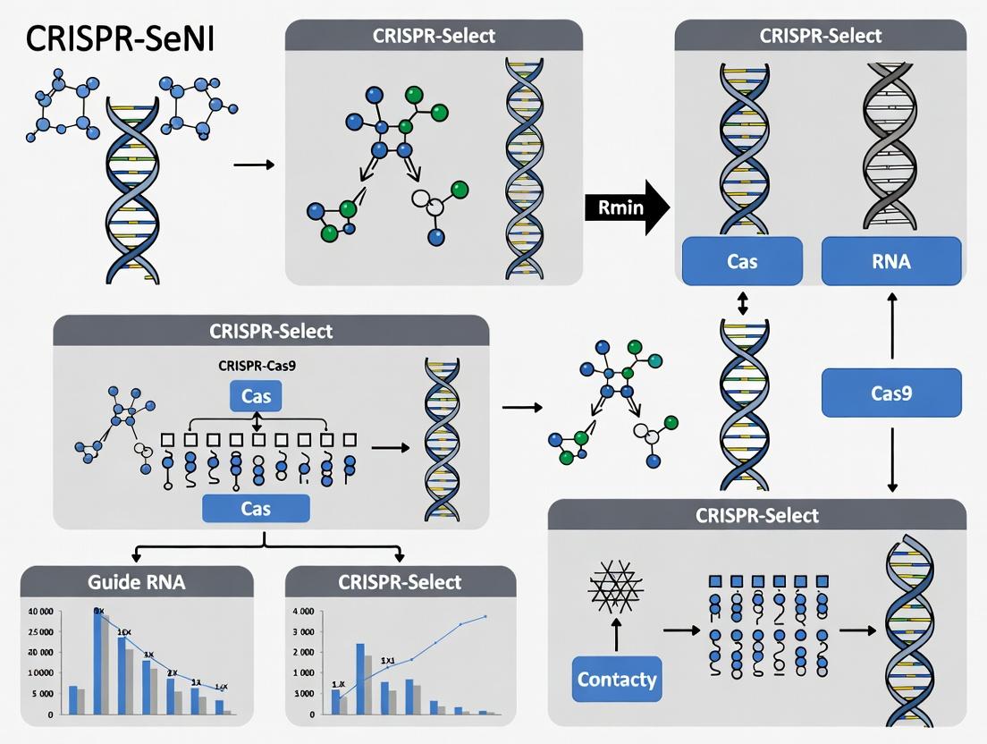
CRISPR-Select: A Comprehensive Guide to Functional Analysis of Genetic Variants for Precision Medicine
This article provides a complete framework for researchers and drug development professionals to implement CRISPR-Select methodologies for the functional annotation of genetic variants.
CRISPR-Select: A Comprehensive Guide to Functional Analysis of Genetic Variants for Precision Medicine
Abstract
This article provides a complete framework for researchers and drug development professionals to implement CRISPR-Select methodologies for the functional annotation of genetic variants. It begins by establishing the critical need to move beyond genomic association to functional understanding in disease research and therapeutic target identification. We then detail the step-by-step workflow of CRISPR-Select, including library design, delivery, and phenotypic screening. The guide addresses common experimental pitfalls and optimization strategies for enhanced sensitivity and specificity. Finally, we present robust validation protocols and compare CRISPR-Select to orthogonal techniques like MPRA and deep mutational scanning, evaluating its advantages and limitations. This resource empowers scientists to confidently apply high-throughput functional genomics to prioritize variants and accelerate the translation of genetic discoveries into actionable insights.
Decoding the Genome's Function: Why CRISPR-Select is Essential for Variant Interpretation
Application Notes
The Functional Genomics Bottleneck in Complex Disease
A central thesis in modern genomics posits that the critical bottleneck in understanding complex diseases is no longer variant discovery, but variant interpretation. Genome-wide association studies (GWAS) have identified hundreds of thousands of statistical associations between single nucleotide polymorphisms (SNPs) and disease phenotypes. However, the vast majority (>90%) of these variants reside in non-coding regions, making their functional impact on gene regulation and protein function obscure. Moving from statistical correlation to biological causation requires systematic functional validation. This is where CRISPR-Select technologies—encompassing base editing, prime editing, and CRISPR-mediated gene regulation—provide a transformative toolkit. By enabling precise, single-nucleotide edits in relevant cellular models, researchers can directly test the causative role of a variant on molecular and cellular phenotypes, deconvoluting the mechanisms that link genetic variation to complex disease etiology.
Strategic Framework for Variant-to-Function Analysis
A robust framework for causal variant analysis integrates computational prediction with empirical functional screening. The process begins with the prioritization of candidate causal variants from linkage disequilibrium (LD) blocks identified by GWAS, using criteria such as regulatory potential (e.g., ENCODE annotations, ATAC-seq peaks) and evolutionary conservation. High-priority variants are then modeled in vitro using CRISPR-Select tools in disease-relevant cell types (e.g., iPSC-derived neurons, cardiomyocytes, or immune cells). The phenotypic readouts are multi-modal, assessing transcriptional changes (single-cell RNA-seq), chromatin accessibility (ATAC-seq), protein expression (CITE-seq, flow cytometry), and disease-relevant cellular behaviors (e.g., cytokine secretion, phagocytosis, contraction). This integrated approach shifts the paradigm from observing association to experimentally establishing causality, a prerequisite for target identification in drug development.
Quantitative Landscape of GWAS Variants and Functional Validation
The table below summarizes the current challenge and the application of CRISPR-based functional analysis.
Table 1: The Variant Interpretation Pipeline: From Association to Causation
| Pipeline Stage | Typical Yield/Data | CRISPR-Select Functional Analysis Role | Key Measurement/Outcome |
|---|---|---|---|
| GWAS Discovery | 100-1000s of trait-associated loci; >90% non-coding. | N/A (Input for prioritization). | Statistical significance (p-value, odds ratio). |
| In Silico Prioritization | 1-10 candidate causal variants per locus. | Guides design for precise editing of each candidate. | Combined Annotation Dependent Depletion (CADD) score, RegulomeDB score. |
| CRISPR-based Saturation Genome Editing | Functional assessment of all possible alleles in a region. | Directly tests variant effect by introducing all possible SNPs in a multiplexed assay. | Functional score (based on cell growth, reporter expression) for each allele. |
| Deep Phenotyping of Isogenic Models | Molecular profiling of 1-3 confirmed causal variants. | Creation of isogenic cell pairs (risk vs. protective allele) for multi-omics. | Differential gene expression (fold-change), pathway enrichment (FDR q-value). |
| Therapeutic Hypothesis Generation | 1 novel drug target or mechanism per 20-50 validated causal variants. | Links variant mechanism (e.g., altered transcription factor binding) to a druggable node. | Target candidate priority score (based on druggability, pathway centrality). |
Protocols
Protocol 1: Design and Cloning of CRISPR Prime Editors for Non-Coding Variant Analysis
Objective: To generate a plasmid expressing a prime editor (PE2 system) and pegRNA for the precise installation of a non-coding candidate causal SNP in human induced pluripotent stem cells (iPSCs).
Materials (Research Reagent Solutions):
- PE2 Plasmid (Addgene #132775): Contains the fusion of Cas9 nickase (H840A) and engineered reverse transcriptase.
- pegRNA Cloning Oligos: Designed using online tools (e.g., pegFinder). Include the ~13-nt primer binding site (PBS) and reverse transcriptase template (RTT) encoding the desired edit.
- High-Efficiency Cloning Kit (e.g., Gibson Assembly, Golden Gate): For seamless assembly of the pegRNA scaffold and target-specific sequence into a U6-expression vector.
- Nucleofection Kit for iPSCs (e.g., Lonza P3 Primary Cell Kit): For delivery of ribonucleoprotein (RNP) complexes or plasmids.
- Next-Generation Sequencing (NGS) Validation Primers: Flanking primers (~300 bp amplicon) for deep sequencing of the edited genomic locus.
Methodology:
- pegRNA Design: For a target genomic coordinate (e.g., chr6:12345678), design a pegRNA with a 30-nt spacer sequence (protospacer adjacent motif (PAM): NGg, where N is any base). The RTT should be ~10-15 nucleotides, with the desired SNP placed in the middle. The PBS should be 8-13 nucleotides complementary to the DNA 3' of the nick.
- Cloning: Synthesize oligos encoding the spacer-RTT-PBS-scaffold sequence. Clone this into a pegRNA expression vector (e.g., pU6-pegRNA-GG-acceptor, Addgene #132777) using a BsaI-based Golden Gate assembly reaction.
- Verification: Transform the assembly reaction into stable cloning bacteria. Isolve plasmid DNA from multiple colonies and validate the insert by Sanger sequencing using a U6 promoter primer.
- Delivery (Plasmid-based): Co-transfect iPSCs (80% confluent in a 24-well plate) with 750 ng of PE2 plasmid and 250 ng of the validated pegRNA plasmid using the nucleofection system according to the manufacturer's protocol for iPSCs.
- Alternative Delivery (RNP-based): For higher specificity, complex purified PE2 protein with in vitro transcribed pegRNA and tracrRNA to form an RNP complex. Deliver via nucleofection.
- Screening and Validation: Allow cells to recover for 72 hours. Extract genomic DNA. Amplify the target region by PCR and subject to Illumina-based amplicon sequencing (2x150 bp). Analyze sequencing data with CRISPResso2 or similar to calculate editing efficiency and precision.
Protocol 2: Multiplexed Functional Screening of Variants in a GWAS Locus using CRISPRi
Objective: To perform a pooled screen to identify which non-coding variants in a linkage disequilibrium block alter the expression of a candidate target gene.
Materials (Research Reagent Solutions):
- dCas9-KRAB Repression Vector (Addgene #71236): For CRISPR interference (CRISPRi)-mediated transcriptional repression.
- Pooled sgRNA Library: A custom-designed library of ~5 sgRNAs per candidate variant (targeting within ±100 bp of SNP) and 100 non-targeting controls. Cloned into a lentiviral vector.
- Lentiviral Packaging System (2nd/3rd Generation): psPAX2 and pMD2.G plasmids for producing viral particles.
- Disease-Relevant Cell Line with Fluorescent Reporter: e.g., A macrophage cell line with a GFP reporter knocked into the 3' UTR of the putative target gene (TNFAIP3, IL6R, etc.).
- FACS Cell Sorter: For isolating cell populations based on reporter signal.
- NGS Library Prep Kit for sgRNA Amplification: To quantify sgRNA abundance from genomic DNA.
Methodology:
- Library Design & Production: Design sgRNAs targeting each candidate regulatory variant. Synthesize the oligo pool, PCR amplify, and clone into the lentiviral sgRNA vector. Produce high-titer lentivirus from HEK293T cells.
- Cell Line Infection and Selection: Infect the reporter cell line at a low multiplicity of infection (MOI ~0.3) to ensure most cells receive one sgRNA. Select with puromycin for 7 days.
- Phenotypic Sorting: After selection, culture cells for 7 more days to allow for gene expression changes. Perform fluorescence-activated cell sorting (FACS) to collect the top 10% (high reporter) and bottom 10% (low reporter) of the GFP expression distribution.
- sgRNA Deconvolution: Extract genomic DNA from each sorted population and the unsorted control. Amplify the integrated sgRNA cassette via PCR, add Illumina adapters and sample barcodes, and sequence on a MiSeq or NextSeq.
- Hit Identification: Align sequences to the sgRNA library reference. For each sgRNA, calculate its log2 fold-change enrichment in the "High" vs. "Low" GFP populations using tools like MAGeCK. sgRNAs targeting functional regulatory variants will be enriched in either the high or low population, identifying variants that causally regulate the target gene's expression.
Diagrams
Functional Genomics Workflow for Variant Causation
Non-coding Variant Alters TF Binding and Signaling
The Scientist's Toolkit: Research Reagent Solutions
Table 2: Essential Reagents for CRISPR-Select Functional Analysis of Variants
| Reagent / Material | Supplier Examples | Function in Variant Analysis |
|---|---|---|
| Prime Editor 2 (PE2) Plasmid | Addgene (#132775) | Core plasmid for precise installation of SNVs without double-strand breaks. |
| pegRNA Cloning Kit | Addgene (pU6-pegRNA vectors) | Modular system for rapid assembly of pegRNA expression constructs. |
| Purified Cas9 & PE2 Protein | Synthego, IDT, Thermo Fisher | For RNP delivery, reducing off-target effects and enabling editing in primary cells. |
| dCas9-KRAB Repression Vector | Addgene (#71236) | For CRISPRi screens to interrogate variant effects on gene regulation. |
| Lentiviral Packaging Mix | Sigma, Takara, Invitrogen | For generating stable cell lines or delivering pooled sgRNA libraries. |
| iPSC Nucleofection Kit | Lonza (P3 Kit) | Enables efficient delivery of CRISPR tools into genetically stable, disease-relevant stem cells. |
| Multiplexed sgRNA Library Synthesis | Twist Bioscience, Agilent | For designing and synthesizing custom pooled libraries targeting many variants in parallel. |
| NGS Amplicon-Seq Kit (Illumina) | KAPA Biosystems | For high-throughput validation of editing efficiency and precision at target loci. |
| Single-Cell Multi-ome Kit (ATAC + Gene Exp.) | 10x Genomics | For deep molecular phenotyping of edited isogenic cell models. |
Application Notes
CRISPR-Select is a novel, high-throughput screening paradigm designed to functionally characterize genetic sequence variants (GSVs), such as single nucleotide variants (SNVs) and indels, in their native genomic and cellular context. This approach is central to a broader thesis on moving beyond variant association to definitive functional annotation, which is critical for interpreting genomes in disease research and therapeutic target identification.
The method leverages pooled CRISPR-Cas9 base-editing or prime-editing platforms to generate allelic variant libraries at specific loci. Instead of merely knocking out genes, CRISPR-Select introduces precise, user-defined variants. The edited cell populations are then subjected to selective pressures (e.g., drug treatment, nutrient stress, tumorigenic conditions), and the enrichment or depletion of specific variants is quantified via next-generation sequencing (NGS). This enables parallel measurement of the functional impact of hundreds to thousands of variants in a single experiment.
Table 1: Key Quantitative Metrics from Representative CRISPR-Select Studies
| Parameter | Study A: Oncogenic SNVs | Study B: Drug Resistance Variants | Study C: Splicing Variants |
|---|---|---|---|
| Library Size (Variants) | 952 | 2,450 | 350 |
| Editing Efficiency (Avg.) | 65% | 58% | 72% |
| Selection Timepoint | 14 days (in vivo) | 21 days (1µM Drug) | 10 days |
| Dynamic Range (Log2 Fold-Change) | -4.8 to +3.5 | -5.2 to +4.1 | -3.0 to +2.8 |
| Identified Functional Variants | 43 (4.5%) | 127 (5.2%) | 28 (8.0%) |
Experimental Protocols
Protocol 1: Design and Cloning of a CRISPR-Select sgRNA-Variant Library
- Target Identification: From GWAS or sequencing data, define a genomic region of interest (e.g., a promoter, an exon, a splicing enhancer).
- sgRNA Design: Design sgRNAs targeting the region using algorithms like CHOPCHOP or CRISPick. For base editing, ensure the target base is within the editing window (e.g., ~positions 4-8 for BE4max).
- Variant Library Oligo Synthesis: Synthesize a pooled oligo library where each oligo encodes the sgRNA sequence and a donor template harboring the desired variant(s) for HDR-mediated editing or, for base editors, the library consists of sgRNAs targeting each variant position.
- Library Cloning: Clone the pooled oligo library into a lentiviral CRISPR vector (e.g., lentiCRISPR v2-BE4max or a prime-editor construct) via Golden Gate assembly. Transform the reaction into electrocompetent E. coli and perform maxiprep to obtain the plasmid library for sequencing validation and virus production.
Protocol 2: Lentiviral Production and Cell Line Generation
- Virus Production: Co-transfect HEK293T cells with the plasmid library, psPAX2 (packaging), and pMD2.G (envelope) plasmids using a PEI transfection reagent. Harvest lentiviral supernatant at 48 and 72 hours post-transfection.
- Transduction and Selection: Transduce the target cell line (e.g., a cancer cell line or iPSCs) at a low MOI (<0.3) to ensure single-integration events. Select transduced cells with appropriate antibiotics (e.g., puromycin) for 5-7 days.
- Editing Validation: Harvest a sample of the polyclonal cell population (~1 week post-selection). Extract genomic DNA from the region of interest and perform NGS to confirm baseline variant representation and editing efficiency.
Protocol 3: Functional Selection and NGS Analysis
- Selection: Split the edited polyclonal cell population into experimental and control arms. Apply the selective pressure (e.g., anticancer drug, hypoxia) for a predetermined duration (typically 2-4 cell doublings). Maintain control cells in standard conditions.
- Genomic DNA Harvest: Harvest genomic DNA from both pre-selection, post-selection experimental, and control populations using a kit-based method.
- Amplicon Sequencing: Perform PCR amplification of the target genomic region from each sample using barcoded primers. Pool amplicons and sequence on an NGS platform (Illumina MiSeq/NovaSeq) to a depth of >500 reads per variant.
- Data Analysis: Align sequences to the reference genome. Quantify the frequency of each variant in each sample. Calculate the enrichment score (e.g., log2 fold-change) for each variant between pre-selection and post-selection populations. Use statistical models (e.g., MAGeCK or edgeR) to identify significantly enriched or depleted variants.
Visualization
CRISPR-Select Screening Workflow
CRISPR-Select Variant Enrichment Logic
The Scientist's Toolkit: Research Reagent Solutions
Table 2: Essential Materials for a CRISPR-Select Screen
| Reagent/Material | Function | Example Product/Type |
|---|---|---|
| Base Editor or Prime Editor Plasmid | Catalytic core for introducing precise point mutations without DSBs. | lentiCMV-BE4max, pPE2. |
| Lentiviral sgRNA Cloning Vector | Backbone for sgRNA library delivery and stable genomic integration. | lentiGuide-Puro, lenti-sgRNA(MS2)_zeo. |
| Pooled Oligonucleotide Library | Defines the specific sgRNA sequences and variant information. | Custom array-synthesized oligo pool. |
| Lentiviral Packaging Plasmids | Required for production of replication-incompetent lentiviral particles. | psPAX2 (packaging), pMD2.G (VSV-G envelope). |
| HEK293T Cells | Highly transfectable cell line for high-titer lentivirus production. | ATCC CRL-3216. |
| Target Cell Line | The cellular model for functional testing (e.g., cancer, iPSC-derived). | HAP1, RPE1, or disease-relevant lines. |
| Next-Generation Sequencer | For deep sequencing of variant libraries pre- and post-selection. | Illumina MiSeq, NovaSeq. |
| gDNA Extraction Kit | High-quality genomic DNA isolation from cell pellets. | DNeasy Blood & Tissue Kit. |
| NGS Library Prep Kit | For preparing amplicon sequencing libraries from target regions. | KAPA HiFi HotStart ReadyMix. |
This application note details the core methodological components for conducting CRISPR-Select functional analysis of genetic sequence variants. This approach enables high-throughput, functional characterization of variant impact on cellular phenotypes, drug response, and fitness within a pooled screening format, critical for target discovery and validation in drug development.
Core Components: Application Notes
gRNA Library Design and Construction
The guide RNA (gRNA) library is the foundation for variant interrogation. Libraries are designed to target not only coding sequences but also regulatory elements and non-coding variants of interest (VOIs).
- Design Principles: For each genetic variant (e.g., SNP, indel), two gRNAs are typically designed: one targeting the reference allele and one targeting the alternate allele. This enables direct comparison of phenotypic consequences.
- Library Diversity: Modern libraries can encompass thousands to hundreds of thousands of gRNAs, covering genome-wide association study (GWAS) hits, somatic mutations from cancer databases, or putative regulatory variants.
- Quantitative Considerations: Essential library parameters must be controlled.
Table 1: Key Quantitative Parameters for gRNA Library Design
| Parameter | Typical Range/Value | Purpose/Rationale |
|---|---|---|
| gRNAs per Variant | 2-6 | Controls for off-target effects and improves statistical confidence. |
| Library Size | 1,000 - 100,000+ gRNAs | Determines screening scale and multiplexing capacity. |
| gRNA Length | 20 nt (SpCas9) | Standard complementarity region for SpCas9. |
| Cloning Vector | Lentiviral backbone (e.g., lentiGuide-puro) | Enables stable genomic integration and selection. |
| Coverage (Depth) | 200-1000x per gRNA | Ensures each gRNA is adequately represented in the population pre- and post-selection. |
Reporter Systems for Phenotypic Readout
Reporter systems translate the molecular consequence of CRISPR editing into a quantifiable signal. Selection-based reporters are paramount for enrichment/depletion screens.
- Fluorescent Reporters (FACS-based): Cells express a fluorescent protein (e.g., GFP) under the control of a pathway element perturbed by the variant. Editing alters fluorescence, enabling sorting.
- Survival/Selective Reporters: The most common format for fitness screens. A resistance gene (e.g., puromycin N-acetyltransferase) is linked to a sensor element. Variant-induced changes in sensor activity modulate resistance gene expression, leading to survival or death under antibiotic pressure.
- Surface Display Reporters (e.g., Lasso-seq): A surface protein (e.g., CD protein) serves as the reporter. Cells are sorted based on expression levels using magnetic-activated cell sorting (MACS) or FACS.
Applying Selective Pressures
The choice of selective pressure defines the biological question. Pressure is applied after library transduction and stable cell line generation.
- Drug Treatment: The primary method for pharmacogenomic studies. Cells are treated with a therapeutic compound at varying concentrations (IC10-IC90). gRNAs targeting variants that confer sensitivity or resistance become depleted or enriched.
- Pathway Activation/Inhibition: Used to probe variant function in specific biological contexts (e.g., growth factor starvation, cytokine stimulation).
- Proliferative Fitness: Culture over multiple passages without exogenous pressure reveals variants impacting baseline cellular growth and survival.
Table 2: Common Selective Pressures and Associated Readouts
| Selective Pressure | Phenotype Interrogated | Typical Duration | Primary Readout Method |
|---|---|---|---|
| Oncogene Inhibitor (e.g., Vemurafenib) | Drug resistance mechanisms | 2-3 weeks | NGS of gRNA abundance |
| Cytotoxic Chemotherapy | DNA repair deficiency, survival | 1-2 weeks | NGS of gRNA abundance |
| Growth Factor Deprivation | Signaling pathway essentiality | 1-3 weeks | NGS of gRNA abundance |
| Hypoxia | Metabolic adaptation, tumor survival | 1-2 weeks | NGS of gRNA abundance |
| None (Proliferation only) | Baseline fitness effect | 3-4 weeks | NGS of gRNA abundance |
Experimental Protocols
Protocol: CRISPR-Select Screen for Variant-Mediated Drug Resistance
Objective: Identify genetic variants that confer resistance to a targeted oncology drug.
Materials: gRNA library plasmid pool, HEK293T or suitable packaging cell line, target cell line (e.g., cancer cell line of interest), lentiviral packaging plasmids (psPAX2, pMD2.G), polybrene, puromycin, drug of interest.
Part A: Library Viral Production & Cell Line Generation
- Day 1: Seed HEK293T cells in a 10cm dish to reach 70-80% confluency the next day.
- Day 2: Transfect cells using PEI or calcium phosphate with: 10 µg gRNA library pool, 7.5 µg psPAX2, 2.5 µg pMD2.G.
- Day 3 & 4: Replace medium with fresh DMEM + 10% FBS.
- Day 5: Harvest viral supernatant, filter through a 0.45µm filter. Aliquot and freeze at -80°C or use immediately.
- Day 5: Infect target cells at a low MOI (~0.3) with viral supernatant plus 8µg/mL polybrene. Include a non-transduced control.
- Day 6: Begin selection with appropriate puromycin concentration (determined by kill curve) for 5-7 days until all control cells are dead.
Part B: Application of Selective Pressure
- Day 0 (Post-selection): Harvest a baseline sample of 5-10 million cells (for gDNA extraction). Seed the remaining cells at a coverage of >500x per gRNA.
- Day 1: Apply drug treatment. Set up two conditions: A) Vehicle control (DMSO), B) Drug at desired concentration (e.g., IC50). Maintain cells in log phase.
- Days 7-21: Passage cells as needed, maintaining coverage and drug/vehicle pressure. Harvest ~10 million cells from each condition every 5-7 days for gDNA extraction.
Part C: gRNA Abundance Quantification by NGS
- gDNA Extraction: Use a column-based kit (e.g., Qiagen Blood & Cell Culture DNA Maxi Kit) to extract high-quality gDNA from all samples.
- PCR Amplification of gRNA Cassettes: Perform a two-step PCR protocol.
- PCR1 (Amplify gRNA region): Use primers binding the constant lentiviral backbone flanking the gRNA. Use a high-fidelity polymerase (e.g., KAPA HiFi). Limit cycles to prevent bias (typically 18-22).
- PCR2 (Add Illumina adaptors & indices): Use 1-5µL of purified PCR1 product as template. Use indexed primers for sample multiplexing.
- Sequencing: Pool PCR2 products, quantify, and sequence on an Illumina NextSeq or HiSeq platform (75bp single-end is sufficient).
- Data Analysis: Align reads to the library reference. Count gRNA reads in each sample. Use statistical packages (e.g., MAGeCK) to compare gRNA abundance between drug-treated and control samples over time, identifying significantly enriched or depleted gRNAs.
Protocol: Fluorescent Reporter Assay for Variant Impact on Promoter Activity
Objective: Quantify how a non-coding variant affects transcriptional activity of a promoter.
Materials: gRNA pairs (Ref/Alt), Cas9-expressing cell line, lentiviral vectors for gRNA delivery, Reporter plasmid (promoter driving GFP), FACS tubes, flow cytometer.
Workflow:
- Reporter Construction: Clone the genomic region containing the reference or alternate variant allele upstream of a minimal promoter and GFP gene in a lentiviral reporter vector.
- Cell Line Preparation: Transduce a Cas9-expressing cell line with the reporter construct. Generate a stable, polyclonal cell line via antibiotic selection.
- Variant Editing: Transduce the reporter cell line with lentivirus carrying either the reference-targeting or alternate-targeting gRNA. Include a non-targeting control (NTC) gRNA.
- Incubation & Analysis: Culture cells for 7-10 days to allow editing and turnover of pre-existing GFP protein. Harvest cells and analyze GFP fluorescence intensity via flow cytometry (10,000+ events per sample).
- Data Interpretation: Compare the median fluorescence intensity (MFI) of the reference-targeted and alternate-targeted populations to the NTC control. A shift in MFI indicates the variant's functional impact on promoter activity.
The Scientist's Toolkit
Table 3: Essential Research Reagent Solutions
| Item | Function in CRISPR-Select | Example Product/Catalog # (Representative) |
|---|---|---|
| Pooled gRNA Library | Targets reference/alternate alleles of variants for functional screening. | Custom synthesized (Twist Bioscience, Agilent). |
| Lentiviral Packaging Plasmids | Required for production of replication-incompetent lentivirus to deliver gRNA library. | psPAX2 (Addgene #12260), pMD2.G (Addgene #12259). |
| Cas9-Expressing Cell Line | Provides the endonuclease effector protein for genome editing. | HEK293T-Cas9, U2OS-Cas9, or custom generated. |
| Polycation Transduction Aid | Enhances lentiviral infection efficiency. | Polybrene (Hexadimethrine bromide), 8 µg/mL working concentration. |
| Selection Antibiotic | Selects for cells successfully transduced with the gRNA vector. | Puromycin dihydrochloride, concentration determined by kill curve. |
| High-Fidelity PCR Kit | Amplifies gRNA sequences from genomic DNA for NGS with minimal bias. | KAPA HiFi HotStart ReadyMix (Roche). |
| Dual-Indexing Primers for NGS | Adds unique sample barcodes and Illumina adaptors during PCR2. | Nextera XT Index Kit v2 (Illumina). |
| gDNA Extraction Kit | Ishes high-molecular-weight genomic DNA from screen samples for PCR. | QIAamp DNA Blood Maxi Kit (Qiagen). |
| Statistical Analysis Software | Identifies significantly enriched/depleted gRNAs from NGS count data. | MAGeCK (Model-based Analysis of Genome-wide CRISPR-Cas9 Knockout). |
Visualizations
Application Notes
Genome-Wide Association Studies (GWAS) identify hundreds of genetic loci associated with diseases, but most are in non-coding regions with unknown functional impact. CRISPR-Select functional analysis enables direct, high-throughput interrogation of these variants to prioritize true causal hits.
Similarly, clinical sequencing generates vast numbers of Variants of Uncertain Significance (VUS) in known disease genes. Functional validation is the critical rate-limiting step in clinical interpretation. CRISPR-Select offers a scalable solution.
By establishing causal variant-to-phenotype relationships, this methodology directly illuminates disease mechanisms, exposing novel proteins and pathways as potential therapeutic targets.
Table 1: Quantitative Outcomes from Recent CRISPR-Based Functional Screens of Non-Coding GWAS Hits
| Disease/Trait | Number of GWAS Loci Screened | Percentage with Regulatory Activity | Key Validated Causal Gene(s) | Primary Functional Readout | Publication Year |
|---|---|---|---|---|---|
| Coronary Artery Disease | 120 | 43% | PCSK9, IL6R, CXCL12 | Gene Expression (scRNA-seq) | 2023 |
| Type 2 Diabetes | 88 | 31% | PPARG, SLC30A8, KCNJ11 | Insulin Secretion (Cell Reporter) | 2024 |
| Inflammatory Bowel Disease | 150 | 52% | IRF1, IL23R, CARD9 | Cytokine Production (Luminex) | 2023 |
| Alzheimer's Disease | 95 | 28% | BIN1, PTK2B, SPI1 | Phagocytosis (High-Content Imaging) | 2022 |
Table 2: Impact of Functional VUS Resolution on Clinical Classification
| Gene Context | Number of VUS Tested | % Reclassified as Likely Pathogenic | % Reclassified as Likely Benign | Standard Method Supplanted | Reference Database |
|---|---|---|---|---|---|
| BRCA1 | 650 | 12% | 41% | ACMG/AMP Guidelines | ClinVar |
| TP53 | 320 | 18% | 35% | Bayesian Prediction Models | IARC TP53 Database |
| KCNH2 (Cardiac) | 155 | 15% | 55% | In silico Tools (REVEL, SIFT) | ClinGen |
Detailed Protocols
Protocol 1: High-Throughput Prioritization of Non-Coding GWAS Variants
Objective: To functionally screen candidate causal SNPs from a GWAS locus using CRISPR-based perturbation and a phenotypic readout.
Materials & Workflow:
- Design: For each target SNP, design two CRISPR guides:
- Allele-Specific gRNAs: One gRNA specific to the reference allele, one to the alternative allele. Use SpCas9-D10A (nickase) to minimize off-targets.
- Common Targeting gRNA: A single gRNA targeting the locus, co-delivered with a donor oligo library containing all haplotype combinations.
- Library Delivery: Electroporate pooled gRNA libraries (with barcodes) and donor oligo pools into relevant human cell lines (e.g., iPSC-derived hepatocytes for lipid traits) at high MOI.
- Phenotypic Selection: Subject cells to a relevant assay (e.g., LDL uptake assay for CAD variants) after 7-14 days. Perform FACS-based selection of top/bottom 20% of the phenotypic distribution.
- Sequencing & Analysis: Extract genomic DNA from pre-selection and selected populations. Amplify gRNA barcodes via PCR and sequence. Enrichment/depletion of specific gRNAs is calculated to identify variants altering the phenotype.
Workflow for GWAS Variant Screening
Protocol 2: Functional Interpretation of a VUS in a Monogenic Disease Gene
Objective: To determine the pathogenicity of a single VUS in a gene of known function using an isogenic cell model and a direct functional rescue assay.
Materials & Workflow:
- Cell Model Generation: Use CRISPR-Cas9 to introduce the specific VUS into a wild-type human induced pluripotent stem cell (iPSC) line. Generate a clonally derived, sequenced-verified isogenic line. The control is the unedited parental line.
- Differentiation: Differentiate both control and VUS iPSC lines into the relevant cell type (e.g., cardiomyocytes for a channelopathy).
- Functional Assay: Perform a gold-standard assay for that gene's function (e.g., patch-clamp electrophysiology for ion channels, enzyme activity assay for a metabolizing enzyme).
- Rescue Experiment: Transduce the VUS cell line with a lentivirus expressing the wild-type cDNA. Re-measure function. Failure to rescue confirms the VUS is loss-of-function.
- Classification: Integrate quantitative functional data (e.g., 70% reduction in current density) with computational predictions for final ACMG/AMP classification.
VUS Resolution via Isogenic Models
Protocol 3: From Causal Variant to Therapeutic Target Identification
Objective: To elucidate the downstream molecular pathway dysregulated by a prioritized causal variant, revealing druggable nodes.
Materials & Workflow:
- Multi-Omics Profiling: Perform RNA-seq and ATAC-seq on isogenic cell pairs (WT vs. Variant) from Protocol 2. Integrate data to define differentially expressed genes and altered regulatory regions.
- Pathway Analysis: Use enrichment tools (GSEA, Ingenuity) on the differentially expressed gene set to identify significantly perturbed signaling pathways (e.g., JAK-STAT, NF-κB).
- CRiNET Screening: Conduct a CRISPR knockout or inhibition screen targeting all genes in the implicated pathway(s) in the variant cell line. Identify genes whose knockout rescues the disease phenotype.
- Druggability Assessment: Cross-reference the "hit" genes from the rescue screen with databases of known drug targets (e.g., DrugBank, ChEMBL) and clinical trial candidates.
Therapeutic Target Discovery Pathway
The Scientist's Toolkit: Research Reagent Solutions
| Item | Function in CRISPR-Select Analysis |
|---|---|
| High-Fidelity Cas9 Variants (e.g., SpCas9-HF1) | Reduces off-target editing, critical for creating clean isogenic models and specific allele perturbations. |
| dCas9-KRAB/VP64 Fusion Proteins | For CRISPR interference (CRISPRi) or activation (CRISPRa) screens to model regulatory variant effects without cutting DNA. |
| Arrayed gRNA Libraries | Pre-defined, individually aliquoted gRNAs for screening in multi-well plates, enabling complex phenotypic readouts (imaging, metabolism). |
| Prime Editing RNP Complexes | Allows precise installation of any SNP or small indel without double-strand breaks, ideal for introducing specific VUS or reverting them. |
| Phenotypic Reporter Cell Lines | Engineered lines with reporters (GFP, Luciferase) under control of a pathway of interest (e.g., NF-κB response element) for rapid screening. |
| Single-Cell Multi-Omic Kits (CITE-seq, ATAC-seq) | Enables simultaneous measurement of transcriptome, surface proteins, and chromatin accessibility in pooled CRISPR screens to deconvolve complex phenotypes. |
| Bioinformatics Pipelines (MAGeCK, PinAPL-Py) | Specialized software for robust statistical analysis of gRNA enrichment/depletion in pooled screen sequencing data. |
This Application Note is framed within the broader thesis of CRISPR-Select functional analysis of genetic sequence variants, a paradigm for functionally annotating variants of uncertain significance (VUS) and non-coding variants. The modern toolkit extends far beyond canonical CRISPR-Knockout (KO), with CRISPR activation (CRISPRa), interference (CRISPRi), and base editing screens now central to establishing causal genotype-phenotype relationships in disease modeling and therapeutic target discovery.
The table below compares the core quantitative outputs, efficiencies, and applications of major CRISPR screening technologies.
Table 1: Comparative Analysis of Advanced CRISPR Screening Platforms
| Screening Modality | Typical Editing Efficiency | Library Size (Guide Count) | Primary Genetic Outcome | Key Application in Variant Analysis |
|---|---|---|---|---|
| CRISPR-KO (Cas9) | 60-90% indels | ~5-10 guides/gene (∼100,000 total) | Frameshift indels, gene knockout | Essential gene identification; loss-of-function (LoF) variant phenocopy. |
| CRISPRa (dCas9-VPR) | 2-10x gene upregulation | 3-5 guides/TSS (∼30,000 total) | Transcriptional activation | Gain-of-function (GoF) simulation; enhancer validation; rescue screens. |
| CRISPRi (dCas9-KRAB) | 70-95% gene repression | 3-5 guides/TSS (∼30,000 total) | Transcriptional repression | Tunable gene suppression; essential gene identification; LoF in diploid cells. |
| CRISPR Base Editing (CBE) | 20-60% base conversion | ~3-10 guides/site (∼50,000 total) | C•G to T•A transition | Saturation mutagenesis of loci; modeling specific SNVs; creating pathogenic or corrective variants. |
| CRISPR Prime Editing | 10-40% edits (with selection) | Varies by target | All 12 base-to-base changes, small insertions/deletions | Precise installation of complex variants for functional assessment. |
Application Notes & Detailed Protocols
Application Note A: CRISPRa/i for Functional Enhancer Validation
Context: Non-coding VUS often reside in putative enhancer regions. A CRISPRa/i tiling screen can functionally map these regions. Objective: To determine if a non-coding sequence variant affects enhancer activity by modulating target gene expression.
Protocol: CRISPRi Tiling Screen for Enhancer De-repression
- Design & Cloning: Synthesize a tiled single-guide RNA (sgRNA) library targeting the genomic region of interest (~200bp windows) with 5-10bp spacing. Clone into a lentiviral dCas9-KRAB backbone (e.g., lentiGuide-KRAB).
- Virus Production & Transduction: Produce lentivirus in HEK293T cells. Transduce the target cell line at an MOI of ~0.3 and 500x coverage. Select with puromycin (2 µg/mL) for 7 days.
- Screen Execution: Harvest cells at baseline (T0) and after 10-14 population doublings (Tend). Extract genomic DNA from both pools.
- Sequencing & Analysis: Amplify the sgRNA cassette via PCR and sequence on a HiSeq platform. Align reads to the reference library. Calculate guide enrichment/depletion using MAGeCK or BAGEL2. Depleted guides indicate regions whose repression reduces cell fitness (putative essential enhancers).
Key Reagent Solutions: See Table 3.
Application Note B: Base Editing Saturation Screen for Variant Functionalization
Context: A locus associated with disease contains many missense VUS. A base editor saturation screen can systematically score their functional impact. Objective: To classify all possible SNVs at a specific amino acid residue as benign, loss-of-function, or gain-of-function.
Protocol: CBE Saturation Mutagenesis at a Codon
- Library Design: Design sgRNAs to target the codon of interest with an NG PAM for BE4max. Generate a library of oligos containing all 64 codon variants (NNS degeneracy) within the sgRNA's activity window (positions 4-8, counting the PAM as 21-23).
- Library Construction & Delivery: Clone the oligo pool into a lentiviral base editor expression system (e.g., BE4max + sgRNA backbone). Produce virus and transduce cells at low MOI (<0.3) to ensure single integrations.
- Phenotypic Selection: Apply relevant selective pressure (e.g., drug treatment, growth factor withdrawal, FACS for a marker) 7 days post-transduction. Harvest genomic DNA from pre-selection and post-selection populations.
- Deep Sequencing & Enrichment Scoring: Perform targeted amplicon sequencing of the edited genomic locus. Use
maseqorCrispRVariantsto quantify the frequency of each variant in input vs. selected pools. Calculate an enrichment ratio (log2 fold-change). Variants depleted upon selection are likely pathogenic (LoF), while enriched variants may confer a selective advantage (GoF).
The Scientist's Toolkit: Research Reagent Solutions
Table 2: Essential Reagents for CRISPR-Select Functional Genomics
| Reagent / Material | Provider Examples | Function in CRISPR-Select Workflow |
|---|---|---|
| dCas9-VPR Lentiviral System | Addgene #114257, TaKaRa | Delivers all components for robust CRISPRa gene activation screens. |
| dCas9-KRAB-MeCP2 Lentiviral System | Addgene #122259 | High-efficiency CRISPRi for potent, consistent gene repression. |
| BE4max-UGI Plasmid | Addgene #112093 | High-efficiency cytosine base editor (CBE) for C•G to T•A saturation screens. |
| All-in-One Prime Editor (PE) | Addgene #174828 | Expresses prime editor and pegRNA for precise variant installation. |
| Genome-Wide CRISPRa Lib. (Calabrese) | Addgene Pooled Library | ~70,000 sgRNA library targeting TSSs for genome-wide activation screens. |
| Brunello CRISPR-KO Lib. | Addgene #73179 | Optimated genome-wide KO library (4 sgRNAs/gene) for essentiality screens. |
| MAGeCK-VISPR Software | Open Source | Comprehensive computational pipeline for analyzing screen read count data. |
| Next-Gen Sequencing Kit (MiSeq) | Illumina | For deep sequencing of sgRNA or target amplicons from screen pools. |
| Lentiviral Packaging Mix (psPAX2, pMD2.G) | Addgene #12260, #12259 | Essential plasmids for producing replication-incompetent lentiviral particles. |
Visualized Workflows & Pathways
Title: Decision Workflow for CRISPR-Select Variant Analysis
Title: CRISPRa/i Tiling Screen for Enhancer Mapping
Title: Base Editing Saturation Screen for Variant Scoring
The CRISPR-Select Protocol: Step-by-Step Guide from Library Design to Hit Calling
Application Notes
This protocol details the initial, in-silico phase for CRISPR-Select, a functional genomics platform designed to interrogate the phenotypic impact of genetic sequence variants (GSVs), such as single nucleotide polymorphisms (SNPs) or coding mutations. The core objective is to construct a dual gRNA library that enables precise, variant-aware cellular perturbations. This strategic design is foundational for downstream pooled screening, allowing researchers to distinguish phenotype drivers from passenger variants in disease contexts like cancer or for pharmacogenomic studies.
The library comprises two primary components:
- Variant-Targeting gRNAs: Designed to complement the specific variant allele (e.g., the mutant or non-reference allele).
- Reference-Targeting gRNAs: Designed to complement the wild-type or reference allele.
When deployed with CRISPR interference (CRISPRi) or activation (CRISPRa), these paired gRNAs facilitate allele-specific transcriptional modulation. A phenotype specific to perturbation of one allele implicates that allele's function in the observed cellular state.
Key Design Parameters & Quantitative Summary
| Parameter | Target Value | Rationale & Considerations |
|---|---|---|
| gRNA Length | 20 nt spacer | Standard length for SpCas9-derived systems, balancing specificity and on-target activity. |
| Protospacer Adjacent Motif (PAM) | NGG (for SpCas9) | Must be present adjacent to target site. Design is adaptable to SaCas9 (NNGRRT) or other engineered Cas variants. |
| Variant Position | Within positions 1-12 of gRNA spacer | Maximizes discriminatory power. Mismatches in the seed region (PAM-proximal 12 bp) severely compromise cleavage/recruitment efficiency. |
| Predicted On-Target Score | > 0.6 (e.g., via Doench-Fusi 2016 rule set) | Filters for high predicted activity. Tools: CRISPRon, CHOPCHOP, or proprietary algorithms. |
| Predicted Off-Target Count | ≤ 3 hits with ≤ 3 mismatches | Minimizes confounding off-target effects. Validate via Bowtie or BLAST against relevant genome build. |
| GC Content | 40-60% | Optimizes gRNA stability and expression. |
| Paired gRNA Distance | Identical genomic locus | Targets the same transcriptional start site, ensuring paired comparison is valid. |
| Control gRNAs | Non-targeting (scrambled) & Essential Gene Targeting | For background and positive control signal normalization. |
Essential Experimental Protocols
Protocol 1: In-Silico Identification of Targetable Variants and gRNA Design
Materials & Reagents:
- Reference Genome FASTA File (e.g., GRCh38.p13).
- Variant Call Format (VCF) File containing GSVs of interest.
- High-Performance Computing Cluster or local workstation with >= 16GB RAM.
- Design Software: CRISPRko: CHOPCHOP, CRISPRscan; CRISPRi/a: CRISPRai, CRISPick.
Methodology:
- Variant Filtering: Load the VCF file into a bioinformatics environment (e.g., Python with
pysam, R withVariantAnnotation). Filter variants to retain those in putative regulatory regions (promoters, enhancers) or coding exons, based on your hypothesis. - PAM Site Identification: For each filtered variant, extract a ~100bp genomic sequence flanking the variant. Programmatically scan both DNA strands for the presence of the appropriate PAM sequence (e.g., NGG) where the variant nucleotide falls within positions 1-12 upstream of the PAM.
- gRNA Spacer Extraction: For each identified PAM site, extract the 20bp sequence immediately 5' to the PAM as the candidate gRNA spacer. Generate two versions: one matching the reference allele and one matching the variant allele.
- Specificity Filtering (Off-Target Prediction): Submit all candidate spacer sequences to an off-target prediction tool. Align each spacer to the reference genome allowing up to 3 mismatches. Discard any spacer with >3 genomic loci with ≤3 mismatches.
- Efficiency Scoring: Calculate a predicted on-target activity score for each passing spacer using a validated algorithm (e.g., DeepHF, Rule Set 2). Rank spacer pairs and select the top-scoring pair for each variant where both reference- and variant-targeting gRNAs have a score > 0.6.
- Cloning Sequence Generation: Append the appropriate 5' and 3' cloning overhangs to each selected 20bp spacer sequence for your chosen delivery vector (e.g., for lentiviral lentiGuide-puro:
CACCG+[20bp spacer]andAAAC+[reverse complement spacer]+C).
Protocol 2: Design and Integration of Control gRNAs
Materials & Reagents:
- Genome Annotation File (GTF/GFF).
- List of Pan-Essential Genes (e.g., from Hart et al., 2015 or DepMap).
Methodology:
- Non-Targeting Controls (NTCs): Generate 50-100 scrambled 20nt sequences with GC content matching your library (40-60%). Ensure they have no significant homology (BLASTn, ≤17bp contiguous identity) to the target genome.
- Positive Controls (Essential Gene Targeting): Design 5-10 high-efficacy gRNAs targeting the coding sequences of pan-essential genes (e.g., RPL9, PSMC1, POLR2D). These serve as a transfection/selection control and a benchmark for strong phenotype (e.g., cell death in a knockout screen).
- Intergenic Negative Controls: Design gRNAs targeting genomically "safe harbor" loci (e.g., AAVS1, ROSA26) or inactive genomic regions, confirmed to have no transcriptional or phenotypic consequence.
The Scientist's Toolkit: Research Reagent Solutions
| Item | Function in CRISPR-Select Phase 1 |
|---|---|
| Lentiviral gRNA Cloning Vector (e.g., lentiGuide, lenti-sgRNA) | Backbone for pooled gRNA library construction; contains resistance marker (puromycin/ blasticidin) for stable cell line generation. |
| CRISPRi/a-Compatible dCas9 Fusion Vector (e.g., dCas9-KRAB for i; dCas9-VPR for a) | Enables transcriptional repression (CRISPRi) or activation (CRISPRa) without DNA cleavage, crucial for studying non-coding variants. |
| High-Fidelity DNA Polymerase (e.g., Q5, KAPA HiFi) | For accurate amplification of gRNA library pools during cloning and preparation for next-generation sequencing (NGS) validation. |
| Next-Generation Sequencing Kit (Illumina-compatible) | For deep sequencing of the cloned library to verify gRNA representation and integrity before screening. |
| Genomic DNA Extraction Kit (Magnetic Bead-Based) | For high-yield, high-quality gDNA extraction from pooled cell populations post-screen for NGS analysis. |
| gRNA Amplification Primers with NGS Adapters | Custom oligonucleotides to add Illumina P5/P7 flow cell adapters and sample indices to gRNA cassettes recovered from screened cells. |
Diagrams
gRNA Library Design & Filtering Workflow
CRISPRi Allele-Specific Targeting & Phenotype Logic
Application Notes
Efficient delivery of CRISPR-Cas9 components is critical for the functional analysis of sequence variants in a CRISPR-Select framework. The choice between lentiviral transduction and electroporation is dictated by cell type, required editing efficiency, and experimental timeline. Lentiviral vectors offer stable genomic integration and are ideal for hard-to-transfect or primary cells, enabling long-term studies. Electroporation provides high-efficiency, transient delivery of ribonucleoprotein (RNP) complexes, minimizing off-target effects and reducing time to analysis. Optimization is non-negotiable; parameters must be tailored to each cell model to balance maximal editing with cell viability.
Quantitative Comparison of Delivery Methods
Table 1: Key Performance Metrics for Lentiviral Transduction vs. Electroporation
| Parameter | Lentiviral Transduction | Electroporation (RNP) |
|---|---|---|
| Typical Editing Efficiency Range | 20-70% (stable pool) | 50-90% (bulk population) |
| Time to Functional Assay | 1-2 weeks post-transduction | 3-7 days post-electroporation |
| Integration Risk | High (random genomic integration) | Very Low (transient presence) |
| Suitability for Primary/Non-dividing Cells | Excellent | Variable (cell-type dependent) |
| Multiplexing Capacity | High (multiple gRNAs) | Moderate |
| Cell Viability Challenge | Low (post-transduction) | Medium to High |
| Optimal Vector/Format | VSV-G pseudotyped, 3rd gen. safety | Cas9-gRNA RNP complex |
Table 2: Common Optimization Parameters
| Method | Key Variable | Typical Test Range | Optimization Goal |
|---|---|---|---|
| Lentiviral Transduction | Multiplicity of Infection (MOI) | 1 - 20 | Balance efficiency & cytotoxicity |
| Polybrene Concentration | 2 - 10 µg/mL | Enhance viral entry | |
| Spinoculation Speed/Time | 600-1200xg, 30-120 min | Increase infection efficiency | |
| Electroporation | Voltage / Pulse Length | Cell-line specific (e.g., 1200-1600V, 20ms) | Maximize RNP delivery & survival |
| RNP Concentration | 10 - 80 pmol Cas9 | Maximize editing, minimize toxicity | |
| Cell Number & Health | 0.5 - 1e6 cells, >90% viability | Ensure consistent outcomes |
Experimental Protocols
Protocol 1: Lentiviral Transduction for Stable Cas9-gRNA Expression
Objective: To generate a polyclonal cell population stably expressing Cas9 and a target-specific gRNA for long-term variant analysis.
Materials:
- HEK293T producer cells
- Lentiviral transfer plasmid (e.g., lentiCRISPRv2)
- Packaging plasmids (psPAX2, pMD2.G)
- Polyethylenimine (PEI) or similar transfection reagent
- Polybrene (hexadimethrine bromide)
- Target cells (e.g., iPSCs, primary fibroblasts)
- Appropriate selection antibiotic (e.g., Puromycin)
Method:
- Day 0: Seed HEK293T cells in a 6-well plate to reach 70-80% confluency the next day.
- Day 1: Co-transfect cells using PEI with the transfer plasmid (1.5 µg), psPAX2 (1.0 µg), and pMD2.G (0.5 µg) per well.
- Day 2: Replace medium with fresh growth medium.
- Day 3 & 4: Harvest viral supernatant at 48h and 72h post-transfection. Filter through a 0.45 µm filter.
- Day 4: Seed target cells. Add filtered viral supernatant supplemented with polybrene (final 4-8 µg/mL). Optionally, perform spinoculation (1000xg, 30-60 min, 32°C).
- Day 5: Replace medium with fresh growth medium.
- Day 6: Begin selection with appropriate antibiotic (e.g., 1-5 µg/mL puromycin) for 3-7 days.
- Day 10+: Validate editing via genomic DNA extraction, PCR, and sequencing (e.g., T7E1 assay or NGS).
Protocol 2: Electroporation of Cas9 RNP Complexes
Objective: To achieve rapid, high-efficiency, footprint-free gene editing for immediate functional assessment of variants.
Materials:
- Recombinant S.p. Cas9 protein (commercial)
- Target-specific synthetic crRNA and tracrRNA (or synthetic sgRNA)
- Electroporation buffer (e.g., Opti-MEM, P3 buffer for 4D-Nucleofector)
- Electroporator (e.g., Neon, 4D-Nucleofector)
- Pre-warmed recovery medium
Method:
- RNP Complex Formation: Anneal equimolar amounts of crRNA and tracrRNA (or use sgRNA) to form guide RNA. Incubate 50 pmol of guide RNA with 50 pmol of Cas9 protein at room temperature for 10-20 minutes to form the RNP complex.
- Cell Preparation: Harvest and count target cells. Wash once with PBS. Resuspend cells in the recommended electroporation buffer at a high density (e.g., 1-2 x 10^7 cells/mL).
- Electroporation: Mix 10 µL of cell suspension with 2-5 µL of pre-formed RNP complex. Transfer to an electroporation cuvette or tip. Apply the pre-optimized electrical pulse (e.g., Neon: 1400V, 10ms, 3 pulses; 4D-Nucleofector: use cell-type specific program).
- Recovery: Immediately transfer cells to pre-warmed recovery medium in a multi-well plate. Incubate at 37°C, 5% CO2.
- Analysis: Assess editing efficiency 48-72 hours post-electroporation by extracting genomic DNA and performing targeted PCR followed by mismatch cleavage assay or Sanger sequencing with decomposition tools (e.g., TIDE, ICE).
Workflow and Pathway Diagrams
Title: Lentiviral Workflow for Stable Cell Line Generation
Title: RNP Electroporation for Rapid Editing
Title: Delivery Phase in CRISPR-Select Workflow
The Scientist's Toolkit
Table 3: Key Research Reagent Solutions for CRISPR Delivery
| Item | Function | Example/Catalog Consideration |
|---|---|---|
| 3rd Generation Lentiviral Plasmids | Ensure biosafety; provide high-titer, replication-incompetent virus. | lentiCRISPRv2, pLX-sgRNA |
| VSV-G Envelope Plasmid (pMD2.G) | Pseudotypes lentivirus for broad tropism. | Essential for packaging. |
| Polybrene | A cationic polymer that neutralizes charge repulsion between virions and cell membrane, increasing infection efficiency. | Use at 4-8 µg/mL. |
| Recombinant Cas9 Protein | High-purity, ready-to-use protein for RNP formation in electroporation. | Alt-R S.p. Cas9, TrueCut Cas9. |
| Synthetic crRNA/tracrRNA | Chemically modified for enhanced stability and RNP activity. | Alt-R CRISPR-Cas9 RNA. |
| Cell-Type Specific Electroporation Kit | Optimized buffer/nucleofector solution for specific cell models (e.g., neurons, iPSCs). | Lonza P3/P4 Kits, Neon Kits. |
| Genomic DNA Cleavage Assay Kit | Rapid validation of editing efficiency post-delivery. | T7 Endonuclease I, Surveyor Assay. |
| Next-Generation Sequencing Library Prep Kit | For deep sequencing of target loci to quantify edits and variant enrichment. | Illumina CRISPR amplicon kits. |
Within the broader context of CRISPR-Select functional analysis of genetic sequence variants, Phase 3 represents the critical experimental execution where selective pressure is applied to edited cell populations. This phase directly tests the functional impact of genetic variants by quantifying changes in cell fitness (survival), specific marker expression (FACS-based), or molecular signaling outputs. The protocols herein detail the implementation of these phenotypic screens, enabling high-resolution attribution of variant effect.
| Phenotype Category | Selective Pressure Method | Typical Assay Duration | Primary Readout | Key Advantage | Key Limitation |
|---|---|---|---|---|---|
| Survival | Chemotherapeutic Agent (e.g., Olaparib) | 10-14 days | Cell Count / Colony Formation | Directly relevant to oncology; clear functional impact. | Confounded by general fitness defects. |
| FACS-Based | Surface Marker Expression (e.g., CD44, PD-L1) | 2-3 days (post-staining) | Fluorescence Intensity Shift | High single-cell resolution; can sort for NGS. | Requires specific, high-quality antibody. |
| Molecular | Pathway Reporter (e.g., NF-κB, STAT3) | 1-2 days | Luminescence/Fluorescence | Direct pathway activity measurement; kinetic possible. | Reporter construct required. |
Table 2: Example Quantitative Outcomes from a Model Screen (BRCA2 Variants + PARPi)
| Variant Class | Normalized Survival Fraction (vs. WT) | 95% Confidence Interval | P-value vs. WT | Phenotype Call |
|---|---|---|---|---|
| Wild-Type (WT) | 1.00 | [0.92, 1.08] | - | Reference |
| Known Pathogenic (p.K3326*) | 0.15 | [0.11, 0.19] | <0.001 | Sensitive |
| VUS (c.7397T>C) | 0.95 | [0.88, 1.02] | 0.18 | Neutral |
| Known Benign (p.S241S) | 1.03 | [0.96, 1.10] | 0.43 | Neutral |
Experimental Protocols
Protocol 3.1: Survival Phenotype – Long-Term Colony Formation Under Drug Selection
Application: Determining if a genetic variant confers sensitivity or resistance to a targeted therapy (e.g., PARP inhibitor in BRCA-mutant context).
Materials:
- CRISPR-edited polyclonal or monoclonal cell populations.
- Appropriate complete cell culture medium.
- Drug of interest (e.g., 1 µM Olaparib in DMSO). Prepare fresh stock solution.
- DMSO vehicle control.
- 6-well tissue culture plates.
- Crystal violet staining solution (0.5% w/v in 25% methanol) or automated cell counter.
- Phosphate-Buffered Saline (PBS).
Procedure:
- Day 0: Seeding
- Harvest edited cells and prepare a single-cell suspension. Determine viable cell count via trypan blue exclusion.
- Seed triplicate wells of a 6-well plate at a low density (e.g., 500-2000 cells/well, optimized for control colony formation) in 2 mL of drug-free medium. Include separate wells for "No Drug" and "Drug" conditions for each cell line/variant.
- Day 1: Drug Application
- After 24 hours, gently aspirate medium from all wells.
- Add 2 mL of fresh medium containing the predetermined IC50-IC90 concentration of drug to "Drug" wells. Add 2 mL of fresh medium with equivalent DMSO concentration to "No Drug" wells.
- Days 1-14: Incubation & Maintenance
- Incubate cells at 37°C, 5% CO₂.
- Refresh medium + drug / + vehicle every 3-4 days to maintain selection pressure.
- Day 14: Fixing & Staining
- Aspirate medium. Gently wash wells with 2 mL PBS.
- Fix cells with 1 mL of 4% paraformaldehyde (PFA) or methanol for 15 minutes at room temperature.
- Aspirate fixative, wash with PBS.
- Stain with 1 mL crystal violet solution for 20 minutes.
- Gently rinse plates under running tap water until background is clear. Air dry.
- Quantification:
- Option A (Manual): Count distinct colonies (>50 cells) per well under a microscope.
- Option B (Digital): Solubilize stain with 1 mL 10% acetic acid, measure absorbance at 590 nm.
- Analysis:
- Calculate plating efficiency (PE) = (colonies counted / cells seeded) for "No Drug".
- Calculate surviving fraction (SF) = (colonies in Drug / cells seeded) / PE.
- Normalize SF of variant lines to the SF of the wild-type control line under the same drug condition.
Protocol 3.2: FACS-Based Phenotype – Cell Surface Marker Intensity Shift
Application: Quantifying variant-induced changes in protein surface expression (e.g., immune checkpoint proteins, receptor levels).
Materials:
- CRISPR-edited cell populations.
- FACS buffer (PBS + 2% FBS + 1 mM EDTA). Keep cold.
- Fluorescently conjugated primary antibody against target antigen (e.g., anti-human CD274(PD-L1)-APC).
- Isotype control antibody (matched to primary antibody).
- Propidium Iodide (PI) or DAPI viability dye.
- 5 mL polystyrene round-bottom FACS tubes.
- Cell strainer (40 µm).
Procedure:
- Day 0: Stimulation (Optional)
- If measuring inducible markers, stimulate cells with appropriate cytokine (e.g., IFN-γ 20 ng/mL for PD-L1) for 24 hours prior to harvesting.
- Day 1: Cell Harvest & Staining
- Harvest cells using gentle dissociation reagent (e.g., enzyme-free). Transfer to a conical tube.
- Wash cells twice with cold FACS buffer by centrifugation (300 x g, 5 min, 4°C).
- Filter cells through a 40 µm strainer into a new tube. Count and aliquot 2-5 x 10⁵ cells per staining condition into FACS tubes.
- Pellet cells and resuspend in 100 µL FACS buffer containing the optimized concentration of fluorescent antibody or isotype control.
- Incubate for 30 minutes in the dark at 4°C.
- Wash & Viability Stain
- Add 2 mL cold FACS buffer, centrifuge (300 x g, 5 min, 4°C). Aspirate supernatant.
- Repeat wash once.
- Resuspend pellet in 200-300 µL FACS buffer containing a viability dye (e.g., 1 µg/mL PI).
- Keep samples at 4°C in the dark until acquisition (within 2 hours).
- FACS Acquisition & Analysis
- Acquire data on a flow cytometer calibrated with appropriate compensation beads.
- Collect a minimum of 10,000 viable, single-cell events per sample.
- Gating Strategy: FSC-A/SSC-A to gate cells → Single cells (FSC-H vs FSC-A) → Viability dye-negative population → Analyze fluorescence intensity in the target channel.
- Compare the Median Fluorescence Intensity (MFI) of the specific antibody stain to the isotype control for each variant population. Calculate fold-change relative to wild-type.
Protocol 3.3: Molecular Phenotype – Luciferase Reporter Pathway Activity
Application: Measuring the impact of variants on specific transcriptional pathway activation (e.g., NF-κB, Wnt/β-catenin).
Materials:
- CRISPR-edited cell line stably harboring a pathway-specific luciferase reporter (e.g., pGL4.32[luc2P/NF-κB-RE/Hygro]).
- Appropriate pathway agonist/inhibitor (e.g., TNF-α for NF-κB).
- Dual-Luciferase Reporter Assay System.
- White-walled, clear-bottom 96-well assay plates.
- Cell culture medium suitable for luminescence.
- Plate-reading luminometer.
Procedure:
- Day 0: Seeding
- Harvest reporter cells and prepare suspension.
- Seed cells in triplicate wells of a 96-well plate at a density ensuring ~80-90% confluence at assay time (e.g., 1-2 x 10⁴ cells/well in 100 µL medium).
- Day 1: Stimulation & Lysis
- After 24 hours, stimulate cells by adding 100 µL of medium containing 2X concentration of agonist (e.g., 20 ng/mL TNF-α) or vehicle control. Incubate for the optimized time (e.g., 6h for NF-κB).
- Equilibrate the Dual-Luciferase reagents to room temperature.
- Remove plate from incubator and let cool to ~22°C for 10 minutes.
- Aspirate medium. Add 20-30 µL of 1X Passive Lysis Buffer (PLB) per well. Rock plate for 15 minutes at room temperature.
- Luminescence Measurement
- Program the luminometer to perform a 2-second measurement delay followed by a 10-second measurement read for each luciferase assay.
- For each well, inject 50-100 µL of Luciferase Assay Reagent II (LAR II), measure firefly luciferase activity (reporter signal).
- Subsequently, inject 50-100 µL of Stop & Glo Reagent, measure Renilla luciferase activity (internal control for normalization).
- Analysis
- For each well, calculate the ratio: Firefly Luciferase RLU / Renilla Luciferase RLU.
- Average the ratios for technical replicates.
- For each variant, calculate fold-induction over baseline (unstimulated) and compare to the wild-type stimulated response.
Visualizations
Title: Phase 3 Selective Pressure Experimental Workflow
Title: NF-κB Pathway & Reporter Readout in Molecular Assay
The Scientist's Toolkit: Research Reagent Solutions
Table 3: Essential Materials for Selective Pressure Screens
| Item Name | Supplier Examples | Function in Phase 3 | Critical Specification/Note |
|---|---|---|---|
| PARP Inhibitor (Olaparib) | Selleckchem, MedChemExpress | Selective pressure agent in survival assays for DNA repair variants. | Use high-purity (>98%) clinical-grade compound for reproducibility. |
| Anti-human CD274 (PD-L1) APC | BioLegend, BD Biosciences | Detection antibody for FACS-based phenotype of immune evasion. | Validate clone for specific cell model; titrate for optimal S/N. |
| Dual-Luciferase Reporter Assay | Promega | Simultaneous measurement of firefly (experimental) and Renilla (control) luciferase. | Enables normalized pathway activity readout in molecular assays. |
| pGL4.32[luc2P/NF-κB-RE] | Promega | NF-κB pathway-specific reporter plasmid for generating stable cell lines. | Contains multiple response elements for sensitive detection. |
| Recombinant Human TNF-α | PeproTech, R&D Systems | Potent agonist for NF-κB pathway induction in molecular assays. | Use carrier protein-free, endotoxin-tested grade. |
| UltraPure DMSO | Thermo Fisher Scientific | Vehicle control for compound dissolution. | Sterile, 0.22 µm filtered to ensure no cellular contamination. |
| Propidium Iodide (PI) | Sigma-Aldrich, BioLegend | Vital dye for excluding dead cells in FACS analysis. | RNase-treated optional; use at low concentration (0.5-1 µg/mL). |
| CellTiter-Glo 2.0 | Promega | Luminescent assay for ATP quantitation (alternative viability readout). | Homogeneous, "add-mix-measure" format for survival screens. |
Application Notes
Within the context of CRISPR-Select functional analysis of genetic sequence variants, precise quantification of guide RNA (gRNA) abundance from pooled screens is critical. This phase directly determines the sensitivity and accuracy in identifying variant-dependent phenotypic effects. Next-Generation Sequencing (NGS) library preparation converts the amplified gRNA cassette into a format compatible with high-throughput sequencing, enabling the counting of each gRNA representation before and after selection. The quality of this step is paramount; biases introduced during library prep can lead to false-positive or false-negative hits in the final variant analysis. Optimized protocols ensure that the sequenced library accurately reflects the true gRNA distribution in the pooled population, a cornerstone for robust statistical analysis in drug development pipelines.
Detailed Experimental Protocols
Protocol 1: Amplified gRNA Cassette Purification and Quality Control
Objective: To purify PCR-amplified gRNA fragments from a pooled CRISPR screen and assess quality prior to library construction.
- Sample: Use gRNA amplicons from Phase 3 (typically 200-300 bp).
- Purification: Perform double-sided size selection using SPRI beads (e.g., AMPure XP). Use a 0.6x bead-to-sample ratio to remove large fragments, then a 0.15x ratio to the supernatant to remove small primers and primer-dimers. Elute in nuclease-free water.
- QC:
- Quantity: Use a fluorometric assay (e.g., Qubit dsDNA HS Assay).
- Quality: Analyze 1 µL on a high-sensitivity Bioanalyzer or TapeStation. A single, sharp peak at the expected size is required.
- Acceptance Criteria: Concentration ≥ 2 nM, no secondary or smeared peaks.
Protocol 2: Illumina-Compatible Adapter Ligation and Indexing
Objective: To attach sequencing adapters and unique dual indices (UDIs) to purified amplicons.
- Tagmentation or Ligation: For high-throughput workflows, use a bead-linked transposase (e.g., Illumina Nextera XT) to fragment and tag amplicons simultaneously. For maximum control, use blunt-end repair, A-tailing, and traditional adapter ligation.
- Indexing PCR: Amplify the tagged fragments using a limited-cycle PCR (typically 12 cycles) with primers that add:
- i5 and i7 index sequences (UDIs to multiplex samples).
- P5 and P7 flow cell binding sequences.
- Sequencing primer binding sites.
- Clean-up: Purify the final library with a 0.9x SPRI bead ratio. Elute in 20-30 µL of resuspension buffer.
Protocol 3: Library Pooling, Quantification, and Normalization
Objective: To accurately pool multiple indexed libraries at equimolar ratios for sequencing.
- Quantification: Use qPCR with library-specific probes (e.g., KAPA Library Quantification Kit) for the most accurate measurement of amplifiable fragments. Cross-check with fluorometry.
- Normalization: Dilute each library to 4 nM based on qPCR concentration.
- Pooling: Combine equal volumes of each 4 nM library into a final pool.
- Final QC: Validate pool size distribution on a Bioanalyzer. A final library pool concentration of ≥ 2 nM is recommended for clustering.
Protocol 4: Sequencing Run Parameters
Objective: To configure the sequencer for optimal gRNA readout.
- Platform: Illumina NextSeq 500/550/2000 (High Output flow cell) or NovaSeq 6000 (SP flow cell).
- Read Length: Paired-end sequencing.
- Read 1: 20-30 cycles (covers the entire gRNA spacer).
- Index 1 (i7): 8-10 cycles.
- Index 2 (i5): 8-10 cycles.
- Read 2: Minimal (0-10 cycles), often not required for simple gRNA counting.
- Coverage: Sequence to a minimum depth of 500 reads per gRNA for the control sample. For complex variant libraries, aim for >1000x coverage to detect low-frequency variants.
Data Presentation
Table 1: Comparison of NGS Library Prep Methods for gRNA Quantification
| Method | Typical Workflow Time | Key Advantage | Key Limitation | Best Suited For |
|---|---|---|---|---|
| Tagmentation (Nextera XT) | ~3 hours | Fast, integrated fragmentation & tagging | Sequence bias potential, cost | High-throughput screens, many samples |
| Ligation-based | ~6 hours | Minimal bias, high reproducibility | Longer protocol, more steps | Critical applications requiring maximal accuracy |
| In-line Amplification | ~4 hours | Single PCR step adds adapters | Primer design critical, risk of bias | Custom, simplified workflows |
Table 2: Recommended Sequencing Specifications
| Parameter | Recommended Specification | Rationale |
|---|---|---|
| Sequencing Depth | 500-1000x per gRNA | Ensures statistical power to detect 2-fold abundance changes. |
| Read Type | Paired-end (Read1 + Indices) | Read1 captures gRNA; dual indices enable robust sample multiplexing. |
| Q30 Score | >85% | Ensures high base-calling accuracy for correct gRNA identification. |
| Cluster Density | Manufacturer's optimal range | Prevents overlap and index misassignment. |
Visualizations
Title: NGS Library Prep & Sequencing Workflow
Title: Data Logic: From NGS Counts to Variant Scoring
The Scientist's Toolkit
Table 3: Research Reagent Solutions for gRNA NGS Library Prep
| Item | Function & Relevance in CRISPR-Select |
|---|---|
| SPRI Size Selection Beads | Paramagnetic beads for precise purification and size selection of gRNA amplicons, crucial for removing primer dimers and ensuring uniform library fragments. |
| High-Fidelity DNA Polymerase | Enzyme for indexing PCR with ultra-low error rates, preventing mutations within the gRNA spacer or index sequences during amplification. |
| Unique Dual Index (UDI) Kits | Provides 96+ unique i5 and i7 index combinations to multiplex hundreds of samples with minimal index hopping, essential for large-scale variant screens. |
| Library Quantification Kit (qPCR-based) | Accurately measures concentration of amplifiable library fragments, enabling precise equimolar pooling for balanced sequencing coverage across samples. |
| High-Sensitivity DNA Analysis Kit | Chip-based capillary electrophoresis (e.g., Agilent Bioanalyzer) to assess library fragment size distribution and quality before costly sequencing. |
| Nextera XT DNA Library Prep Kit | Enables fast, simultaneous fragmentation and tagging of gRNA amplicons via tagmentation, streamlining workflow for high sample numbers. |
Within the broader thesis on CRISPR-Select functional analysis of genetic sequence variants, this phase focuses on the computational frameworks essential for interpreting high-throughput CRISPR screening data. The transition from raw sequencing reads to high-confidence genetic hits relies on robust bioinformatic pipelines. This section details the application of MAGeCK and CERES algorithms and the critical establishment of significance thresholds for identifying variants with functional impact in disease models and therapeutic contexts.
Core Algorithms: Principles and Applications
MAGeCK (Model-based Analysis of Genome-wide CRISPR-Cas9 Knockout)
MAGeCK is designed to identify positively and negatively selected sgRNAs and genes from CRISPR knockout screens. It uses a negative binomial model to account for read count variance and a robust ranking algorithm (RRA) to prioritize genes.
Key Statistical Model:
The count of sgRNA i in sample j is modeled as a negative binomial distribution:
K_ij ~ NB(μ_ij, σ^2_ij)
where the mean μ_ij is estimated from control sgRNAs or sample normalization, and variance σ^2 is modeled as a function of the mean.
CERES (CRISPR Effect Robust Estimation and Selection)
CERES is specifically developed for CRISPR knockout screens in cancer cell lines to correct for copy-number-specific false positives and false negatives. It models the dependency of sgRNA efficacy on the genomic copy number at the target site.
CERES Correction Model:
The observed depletion y_g for gene g is modeled as:
y_g = B_g + β_g * f(CN_g) + ε
where B_g is the gene-specific knockout effect, f(CN_g) is a function of the copy number CN_g, β_g is a scaling parameter, and ε is noise.
Table 1: Comparison of MAGeCK and CERES Algorithms
| Feature | MAGeCK | CERES | Primary Use Case in Variant Analysis |
|---|---|---|---|
| Core Function | Identifies enriched/depleted sgRNAs/genes | Corrects for copy-number confounding effects | MAGeCK: Initial hit calling; CERES: Hit refinement in aneuploid genomes |
| Statistical Model | Negative Binomial + Robust Rank Aggregation (RRA) | Bayesian hierarchical model with copy-number kernel | |
| Key Output | β-score (log2 fold-change), p-value, FDR | CERES score (predicted gene effect), p-value | β-score/CERES score quantifies variant functional impact |
| Handles Copy Number? | No (requires pre-filtering) | Yes, explicitly models and corrects for it | CERES is critical for screens in cancer cell lines with prevalent CNVs |
| Typical FDR Threshold | 0.05 - 0.25 | 0.05 - 0.25 | Adjusted based on screen size and biological validation capacity |
| Input Requirements | sgRNA count matrix, sample phenotype labels | sgRNA count matrix, sample labels, genomic copy number data | Copy number data can be derived from same sequencing or external arrays |
Table 2: Recommended Hit Significance Thresholds for CRISPR-Select Variant Screens
| Screen Type & Goal | Primary Metric | Significance Threshold (FDR) | Magnitude Threshold (Score) | Rationale | ||
|---|---|---|---|---|---|---|
| Discovery/Genome-wide | Gene-level p-value (RRA) | < 0.25 | Maximizes sensitivity for novel variant hits; requires stringent validation. | |||
| Focused/Validation | Gene-level p-value (RRA) | < 0.05 - 0.1 | Balances discovery with false positive control for defined variant libraries. | |||
| Essential Gene ID (CERES) | CERES Score | < 0.01 | ≤ -0.5 | Strong, confident essential genes. CERES score < 0 suggests depletion. | ||
| Druggable Target ID | CERES Score & FDR | < 0.05 | ≤ -0.25 | Identifies genes whose knockout inhibits growth/survival. | ||
| Context-Specific Essentiality | Differential β/CERES (Δ) | < 0.1 | Δ | > 1 | Identifies variants essential only in specific genetic backgrounds (e.g., oncogenic variant presence). |
Experimental Protocols
Protocol: MAGeCK Analysis Workflow for Variant-Specific Screening
Objective: To identify genetic sequence variants that confer vulnerability (essentiality) or resistance upon knockout.
Materials: High-performance computing cluster or server with ≥ 16 GB RAM; Linux/macOS environment; Python (≥3.7); R (≥4.0); MAGeCK software.
Procedure:
Data Preparation:
- Generate a raw count matrix file (
counts.txt) with columns: sgRNA sequence, gene/variant identifier, and read counts for each sample (T0, Tfinal, controls). - Prepare a sample annotation file (
samples.txt) linking each count column to a sample group (e.g., "initial," "treatment," "control"). - Prepare a library file (
library.csv) specifying sgRNA, target gene, and variant identifier.
- Generate a raw count matrix file (
Quality Control (mle mode):
- Run MAGeCK mle to assess screen quality.
- Command:
mageck mle -k counts.txt -d designmatrix.txt -n analysis_output --control-sgrna control_guides.txt - Output: QC plots (sgRNA count distribution, Gini index, PCA). Proceed if negative control sgRNAs show no depletion.
Test for Positive/Negative Selection (test mode):
- Run MAGeCK test to compare conditions (e.g., final vs initial).
- Command:
mageck test -k counts.txt -t treatment_sample -c control_sample -n output_prefix --norm-method median --gene-lfc-method median - Critical for Variants: Use
--gene-idflag to specify the column in the library file containing the variant identifier (e.g., "Gene_Variant123") instead of just the gene name.
Pathway/Enrichment Analysis (pathway):
- Run MAGeCK pathway on significant hits to understand biological functions.
- Command:
mageck pathway -g gene_summary.txt -o pathway_results --database KEGG_2021_Human
Protocol: CERES Analysis for Correcting Copy-Number Effects
Objective: To accurately quantify gene knockout effects in genetically unstable (e.g., cancer) cell lines, removing false positives/negatives driven by local copy number alterations.
Materials: Python environment with CERES package (Avana or Brunello model files). Genomic segmentation file (.seg) or gene-level copy number matrix for your cell lines.
Procedure:
Data Preparation:
- Format sgRNA count data into a pandas DataFrame (genes x samples).
- Format copy number data into a DataFrame with matching gene identifiers and samples.
CERES Model Fitting:
- Initialize the CERES model for the appropriate library (e.g., 'Avana').
- Fit the model using the count and copy number data.
- Python Code:
Output Generation and Interpretation:
- Extract the CERES gene effect scores. A score ≤ 0 indicates gene loss reduces fitness (essential). More negative scores imply stronger essentiality.
- Compare CERES scores to uncorrected (MAGeCK) scores to identify genes whose significance is driven by copy number.
Diagrams
CRISPR-Select Bioinformatic Pipeline Workflow
CERES Model Logic for Hit Calling
The Scientist's Toolkit
Table 3: Essential Research Reagent Solutions for CRISPR-Select Bioinformatics
| Item | Function/Description | Example/Supplier |
|---|---|---|
| Curated sgRNA Library File | Maps each sgRNA sequence to its target gene and specific variant (e.g., SNP ID, mutant allele). Critical for MAGeCK/CERES analysis. | Custom-designed for thesis variant set; format: sgRNA, Gene, Variant_ID. |
| Genomic Copy Number Data | Gene-level or segmental copy number estimates for each screened cell line. Required for CERES correction. | Derived from whole-exome/genome sequencing of cell lines or platforms like OncoScan. |
| Negative Control sgRNA Set | Non-targeting sgRNAs or targeting safe genomic loci. Used for normalization and background estimation in MAGeCK. | Included in commercial libraries (e.g., Brunello) or designed in-house. |
| Positive Control sgRNA Set | sgRNAs targeting known essential genes (e.g., ribosomal proteins). Used for QC and assay performance monitoring. | Included in commercial libraries or selected from core essential genes. |
| MAGeCK Software Package | Comprehensive toolkit for CRISPR screen analysis from count to hit calling. | Available on GitHub: https://github.com/liulab-dfci/jacksta |
| CERES Python Package | Implements the CERES algorithm for copy-number effect correction. | Available on GitHub: https://github.com/broadinstitute/ceres |
| High-Performance Compute (HPC) Access | Necessary for processing large sequencing datasets and running iterative statistical models. | Local university cluster or cloud computing (AWS, Google Cloud). |
| Gene Set Enrichment Database | Collections of annotated gene sets (pathways, GO terms) for functional interpretation of hits. | MSigDB, KEGG, Reactome. Integrated into MAGeCK-pathway. |
This application note is framed within a broader thesis on the CRISPR-Select functional analysis of genetic sequence variants. The thesis posits that high-throughput, precise genome editing, combined with selective pressure, is paramount for functionally annotating variants of unknown significance (VUS), particularly in non-coding genomic regions. This case study demonstrates the application of the CRISPR-Select platform to identify and validate non-coding variants that act as oncogenic drivers by modulating gene expression.
Core Methodology: CRISPR-Select Platform
CRISPR-Select is a pooled screening approach that integrates saturating mutagenesis of target genomic regions with phenotypic selection. It enables the functional assessment of thousands of variants in parallel within a biologically relevant context.
Experimental Workflow
Title: CRISPR-Select Workflow for Non-Coding Variant Screening
Application Notes: Identifying an Oncogenic Enhancer Variant
Target Selection & Library Design
We targeted a 5kb non-coding region upstream of the MYC oncogene, previously implicated in lymphoma by GWAS. The library was designed to introduce all possible single-nucleotide variants (SNVs) and small indels across this region.
Table 1: CRISPR-Select Library Statistics for MYC Enhancer Region
| Parameter | Value |
|---|---|
| Genomic Region Coordinates (hg38) | chr8:127,735,000-127,740,000 |
| Targeted Region Size | 5,000 bp |
| Number of Designed sgRNAs | 12,500 |
| Average Coverage (variants/sgRNA) | 5x |
| Predicted SNVs Generated | ~15,000 |
| Cell Line Used | P493-6 B-cell line (MYC-inducible) |
| Selection Phenotype | Cellular Proliferation |
Key Protocol: Pooled Library Transduction and Selection
Protocol 3.2.1: Generation of Saturated Mutagenesis Pool
- Lentivirus Production: HEK293T cells were co-transfected with the sgRNA library plasmid (lentiCRISPR-v2 backbone), psPAX2, and pMD2.G using PEI transfection reagent. Virus was harvested at 48h and 72h.
- Cell Transduction: P493-6 cells were transduced at an MOI of ~0.3 to ensure most cells received a single sgRNA. Cells were selected with puromycin (1 µg/mL) for 72h.
- Selection Passaging: Post-selection, 50 million cells were passaged continuously for 21 days. A reference sample (Day 0) was harvested before selection. Cells were counted and re-seeded to maintain constant library representation.
- Genomic DNA Extraction: gDNA was harvested from ~20 million cells at Day 0 and Day 21 using the Qiagen Blood & Cell Culture DNA Maxi Kit.
- NGS Library Prep: The integrated sgRNA cassette was PCR-amplified using indexed primers. Amplicons were sequenced on an Illumina NovaSeq (150bp single-end).
Data Analysis & Hit Calling
sgRNA abundances from Day 0 and Day 21 were compared. Enrichment scores (log2 fold-change) were calculated for each sgRNA. Variants were scored by aggregating data from all sgRNAs generating that variant.
Table 2: Top Enriched Oncogenic Candidate Variants from Screen
| Variant Position (hg38) | Reference Allele | Altered Allele | Log2 Fold-Change (Day21/Day0) | p-value (FDR corrected) | Putative Functional Element |
|---|---|---|---|---|---|
| chr8:127,736,822 | G | A | +4.2 | 1.5e-7 | TEAD1 Transcription Factor Motif |
| chr8:127,737,450 | T | C | +3.8 | 4.2e-6 | Enhancer Open Chromatin Region |
| chr8:127,738,101 | AAAG | - (Deletion) | +3.5 | 8.9e-5 | Potential Insulator Sequence |
Validation: Pathway Analysis of Confirmed Hit
The top hit (chr8:127,736,822 G>A) was validated by introducing it via HDR in a monoclonal cell line. This variant increased MYC expression by 3.5-fold and enhanced proliferation in soft agar assays. ChIP-qPCR confirmed increased TEAD1 and EP300 binding at the mutant locus.
Title: Signaling Pathway of Validated Oncogenic Enhancer Variant
The Scientist's Toolkit: Research Reagent Solutions
Table 3: Essential Reagents for CRISPR-Select Screen on Non-Coding Variants
| Item | Function & Role in Protocol | Example Product/Catalog |
|---|---|---|
| Saturated sgRNA Library | Delivers all desired mutations to target region via NHEJ/HDR. Custom-designed. | Custom Array-synthesized oligo pool (Twist Biosciences) |
| Lentiviral Packaging Plasmids | Required for production of infectious lentiviral particles carrying sgRNA library. | psPAX2 (Addgene #12260), pMD2.G (Addgene #12259) |
| High-Sensitivity DNA/RNA Kit | Critical for high-quality gDNA extraction from limited cell pellets post-selection. | Qiagen DNeasy Blood & Tissue Kit |
| Next-Gen Sequencing Kit | For preparing sgRNA amplicon libraries from harvested genomic DNA. | Illumina Nextera XT DNA Library Prep Kit |
| Phenotype-Specific Selection Reagent | Applies selective pressure to isolate functional variants (e.g., chemotherapeutic agent). | Puromycin, G418, or targeted inhibitor (e.g., Trametinib) |
| CRISPR HDR Donor Template | For precise validation of single-nucleotide hits in monoclonal cell lines. | Single-stranded DNA oligo (Ultramer, IDT) |
| ChIP-Validated Antibodies | For confirming molecular mechanism of hits (e.g., TF binding, histone marks). | Anti-TEAD1 (Cell Signaling #12292), Anti-H3K27ac (Active Motif #39133) |
| Cell Viability/Proliferation Assay | Quantifying phenotypic impact of validated variants. | CellTiter-Glo Luminescent Assay (Promega) |
Optimizing Your CRISPR-Select Screen: Solving Common Pitfalls and Boosting Signal-to-Noise
Troubleshooting Low Infection Efficiency and Library Representation
Within the broader thesis on CRISPR-Select functional analysis of genetic sequence variants, a critical technical hurdle is achieving high-efficiency viral delivery of the CRISPR library to ensure comprehensive and unbiased variant representation. Low infection efficiency and skewed library representation introduce confounding variables, obscuring the true functional impact of variants in pooled screening. This document provides application notes and protocols to diagnose and rectify these issues.
Diagnosis: Quantitative Assessment of the Problem
The first step is to quantify the bottleneck using the following assays.
Table 1: Diagnostic Assays for Infection & Representation
| Assay | Purpose | Target Metric | Acceptable Range |
|---|---|---|---|
| Functional Titer (TU/mL) | Measure infectious virus particles capable of transducing cells. | Transducing Units per mL | >1 x 10^8 TU/mL for pooled libraries. |
| Infection Efficiency (% GFP+) | Assess percentage of target cells successfully transduced. | % Fluorescent or Selected Cells | >80% for arrayed; >30-40% MOI~0.3-0.4 for pooled. |
| Library Coverage (Sequencing) | Determine if all library elements are present post-infection. | % of gRNAs/Constructs Detected | >90% of library at >100x read depth per element. |
| Population Skew (PCR + NGS) | Evaluate relative abundance of constructs pre- vs post-infection. | Pearson Correlation (Pre/Post) | R > 0.9 indicates minimal skew. |
| Cell Viability Post-Infection | Rule out cytotoxicity from virus or transduction reagents. | % Viability (vs. Untreated) | >80% viability. |
Protocol 2.1: Functional Titer Determination (by Flow Cytometry)
Materials: HEK293T or equivalent permissive cells, polybrene (8 µg/mL), serial dilutions of lentiviral supernatant, flow cytometer. Method:
- Seed 1e5 cells/well in a 24-well plate.
- Prepare 5-fold serial dilutions of viral supernatant in complete medium + polybrene.
- Add dilutions to cells in duplicate. Include a no-virus control.
- Refresh medium after 24 hours.
- At 72 hours post-infection, harvest cells and analyze the percentage of GFP+ (or other marker) cells by flow cytometry.
- Calculate TU/mL:
TU/mL = (%GFP+ cells / 100) * (Number of cells at infection) * (Dilution Factor) / (Volume of virus in mL). Note: Use the dilution where %GFP+ is between 5% and 20% for accurate calculation.
Protocol 2.2: Library Representation Check by NGS
Materials: Miniprep kit for plasmid DNA, QIAamp DNA Blood Mini Kit for genomic DNA, PCR primers with Illumina adapters, high-fidelity polymerase. Method:
- Pre-infection Library Plasmid Prep: Isolate plasmid DNA from the pooled CRISPR library stock. Amplify the gRNA region via PCR (18-25 cycles) and submit for NGS.
- Post-infection Genomic DNA Prep: Infect cells at low MOI (<0.3) to ensure most cells receive one viral construct. After selection (e.g., puromycin for 5-7 days), harvest 1e7 cells. Extract gDNA.
- Amplify Integrated Constructs: Perform a two-step PCR. Step 1: Amplify the integrated gRNA cassette from 5-10 µg of gDNA (25 cycles). Step 2: Add sample-indexing and sequencing adapters (8-10 cycles).
- Sequencing & Analysis: Sequence on an Illumina platform. Align reads to the library manifest. Calculate read counts per gRNA. The correlation between pre-virus plasmid and post-integration gDNA read counts indicates representation skew.
Troubleshooting and Optimization Protocols
Table 2: Common Causes and Solutions
| Problem Area | Potential Cause | Recommended Solution |
|---|---|---|
| Viral Production | Low-quality plasmid prep, inefficient transfection, poor harvest timing. | Use endotoxin-free maxiprep kits. Optimize transfection reagent ratios. Harvest supernatant at 48h and 72h post-transfection. |
| Target Cells | Low divisibility, innate antiviral defenses, inappropriate cell type. | Use early-passage, actively dividing cells. Consider VSV-G pseudotyped virus for broad tropism. Titrate polybrene or use protamine sulfate (2-5 µg/mL). |
| Transduction | Suboptimal enhancers, high cell density, incorrect viral volume. | Test transduction enhancers (e.g., LentiBooster, Polybrene, Spinoculation at 1000xg for 30-60 mins). Infect at 30-50% confluency. |
| Library Handling | Over-amplification of plasmid library, freeze-thaw cycles of virus. | Always transform/library amplify at high colony count (>> library complexity). Aliquot viral supernatant; avoid >2 freeze-thaw cycles. Use fresh or snap-frozen virus. |
| Selection Pressure | Inappropriate antibiotic concentration or duration, high MOI. | Titrate selection agent (e.g., puromycin 0.5-5 µg/mL) to kill all uninfected cells in 3-5 days. For pooled screens, use MOI ~0.3 and ensure >500x cell representation per gRNA. |
Protocol 3.1: Enhanced Lentiviral Production (Lenti-X System)
Materials: Lenti-X 293T cells, Xfect Transfection reagent, pSPAX2, pMD2.G, CRISPR library plasmid, 0.45 µm PVDF filter. Method:
- Day 1: Seed 6e6 Lenti-X cells in 10 mL medium in a 10cm dish.
- Day 2 (Cells ~80% confluent): Prepare transfection mix in separate tubes: Tube A: 1.5 mL Opti-MEM + 18 µL Xfect Polymer. Tube B: 1.5 mL Opti-MEM + 9 µg library plasmid + 6.75 µg pSPAX2 + 2.25 µg pMD2.G. Combine A and B, incubate 10 min, add dropwise to cells.
- Day 3 (24h post-transfection): Replace with 6 mL fresh, warm medium.
- Day 4 & 5 (48h & 72h post-transfection): Harvest supernatant, filter through 0.45 µm PVDF filter. Pool harvests or keep separate. Concentrate if needed using centrifugal concentrators. Aliquot and freeze at -80°C.
Protocol 3.2: Spinoculation for Difficult-to-Transduce Cells
Materials: Retronectin-coated plates, polybrene, centrifuge with plate adapters. Method:
- Pre-coat plates with RetroNectin (16 µg/mL in PBS) for 2h at room temp.
- Block with 2% BSA for 30 min. Wash with PBS.
- Plate target cells in viral supernatant supplemented with polybrene (4-8 µg/mL).
- Centrifuge plate at 1000 x g for 30 minutes at 32°C.
- Incubate at 37°C for an additional 3-4 hours.
- Carefully replace with fresh, pre-warmed medium. Continue standard culture.
The Scientist's Toolkit: Research Reagent Solutions
| Reagent / Material | Function & Purpose |
|---|---|
| Endotoxin-Free Maxiprep Kit | Purifies high-quality plasmid DNA for transfection, reducing cellular toxicity and improving viral titer. |
| VSV-G Pseudotyping Plasmid (pMD2.G) | Provides broad tropism for infecting a wide range of mammalian cell types via the LDL receptor. |
| Polybrene or Hexadimethrine Bromide | A cationic polymer that neutralizes charge repulsion between virions and cell membrane, enhancing transduction. |
| RetroNectin (Recombinant Fibronectin) | Coats plates, co-localizing viral particles and target cells to dramatically improve infection efficiency. |
| Lenti-X Concentrator | PEG-based solution for gently concentrating lentivirus, increasing functional titer 100-fold with good recovery. |
| Puromycin Dihydrochloride | Selection antibiotic for cells transduced with puromycin resistance-containing vectors; kills non-transduced cells. |
| Transduction Enhancers (e.g., LentiBooster) | Proprietary formulations that block innate antiviral responses, boosting transduction in refractory cells. |
| High-Fidelity PCR Kit (e.g., Q5, KAPA HiFi) | Accurately amplifies gRNA regions from genomic DNA for NGS library prep, minimizing amplification bias. |
Visualizing Workflows and Relationships
Diagram 1: Troubleshooting Low Infection and Library Representation
Diagram 2: Lentiviral Pathway and Key Factors Affecting Efficiency
Mitigating Off-Target Effects and False Positives in gRNA Design
This document provides detailed Application Notes and Protocols for mitigating off-target effects and false positives in guide RNA (gRNA) design. This work is integral to the broader thesis on CRISPR-Select functional analysis of genetic sequence variants, which aims to establish robust, high-fidelity methodologies for discerning the functional impact of genetic variants. Reliable gRNA design is paramount to ensure that observed phenotypic outcomes are due to the intended on-target modification and not confounded by off-target edits or experimental artifacts.
Core Principles and Quantitative Benchmarks
Effective mitigation relies on understanding and quantifying key parameters. The following tables summarize current quantitative benchmarks for high-fidelity gRNA design.
Table 1: Key Parameters for On-target vs. Off-target Prediction
| Parameter | Optimal Range for On-target | High Risk for Off-target | Measurement Tool/Method |
|---|---|---|---|
| Doench '16 CFD Score | >0.6 | <0.3 | Azimuth 2.0 / inDelphi |
| MIT Specificity Score | >70 | <50 | CRISPR Design Tool (Broad) |
| Off-target Mismatch Tolerance | N/A | >3 mismatches in seed region (bp 1-12) | Cas-OFFinder, CHOPCHOP |
| Genomic Copy Number (approx.) | 1 (unique) | >5 highly homologous loci | BLAST, UCSC In-Silico PCR |
| Predicted Cutting Frequency (CFD) | High | Medium/High at off-target site | CCTop, Cas-OFFinder |
Table 2: Comparative Performance of High-Fidelity Cas Variants
| Cas Nuclease | On-target Efficiency (Relative to SpCas9) | Off-target Rate (Relative to SpCas9) | Key Trade-off | Primary Use Case |
|---|---|---|---|---|
| Wild-type SpCas9 | 1.0 (Baseline) | 1.0 (Baseline) | High off-targets | Initial screening, non-therapeutic |
| SpCas9-HF1 | 0.7 - 0.9 | 10-100x lower | Slightly reduced on-target | High-precision editing |
| eSpCas9(1.1) | 0.7 - 0.9 | 10-100x lower | Slightly reduced on-target | High-precision editing |
| HypaCas9 | ~0.8 | >100x lower | Minimal reduction | Functional genomics, therapeutics |
| Cas12a (Cpf1) | ~0.5 - 0.8 (varies) | Demonstrated higher fidelity | Different PAM (TTTV), staggered cuts | AT-rich regions, multiplexing |
Detailed Experimental Protocols
Protocol 3.1: Comprehensive In-Silico gRNA Design and Off-target Prediction
Objective: To design gRNAs with maximal on-target activity and minimal predicted off-target effects for CRISPR-Select variant analysis. Materials: Computer with internet access, target genomic sequence (FASTA), reference genome (e.g., hg38). Procedure:
- Define Target Region: Identify the 20-30bp genomic sequence flanking the variant of interest for CRISPR-Select interrogation.
- Identify Candidate gRNAs: Use a primary design tool (e.g., CHOPCHOP, Benchling, IDT) to generate all possible gRNAs targeting the region. Filter for those with a canonical NGG PAM (for SpCas9) proximal to the variant.
- Score On-target Efficiency: For each candidate, retrieve the Doench '16 CFD score and MIT specificity score. Prioritize gRNAs with scores >0.6 and >70, respectively.
- Perform Genome-Wide Off-target Search: a. Input each candidate's 20bp spacer sequence into Cas-OFFinder. b. Set parameters: Genome = appropriate assembly (hg38/mm39), Mismatches = up to 4, DNA Bulge = 1, RNA Bulge = 1. c. Execute search. The tool will return a list of all genomic loci with ≤4 mismatches/bulges.
- Annotate and Rank Off-targets: a. For each potential off-target site, note the number and position of mismatches (seed region mismatches are particularly deleterious). b. Cross-reference with public databases (e.g., Ensembl) to determine if the site falls within an exon, intron, promoter, or non-coding region. Off-targets in coding exons or regulatory elements are high-risk. c. Use the Cutting Frequency Determination (CFD) score from the output to estimate relative cleavage probability.
- Final Selection: Select the gRNA with the highest composite on-target score and the fewest high-risk off-targets (especially those with CFD > 0.1). If necessary, move the target window 50-100bp upstream/downstream and repeat.
Protocol 3.2: Experimental Validation of Off-target Editing (GUIDE-seq)
Objective: Empirically identify and quantify off-target cleavage sites for a given gRNA/Cas9 complex in your cellular model. Materials: Cells for transfection, SpCas9 expression plasmid or RNP, gRNA, GUIDE-seq oligonucleotide duplex, transfection reagent, genomic DNA extraction kit, NGS library prep kit, bioinformatics pipeline. Procedure:
- Design and Order: Synthesize the double-stranded, phosphorothioate-protected GUIDE-seq oligo as described (Tsai et al., Nat. Biotechnol., 2015).
- Co-transfection: Co-transfect cells with:
- SpCas9 expression plasmid (or Cas9 RNP)
- In vitro transcribed or synthesized target gRNA
- GUIDE-seq oligonucleotide duplex (at an optimized concentration, e.g., 50-100 pmol per transfection) Use an appropriate negative control (e.g., Cas9 only, irrelevant gRNA).
- Harvest and Extract: 72 hours post-transfection, harvest cells and extract genomic DNA.
- NGS Library Preparation: a. Fragment gDNA (e.g., via sonication) to ~500bp. b. Perform end-repair, A-tailing, and ligation of indexed Illumina adapters. c. Perform two successive nested PCRs using primers specific to the GUIDE-seq oligo and the Illumina adapters to enrich for integration events.
- Sequencing and Analysis: Sequence libraries on an Illumina platform. Process reads using the GUIDE-seq analysis software (available on GitHub) to map double-strand break sites tagged with the oligo. This generates a ranked list of empirically determined off-target sites.
- Integration: If high-risk, validated off-targets are found, redesign the gRNA or switch to a high-fidelity Cas variant (see Table 2).
Protocol 3.3: Confirmation of On-target Editing in CRISPR-Select Workflow
Objective: To rigorously verify that a phenotypic readout in a CRISPR-Select assay is linked to precise on-target editing at the variant locus, minimizing false positives from random integration or selection bias. Materials: Clonally derived cell populations (post-selection/ screening), PCR primers flanking target, Sanger sequencing reagents, T7 Endonuclease I or TIDE analysis software. Procedure:
- Clonal Isolation: Following the CRISPR-Select functional screen/enrichment, single-cell clone the population of interest to isolate individual edited genomes.
- Genotype Each Clone: a. Perform PCR amplification of the target locus from clonal genomic DNA. b. For initial screening, use the T7 Endonuclease I assay on the PCR product to confirm the presence of indels. c. Sanger sequence PCR products from T7E1-positive clones. Align sequences to the wild-type reference using software like SnapGene or TIDE.
- Phenotype-Genotype Correlation: Correlate the specific genotype (heterozygous edit, homozygous edit, compound heterozygous, etc.) with the observed functional phenotype (e.g., drug resistance, reporter expression) for each clone.
- Exclude False Positives: Clones that show the phenotype but lack editing at the target locus indicate a false positive (e.g., from an unrelated genomic rearrangement or selection artifact). These should be excluded from final analysis.
- Final Validation: For key clones, perform amplicon deep sequencing of the target locus to rule out low-frequency mosaicism and precisely quantify editing efficiency.
Visualization: Workflows and Relationships
Title: gRNA Design & Validation Workflow for CRISPR-Select
The Scientist's Toolkit: Research Reagent Solutions
| Item | Function in Mitigating Off-targets/False Positives | Example Vendor/Product |
|---|---|---|
| High-Fidelity Cas9 Nuclease | Engineered protein variant with reduced non-specific DNA binding, drastically lowering off-target cleavage. | IDT Alt-R S.p. HiFi Cas9 Nuclease V3; Thermo Fisher TrueCut HiFi Cas9 Protein. |
| Chemically Modified sgRNA | Synthetic gRNAs with phosphorothioate modifications and 2'-O-methyl analogs increase stability and can enhance specificity. | Synthego sgRNA EZ Kit; IDT Alt-R CRISPR-Cas9 sgRNA. |
| GUIDE-seq Oligo Duplex | A defined double-stranded oligo that integrates at double-strand breaks, enabling unbiased, genome-wide off-target discovery via NGS. | Custom synthesis (Tsai et al. design). |
| Off-target Prediction Software | In-silico tools to predict and rank potential off-target sites for a given gRNA sequence. | Cas-OFFinder, CHOPCHOP, CCTop, Benchling. |
| T7 Endonuclease I | Enzyme that cleaves heteroduplex DNA formed by annealing wild-type and edited strands; cost-effective initial check for editing. | NEB T7EI (M0302S). |
| Amplicon Deep Sequencing Kit | Enables high-throughput sequencing of the target locus from a pooled population to quantify editing efficiency and profile indels. | Illumina DNA Prep; Paragon Genomics CleanPlex. |
| Analysis Software (TIDE, ICE) | Web-based tools for deconvoluting Sanger sequencing traces to quantify editing efficiency and identify major indel sequences. | TIDE (trackindels.nl); ICE (Synthego). |
| Clonal Isolation Medium | Reagents for reliable single-cell derivation and outgrowth to establish genetically pure lines for phenotype-genotype linkage. | Thermo Fisher CloneR; STEMCELL Technologies CloneR-1. |
1. Introduction & Thesis Context Within a broader thesis on CRISPR-Select functional analysis of genetic sequence variants, establishing robust selection parameters is critical. CRISPR-Select (also known as "co-selection" or "co-CRISPR") leverages a selectable phenotype, such as drug resistance or fluorescence, linked to the editing event to enrich for cells harboring a specific genetic variant of interest. A core challenge is optimizing the stringency (e.g., drug concentration) and duration of selection to achieve maximal separation between isogenic cell populations differing only by the sequence variant under study, without inducing excessive non-specific cell death. This protocol details a systematic approach for this optimization.
2. Key Experimental Parameters & Quantitative Data Summary Data from recent literature and internal studies highlight the interdependence of selection agent, concentration, duration, and cell type. The following tables summarize critical quantitative benchmarks.
Table 1: Common Selection Agents for CRISPR-Select Applications
| Selection Agent | Typical Target Gene | Effective Concentration Range (Common Cell Lines) | Mechanism for Co-Selection |
|---|---|---|---|
| Puromycin | PAC (Puromycin N-acetyltransferase) | 0.5 - 5.0 µg/mL | Resistant cells inactivate the antibiotic via acetylation. |
| G418 (Geneticin) | neo (Aminoglycoside 3’-phosphotransferase) | 200 - 1000 µg/mL | Resistant cells phosphorylate and inactivate the antibiotic. |
| Hygromycin B | hph (Hygromycin B phosphotransferase) | 50 - 300 µg/mL | Resistant cells phosphorylate and inactivate the antibiotic. |
| Blasticidin S | bsr (Blasticidin S deaminase) | 2 - 20 µg/mL | Resistant cells deaminate and inactivate the antibiotic. |
| 6-Thioguanine (6-TG) | HPRT1 (Hypoxanthine phosphoribosyltransferase 1) | 5 - 40 µM | Wild-type HPRT incorporates toxic nucleotide analogs; mutant cells survive. |
Table 2: Optimization Matrix for Selection Stringency (Example: Puromycin Selection)
| Cell Line (Example) | Baseline Viability IC50 (µg/mL) | Recommended Start Concentration for Kill Curve (µg/mL) | Typical Optimal Duration for Clear Separation | Key Phenotypic Readout |
|---|---|---|---|---|
| HEK293T | ~1.0 | 0.5, 1.0, 2.0, 4.0, 8.0 | 3-5 days | % Confluency, Fluorescence (if linked) |
| HAP1 | ~0.7 | 0.25, 0.5, 1.0, 2.0, 4.0 | 4-7 days | Colony Formation, Metabolic Activity |
| iPSC-derived Cardiomyocytes | ~0.3 | 0.1, 0.25, 0.5, 1.0, 2.0 | 7-10 days* (with careful media change) | Microscopy, Flow Cytometry |
*Note: Duration for sensitive primary-like cells often requires slower, pulsed selection.
3. Detailed Experimental Protocols
Protocol 3.1: Determination of Baseline Selection Agent Sensitivity (Kill Curve) Objective: Establish the minimum concentration and duration required to kill 100% of non-resistant parental cells. Materials: See "Scientist's Toolkit" below. Procedure:
- Seed parental (non-transfected/non-transduced) cells in a 96-well plate at 30-50% confluency. Include triplicates for each condition.
- After 24 hours, replace medium with fresh medium containing a serial dilution of the selection agent (e.g., 0, 0.5, 1, 2, 4, 8 µg/mL puromycin).
- Refresh selection media every 2-3 days for adherent cells.
- Monitor cell death daily via microscopy. Quantify viability at Day 3, 5, and 7 using a metabolic assay (e.g., CellTiter-Glo).
- Analysis: The concentration that results in >99% cell death by Day 5 is the minimum lethal concentration (MLC). This MLC serves as the starting point for co-selection experiments.
Protocol 3.2: Co-Selection Stringency Titration for Variant Separation Objective: Identify the selection window that maximizes enrichment of edited cells while maintaining viability of the desired variant population. Materials: Isogenic cell pools: (A) Parental/Wild-Type, (B) Edited with Neutral Variant + Resistance, (C) Edited with Pathogenic Variant + Resistance. Procedure:
- Seed all three cell pools in parallel 12-well plates at identical densities.
- Apply a range of selection agent concentrations, from 0.5x to 2x the previously determined MLC, in separate wells.
- Maintain selection for a fixed duration (e.g., 5 days), refreshing media as needed.
- On Day 5, release all wells from selection (switch to normal media).
- Allow cells to recover and proliferate for 3-5 days.
- Analysis: Harvest cells and perform a functional assay relevant to the variant (e.g., enzyme activity, reporter signal, contractility). Calculate the "Separation Index" as (Mean Signal of Pathogenic Pool / Mean Signal of Neutral Pool) under each selection condition. The condition yielding the highest Separation Index with acceptable cell yield for Pool B is optimal.
Protocol 3.3: Time-Course Analysis of Phenotypic Separation Objective: Determine the minimal required selection duration to achieve stable phenotypic separation. Procedure:
- Set up the optimal selection concentration determined in Protocol 3.2 for the three cell pools.
- Harvest replicate wells at defined time points (e.g., Day 1, 3, 5, 7, 10).
- At each time point: (i) Count viable cells, (ii) Extract genomic DNA for droplet digital PCR (ddPCR) to quantify editing efficiency, (iii) Perform the key phenotypic assay.
- Analysis: Plot viable cell count, % edited alleles, and phenotypic score over time. The optimal duration is the point after which the phenotypic score for the variant pool plateaus and the unedited allele fraction in that pool becomes minimal.
4. Visualizing Workflows and Pathways
Title: Workflow for Optimizing Selection Stringency and Duration
Title: CRISPR-Select Links Resistance to Variant Phenotype
5. The Scientist's Toolkit: Key Research Reagent Solutions
| Item | Function in Optimization | Example/Notes |
|---|---|---|
| Validated Selection Agents | Induce death in non-edited cells; core of stringency control. | Puromycin dihydrochloride, G418 sulfate. Use high-purity, cell culture-tested grades. |
| CRISPR-Select Vector System | Delivers both sgRNA for variant editing and resistance cassette. | All-in-one plasmids (e.g., pXPR series with Puromycin R) or dual-vector systems. |
| Cell Viability Assay Kit | Quantifies kill curve and survival post-selection. | CellTiter-Glo 2.0 (luminescence), PrestoBlue (fluorescence). |
| ddPCR Master Mix & Assays | Precisely quantifies editing efficiency and allelic fraction over time. | Bio-Rad ddPCR Supermix for Probes + custom TaqMan assays for variant/indel. |
| Flow Cytometry Antibodies | Measures surface or intracellular phenotypic markers for separation. | Critical if phenotype is protein localization or abundance. Validate for fixation. |
| Live-Cell Imaging System | Monitors confluence, morphology, and fluorescent reporters over time. | Enables non-destructive, kinetic tracking of selection progression. |
| Cloning-Ready Isogenic Pairs | Starting cell lines with and without the variant of interest. | Can be generated via CRISPR editing followed by single-cell cloning and validation. |
Addressing PCR and Sequencing Biases in NGS Readout
In CRISPR-Select functional genomic screens, the precision of quantifying variant enrichment or depletion hinges on unbiased Next-Generation Sequencing (NGS) readout. PCR amplification and sequencing steps introduce systematic biases—such as GC-content effects, amplification efficiency differences, and sequence-specific errors—that can distort the true representation of genetic variant frequencies. This compromises the accuracy of conclusions regarding variant function, fitness, or drug response. These Application Notes provide detailed protocols to identify, measure, and mitigate these biases to ensure data integrity for downstream analysis in therapeutic target validation and drug development.
The following tables summarize key sources and magnitudes of bias documented in recent literature.
Table 1: Primary Sources of PCR & Sequencing Bias in NGS Library Prep
| Bias Type | Primary Cause | Typical Impact on Variant Frequency | Relevant Stage |
|---|---|---|---|
| GC-Content Bias | Differential melting temps & polymerase efficiency. | Up to 5-fold under/over-representation. | PCR Amplification |
| Amplicon Length Bias | Favored amplification of shorter fragments. | Up to 10-fold difference. | PCR Amplification |
| Sequence-Specific Bias | Polymerase pausing, secondary structures. | Variant-specific; hard to predict. | PCR & Sequencing |
| Cluster Amplification Bias | Inequities in bridge PCR on flow cell. | Moderate skew in read counts. | Sequencing (Illumina) |
| Duplication Bias | Over-amplification of identical molecules. | Inflates library complexity. | PCR & Sequencing |
Table 2: Performance Comparison of High-Fidelity Polymerases
| Polymerase | Error Rate (mutations/bp) | GC-Bias Handling | Recommended Use Case |
|---|---|---|---|
| Phusion HF | 4.4 x 10^-7 | Moderate | Standard amplicon prep. |
| KAPA HiFi | 2.8 x 10^-7 | Low (Best) | Complex or GC-rich templates. |
| Q5 Hot Start | 2.8 x 10^-7 | Low | High-accuracy NGS libraries. |
| Herculase II | ~1 x 10^-6 | Moderate | Long-amplicon generation. |
Detailed Protocols
Protocol 1: Assessing PCR Amplification Bias Using Spike-In Controls
Objective: Quantify sequence-specific amplification bias introduced during library preparation.
Materials:
- Genomic DNA sample.
- Spike-in Control: Commercially available, equimolar pool of synthetic oligonucleotides with known sequences covering a range of GC contents and lengths (e.g., from 150-500bp).
- High-fidelity DNA polymerase (e.g., KAPA HiFi HotStart ReadyMix).
- PCR purification kit.
- Bioanalyzer/TapeStation.
Method:
- Spike-in Addition: Add 0.1% (by mass) of the equimolar spike-in control oligonucleotide pool to your genomic DNA sample prior to any PCR.
- Library Preparation: Proceed with your standard NGS library prep protocol, including adapter ligation and PCR amplification (limit to ≤12 cycles).
- Sequencing: Sequence the final library on an appropriate NGS platform.
- Data Analysis:
- Map reads to a combined reference (genome + spike-in sequences).
- Calculate the observed read count for each spike-in sequence.
- Compute the Amplification Bias Factor (ABF) for each spike-in i:
ABF_i = (Observed Read Count_i / Expected Read Count_i) - Expected read count is based on the known equimolar input. Plot ABF against GC% and amplicon length to identify bias patterns.
Protocol 2: Duplex Unique Molecular Identifier (UMI) Tagging for Bias Correction
Objective: Eliminate biases from PCR duplication and differential amplification by tracking original molecules.
Materials:
- Sample DNA.
- Duplex UMI Adapters: Adapters containing double-stranded, random molecular barcodes (e.g., 12bp x2).
- Enzymatic fragmentation mix (if needed).
- End-repair, A-tailing, and ligation reagents.
- UMI-aware analysis pipeline (e.g., fgbio, umi-tools).
Method:
- Library Construction with UMIs: Following fragmentation and end-prep, ligate the duplex UMI adapters to your DNA fragments. Do not perform PCR amplification at this stage.
- Target Enrichment (if needed): Perform hybrid capture for your target regions. Keep PCR cycles minimal.
- Minimal PCR Amplification: Amplify the captured/library DNA using 4-8 cycles to generate sufficient material for sequencing.
- Sequencing: Sequence with paired-end reads, ensuring full read-through of both UMI sequences.
- Data Processing:
- Group reads by their genomic coordinates and UMI sequence.
- Perform Duplex Consensus Calling: For each original double-stranded molecule, require complementary single-strand UMIs. Create a consensus sequence from reads sharing the same duplex UMI family, correcting for PCR and sequencing errors.
- Final variant frequency is calculated from the count of consensus molecules, not raw reads, eliminating amplification noise.
Visualization of Workflows and Relationships
Title: NGS Bias Impact on CRISPR-Select Data Flow
Title: Two Complementary Experimental Protocols
The Scientist's Toolkit: Research Reagent Solutions
Table 3: Essential Materials for Bias-Aware NGS Library Prep
| Item | Example Product(s) | Function in Bias Mitigation |
|---|---|---|
| High-Fidelity, Low-Bias Polymerase | KAPA HiFi HotStart, Q5 Hot Start | Minimizes sequence-specific amplification errors and GC-bias during PCR. |
| Spike-In Control Libraries | Illumina PhiX, External RNA Controls Consortium (ERCC) spikes, Custom oligo pools | Provides internal standards to quantify and correct for technical bias. |
| Duplex UMI Adapters | IDT Duplex Sequencing Adapters, Twist Unique Dual Index UMI adapters | Uniquely tags original DNA molecules to enable consensus calling and remove PCR duplicate bias. |
| PCR-Free Library Prep Kit | Illumina TruSeq DNA PCR-Free, Nextera Flex | Eliminates amplification bias entirely for sufficient input DNA. |
| Bias-Correcting Data Analysis Software | fgbio (Duplex Consensus), umi-tools, Picard Tools | Implements algorithms to process UMIs and generate accurate molecular counts. |
This protocol details advanced CRISPR-Cas9 and CRISPR-Cas12a (Cpf1) strategies for the functional analysis of non-coding genetic sequence variants, a core component of the CRISPR-Select research paradigm. Moving beyond single-guide knockout, these methods enable precise genomic deletions, inversions, and the interrogation of variants within their native epigenetic landscape. Applications include dissecting regulatory element function, modeling structural variants, and assessing variant impact in disease-relevant cellular contexts for drug target validation.
Application Notes & Strategic Considerations
Paired gRNA Designs for Structural Alterations
Paired gRNA strategies direct Cas9 to two genomic loci simultaneously, generating a double-strand break (DSB) at each site. The ensuing repair via non-homologous end joining (NHEJ) results in deletion of the intervening sequence or, depending on gRNA orientation, inversion.
- Key Design Parameter: Optimal spacing between gRNAs. Larger deletions (>10kb) are less efficient. Internal controls (single-gRNA cuts) are essential.
- Quantitative Data Summary:
Table 1: Efficiency of Paired-gRNA Deletions by Size
| Genomic Deletion Size | Approximate NHEJ Efficiency Range* | Primary Application in CRISPR-Select |
|---|---|---|
| 100 bp - 1 kb | 5% - 20% | Fine-mapping enhancers; removing small protein domains. |
| 1 kb - 10 kb | 2% - 10% | Deleting full regulatory modules (enhancers/silencers). |
| 10 kb - 100 kb | 0.5% - 5% | Modeling structural variants; locus control region analysis. |
| > 100 kb | < 1% | Chromosomal rearrangement modeling. |
*Efficiency measured as % of alleles modified in bulk transfected HEK293T cells. Efficiency is cell-type and locus dependent.
Dual-Guide Systems for Enhanced Specificity & Activation
"Dual-guide" herein refers to two distinct systems: (a) paired nickases (Cas9-D10A) for reduced off-target effects, and (b) synergistic transcriptional activation using dCas9-VPR paired guides.
- Paired Nickases: Two adjacent offset gRNAs (e.g., +- 50-100 bp) each guide a Cas9 nickase to generate a staggered DSB, dramatically improving specificity.
- Dual-guide Activation: Two gRNAs targeting the same enhancer or promoter, co-delivered with dCas9-VPR, show synergistic (>10-fold) gene activation over single guides, critical for gain-of-function variant analysis.
Table 2: Comparison of Dual-Guide Strategies
| System | Cas Nuclease | gRNA Spacing/ Target | Purpose | Key Benefit |
|---|---|---|---|---|
| Paired Deletion | Wild-type Cas9 | 100 bp - 100 kb apart | Create deletions/inversions | Structural variant modeling |
| Paired Nicking | Cas9-D10A mutant | < 100 bp, opposite strands | Knock-in or precise knockout | Ultra-high specificity; reduced off-targets |
| Synergistic Activation | dCas9-VPR | Same enhancer/promoter | Gene upregulation | Enhanced, more predictable transcriptional activation |
Incorporating Epigenetic Context
Functional impact of a non-coding variant is often conditional on chromatin state. Strategies to account for this include:
- Epigenetic Priming: Transient pre-treatment with small molecule inhibitors (e.g., DNMTi, HDACi) to alter chromatin accessibility prior to CRISPR screening.
- Contextual Delivery: Performing CRISPR-Select in primary cells or engineered cell lines with disease-relevant epigenomes, rather than standard immortalized lines.
- gRNA Design Filtering: Utilizing ATAC-seq and ChIP-seq data (H3K27ac, H3K4me3) to prioritize gRNAs targeting open, regulatory chromatin, increasing activity.
Experimental Protocols
Protocol 3.1: Paired gRNA Deletion/Inversion and Analysis
Objective: Generate and quantify a 5kb genomic deletion encompassing a candidate enhancer variant. Materials: See "Scientist's Toolkit" (Section 5). Workflow:
- Design: Using a reference genome (GRCh38), design two gRNAs flanking the 5kb target region. Ensure minimal off-target potential (validate via CRISPRseek or similar).
- Cloning: Clone each gRNA sequence into a U6-driven expression plasmid (e.g., pX459 or separate U6 plasmids). A single plasmid expressing both gRNAs is preferred.
- Transfection: Co-transfect 500ng of each gRNA plasmid (or 1μg of dual-expression plasmid) into 2e5 HEK293T cells in a 24-well plate using lipofectamine 3000.
- Harvest: At 72 hours post-transfection, harvest genomic DNA.
- Analysis (PCR & Sequencing):
- Perform a junction PCR with primers outside the deletion boundaries.
- Use a T7 Endonuclease I (T7E1) or Surveyor assay on the PCR product to detect heterogeneous deletions.
- Clone PCR products and Sanger sequence to confirm precise deletion boundaries.
- Phenotyping: Perform RNA extraction and qRT-PCR on genes within or near the deleted region to assess transcriptional consequence.
Diagram 1: Paired gRNA deletion workflow
Protocol 3.2: Epigenetically-Informed Functional Screening
Objective: Assess the functional impact of sequence variants within a primed chromatin context. Materials: HDAC inhibitor (Trichostatin A, TSA), DNMT inhibitor (5-Azacytidine), ATAC-seq data. Workflow:
- Priming: Treat target cell line (e.g., primary fibroblasts) with 500nM TSA and 1μM 5-Azacytidine for 48 hours.
- gRNA Design Filter: Filter your library of variant-targeting gRNAs, selecting only those with target sites falling in ATAC-seq peaks present in your primed cells.
- Transduction: Transduce primed cells with lentiviral library of epigenetically-filtered gRNAs at low MOI (<0.3) to ensure single copy integration. Include non-targeting control gRNAs.
- Selection & Harvest: Apply appropriate selection (e.g., puromycin) for 5 days. Harvest genomic DNA from the pooled population at the start (T0) and end (T14) of the experiment.
- NGS & Analysis: Amplify the integrated gRNA cassette via PCR and subject to NGS. Quantify gRNA abundance fold-change (T14 vs T0) to identify variants where perturbation confers a selective growth or transcriptional advantage.
Diagram 2: Epigenetic screening workflow
Visualization of Logical Relationships
Diagram 3: Advanced CRISPR strategies logic
The Scientist's Toolkit
Table 3: Essential Research Reagent Solutions
| Item | Function & Relevance to Protocol | Example Product/Catalog |
|---|---|---|
| High-Efficiency Cas9 Vector | Expresses Cas9 nuclease; backbone for gRNA cloning. Essential for all editing. | pSpCas9(BB)-2A-Puro (pX459) |
| Dual gRNA Expression Vector | Single plasmid expressing two U6-driven gRNAs. Simplifies paired gRNA delivery. | pX458-Dual (Addgene) |
| dCas9-VPR Activation Plasmid | For synergistic transcriptional activation studies in dual-guide systems. | dCas9-VPR (Addgene #63798) |
| T7 Endonuclease I | Detects indels and small deletions by cleaving heteroduplex DNA. For initial deletion screening. | NEB, #M0302S |
| Lipofectamine 3000 | High-efficiency transfection reagent for plasmid delivery into immortalized cell lines. | Thermo Fisher, L3000015 |
| HDAC & DNMT Inhibitors | For epigenetic priming to alter chromatin context prior to screening. | Trichostatin A (TSA), 5-Azacytidine |
| Next-Generation Sequencing Kit | For deep sequencing of gRNA libraries or amplicons to quantify abundance. | Illumina Nextera XT |
| Validated Control gRNAs | Non-targeting and positive targeting (e.g., essential gene) gRNAs for normalization. | Dharmacon Edit-R Controls |
| Cell Line-Specific Growth Media | Primary or engineered cell lines with disease-relevant epigenetic backgrounds. | ATCC, various |
Benchmarking CRISPR-Select: Validation Strategies and Comparison to Orthogonal Functional Assays
Within the broader thesis on CRISPR-Select functional analysis of genetic sequence variants, a critical step following high-throughput screening is rigorous post-screen validation. This phase confirms that observed phenotypes are directly attributable to the target genetic variant and not to off-target effects or screening artifacts. This Application Note details a dual-approach framework: orthogonal CRISPR editing to reintroduce the variant via a distinct mechanism, and focused, individual phenotypic assays to measure specific functional consequences.
The Validation Framework: Principles and Workflow
Core Principles
- Orthogonality: Utilizing a different CRISPR system or delivery method than the primary screen to edit the locus minimizes the risk of validating artifacts from the initial screening tool.
- Isogenic Control: Generating clonal cell lines where the only genetic difference is the variant of interest.
- Multi-Phenotypic Interrogation: Moving beyond the screening readout to assess specific, biologically relevant phenotypes predicted to be affected by the variant.
The following workflow diagram outlines the sequential and parallel processes involved in this validation pipeline.
Diagram Title: Post-CRISPR Screen Validation Workflow
Application Notes & Protocols
Part 1: Orthogonal CRISPR Editing Protocol
Objective: To reintroduce or correct the candidate sequence variant using an editing tool orthogonal to the primary screen (e.g., using Cas12a if the screen used SpCas9, or using ribonucleoprotein (RNP) electroporation if the screen used lentiviral delivery).
Key Considerations:
- Editing Efficiency: Requires optimization for each locus. Using chemically modified synthetic sgRNAs and high-fidelity Cas variants can improve efficiency and specificity.
- Delivery: RNP nucleofection is recommended for orthogonal validation to avoid DNA integration events and achieve rapid, transient editing.
Protocol: Design and Synthesis of Orthogonal CRISPR Reagents
- gRNA Design: For the target variant locus, design a new gRNA using the orthogonal nuclease's preferred PAM sequence. The cut site should be within 10-15 bp of the variant.
- ssODN Template Design: Synthesize a single-stranded oligodeoxynucleotide (ssODN) homology-directed repair (HDR) template. It must contain:
- The desired variant (and potentially a silent restriction site or tag for screening).
- Homology arms of 90-120 nucleotides flanking the cut site.
- Phosphorothioate modifications on the 5' and 3' ends to enhance stability.
Protocol: Cell Editing and Clonal Isolation
- RNP Complex Formation: Complex 30 pmol of high-fidelity Cas protein with 36 pmol of synthetic gRNA in nucleofection buffer. Incubate at room temperature for 10 minutes.
- Cell Nucleofection: Harvest 1e5 - 2e5 target cells (e.g., HEK293T, HAP1, or relevant cell line). Resuspend cell pellet in the RNP complex mix + 1 nmol of ssODN template. Electroporate using optimized program (e.g., Lonza SE-113).
- Recovery and Expansion: Transfer cells to pre-warmed medium. After 48-72 hours, harvest a sample for bulk genomic DNA analysis to assess editing efficiency via next-generation sequencing (NGS) or T7E1 assay.
- Single-Cell Cloning: Serial dilute edited cells to 0.5 cells/well in a 96-well plate. Expand clones for 2-3 weeks.
- Genotypic Validation: Isolate genomic DNA from expanded clones. Perform PCR amplification of the target locus and confirm the genotype by Sanger sequencing or targeted NGS. Select 2-3 positive clones (variant) and 2-3 wild-type clones from the same editing experiment as isogenic controls.
Part 2: Individual Phenotypic Assays
Objective: To quantitatively measure specific functional deficits conferred by the validated genetic variant using tailored assays.
Selected Phenotypic Assays for Functional Variant Analysis
| Phenotype Category | Example Assay | Key Readout | Typical Assay Window | Z'-Factor Benchmark |
|---|---|---|---|---|
| Cell Proliferation & Viability | Real-Time Cell Analysis (RTCA) | Cell Index over time | 72-96 hours | >0.4 |
| DNA Damage Response | γH2AX Flow Cytometry | % γH2AX positive cells | 24 hours post-IR (2-4 Gy) | >0.5 |
| Transcriptional Activity | Dual-Luciferase Reporter Assay | Firefly/Renilla Luminescence Ratio | 48 hours post-transfection | >0.6 |
| Protein Localization | High-Content Imaging (HCI) | Nucleus/Cytoplasm Intensity Ratio | 24-48 hours | >0.5 |
| Metabolic Activity | Seahorse Glycolysis Stress Test | Extracellular Acidification Rate (ECAR) | 90-minute assay | >0.4 |
Protocol: High-Content Imaging for Protein Localization/Mis-localization
- Cell Seeding: Seed isogenic variant and wild-type control cells in a collagen-coated 96-well imaging plate at 8,000 cells/well. Incubate for 24 hours.
- Fixation and Staining: Wash with PBS and fix with 4% paraformaldehyde for 15 minutes. Permeabilize with 0.2% Triton X-100, block with 3% BSA, and incubate with primary antibody (e.g., against the protein of interest) overnight at 4°C. Incubate with fluorescent secondary antibody and nuclear stain (Hoechst 33342) for 1 hour at room temperature.
- Image Acquisition: Acquire 20x images across 5-10 fields per well using an automated high-content imager (e.g., ImageXpress).
- Image Analysis: Use analysis software (e.g., CellProfiler, IN Carta) to:
- Identify nuclei based on the Hoechst signal.
- Define a cytoplasmic ring around each nucleus.
- Measure mean fluorescence intensity of the target protein in the nucleus and cytoplasm.
- Calculate the Nuc/Cyt ratio for each cell. Analyze 500-1000 cells per clone per condition.
Data Analysis: Compare the distribution of the Nuc/Cyt ratio between isogenic variant and wild-type control clones using a non-parametric Mann-Whitney U test. A significant shift (p < 0.01) indicates a variant-induced mis-localization phenotype.
The Scientist's Toolkit: Research Reagent Solutions
| Item | Function in Validation | Example/Note |
|---|---|---|
| High-Fidelity Cas Nuclease | Orthogonal editing enzyme with reduced off-target activity. | HiFi SpCas9, AsCas12a, enAsCas12a. |
| Chemically Modified Synthetic gRNA | Enhances stability and editing efficiency in RNP format. | 2'-O-methyl 3' phosphorothioate at first 3 and last 3 bases. |
| ssODN HDR Template | Precise template for introducing the specific nucleotide variant. | Ultramer DNA Oligos (IDT), 200nt length recommended. |
| Electroporation System | Efficient, transient delivery of RNP complexes. | Lonza 4D-Nucleofector, Neon Transfection System. |
| Clonal Isolation Medium | Supports single-cell survival and growth. | Conditioned medium or commercial supplements (e.g., CloneR). |
| NGS Library Prep Kit (Targeted) | Validates editing and confirms clonal genotype. | Illumina DNA Prep with enrichment (Illumina), AmpliSeq (Thermo). |
| Real-Time Cell Analyzer (RTCA) | Label-free, kinetic monitoring of cell proliferation/viability. | xCELLigence (Agilent) or Incucyte S3 (Sartorius). |
| Extracellular Flux Analyzer | Measures metabolic phenotypes (glycolysis, respiration). | Seahorse XF (Agilent). |
| High-Content Imager | Automated, quantitative imaging of subcellular phenotypes. | ImageXpress (Molecular Devices), Opera Phenix (Revvity). |
| Analysis Software | Quantifies complex phenotypic data from images or traces. | CellProfiler (Open Source), IN Carta (Sartorius), FlowJo. |
Integrated Data Interpretation
The pathway below illustrates how data from orthogonal genotyping and multiple phenotypic assays converge to confirm a variant's functional impact within a specific biological context, such as DNA damage signaling.
Diagram Title: Data Integration for Functional Impact Conclusion
Within the broader thesis on CRISPR-Select functional analysis, evaluating the optimal method for high-throughput functional validation of non-coding variants is paramount. This Application Note provides a direct comparison between the newer CRISPR-Select (also known as CRISPRi/a screening or variant-SCAN) and the established Massively Parallel Reporter Assay (MPRA) frameworks, detailing protocols and applications for research and drug development.
Table 1: Core Methodological Comparison
| Feature | CRISPR-Select | MPRA |
|---|---|---|
| Genomic Context | Endogenous, native chromatin | Episomal (plasmid), limited chromatin context |
| Variant Testing | Direct genome editing (SNPs, indels) | Cloned oligonucleotide libraries |
| Regulatory Output | Measures endogenous gene expression (mRNA/protein) | Measures reporter gene (e.g., GFP) expression |
| Throughput | Ultra-high (10^5 - 10^6 variants/screen) | High (10^4 - 10^5 variants/assay) |
| Multiplexing | Yes, via pooled screening | Yes, via barcoded reporters |
| Perturbation Type | CRISPR interference/activation (CRISPRi/a) or base editing | Transcriptional reporter construct |
| Key Readout | Sequencing-based census (e.g., scRNA-seq, survival) | Barcode sequencing (RNA vs. DNA) |
Table 2: Performance Metrics from Recent Studies (2023-2024)
| Metric | CRISPR-Select | MPRA | Notes |
|---|---|---|---|
| Dynamic Range | ~10-100 fold (CRISPRi/a) | ~100-1000 fold | MPRA often has higher fold-change. |
| Validation Rate (vs. GWAS) | 60-80% | 40-60% | CRISPR-Select shows higher validation in native context. |
| False Positive Rate (est.) | Low-Medium | Medium-High | MPRA prone to plasmid integration site effects. |
| Tiling Screen Density | 1 guide/variant | 1-5 barcodes/variant | Both use redundancy for robustness. |
| Turnaround Time (Library to Data) | 4-6 weeks | 2-3 weeks | MPRA is typically faster. |
| Cost per Variant Tested | ~$0.50 - $1.00 | ~$0.10 - $0.30 | MPRA is more cost-effective for pure enhancer testing. |
Experimental Protocols
Protocol A: CRISPR-Select for Enhancer Variant Screening
Objective: To functionally assess thousands of non-coding variants by modulating their endogenous regulatory activity and measuring effects on target gene expression. Workflow:
- Library Design: Design and clone a pooled sgRNA library targeting putative regulatory elements. Include >3 sgRNAs per variant (CRISPRi for repression, CRISPRa for activation) and non-targeting controls.
- Cell Preparation & Transduction: Lentivirally transduce the sgRNA library into a stable cell line expressing dCas9-KRAB (for i) or dCas9-VPR (for a) at low MOI (<0.3) to ensure single integration. Maintain >500x coverage per sgRNA.
- Selection & Harvest: Apply puromycin selection (2 µg/mL, 5-7 days). Harvest cells at a minimum coverage of 1000x per guide. Split population for genomic DNA (gDNA) and RNA/protein extraction.
- Readout & Sequencing:
- For survival-based screens, extract gDNA (Qiagen) and PCR-amplify sgRNA regions for NGS.
- For expression-based screens, perform single-cell RNA sequencing (10x Genomics). Use CRISPR guide capture (CROP-seq or similar) to link sgRNA identity to transcriptional phenotypes.
- Analysis: For NGS data, use MAGeCK or similar to calculate sgRNA enrichment/depletion. For scRNA-seq, align reads, call cells, assign sgRNAs, and compute differential expression of the target gene(s) for each sgRNA/variant.
Protocol B: MPRA for Enhancer Activity Quantification
Objective: To measure the transcriptional activity of thousands of oligonucleotide sequences containing reference and alternative alleles in a parallel reporter assay. Workflow:
- Oligo Library Design & Cloning: Synthesize an oligonucleotide pool containing putative regulatory sequences (150-200 bp) centered on the variant. Each sequence is associated with a unique 15-20 bp barcode. Clone this pool into a plasmid vector upstream of a minimal promoter and a reporter gene (e.g., GFP) and downstream of a barcode region. Critical: Clone in E. coli with >500x coverage per variant.
- Transfection & RNA Harvest: Transfect the pooled plasmid library (1-5 µg) into relevant cell lines (e.g., HepG2, K562) in biological triplicate using a high-efficiency method (e.g., lipofection). Harvest cells 24-48 hours post-transfection. Extract total RNA and treat with DNase I.
- Library Preparation for Sequencing:
- DNA Census: PCR amplify the barcode region from the plasmid pool (input library).
- RNA Census: Convert total RNA to cDNA. PCR amplify the barcode region from cDNA.
- Purify amplicons and sequence on an Illumina platform (150 bp single-end).
- Data Analysis: Count barcodes in DNA and RNA samples. Normalize RNA barcode counts to DNA barcode counts for each construct to control for differences in cloning efficiency. Calculate the activity of each allele as the log2(RNA/DNA) ratio. Use statistical testing (e.g., t-test) to compare allelic activity.
The Scientist's Toolkit
Table 3: Essential Research Reagent Solutions
| Item | Function in CRISPR-Select | Function in MPRA |
|---|---|---|
| dCas9-KRAB/dCas9-VPR Stable Cell Line | Provides the essential CRISPRi/a effector protein constitutively. | Not required. |
| Lentiviral sgRNA Packaging System | Produces the pooled, infectious viral library for CRISPR-Select delivery. | Not required. |
| Pooled Oligonucleotide Library | The source of variant-targeting sgRNA sequences. | The source of variant-containing regulatory elements and barcodes. |
| Minimal Promoter Reporter Plasmid | Not typically used. | Backbone for cloning oligo library; drives reporter expression. |
| High-Efficiency Transfection Reagent | Used during stable line generation. | Critical for delivering plasmid library into cells for MPRA. |
| Barcode Extraction Primers | For amplifying sgRNA regions from gDNA. | For amplifying barcode regions from plasmid DNA and cDNA. |
| Single-Cell RNA-seq Kit (e.g., 10x Genomics) | Key readout for linking regulatory perturbation to transcriptome. | Not typically used. |
| Next-Generation Sequencing Platform | For sequencing sgRNAs or single-cell libraries. | For sequencing barcode counts from DNA and RNA. |
CRISPR-Select excels in physiological relevance by testing variants in their native genomic and chromatin context, directly linking them to endogenous gene expression—a cornerstone of the thesis on functional variant analysis. MPRA remains a powerful, rapid, and cost-effective tool for high-throughput assessment of pure enhancer activity in a controlled, but artificial, setting. The choice depends on the research question: prioritizing biological context (CRISPR-Select) versus throughput and speed for element discovery (MPRA).
This application note is framed within a broader thesis arguing that CRISPR-Select—an integrated platform combining precise CRISPR-Cas editing with high-throughput phenotypic selection—represents a transformative approach for the functional analysis of genetic sequence variants. While Deep Mutational Scanning (DMS) has been the cornerstone for mapping genotype-phenotype relationships, CRISPR-Select offers a more direct, physiologically relevant, and scalable pathway for studying coding variants in their native genomic context, accelerating functional genomics and variant interpretation for drug discovery.
Deep Mutational Scanning (DMS): A method that involves creating a comprehensive library of all possible single amino acid substitutions (or nucleotide changes) within a gene of interest via in vitro mutagenesis. This variant library is then introduced into a cellular model (often via plasmid transfection/lentiviral integration) and subjected to a functional selection or screen. High-throughput sequencing pre- and post-selection quantifies the enrichment or depletion of each variant, revealing its functional impact.
CRISPR-Select: A targeted, in situ genome editing platform. It utilizes pools of synthetic single-guide RNAs (sgRNAs) designed to introduce specific single-nucleotide variants (SNVs) or short edits directly into the endogenous genomic locus via homology-directed repair (HDR). Edited cells are then subjected to a selective pressure (e.g., drug treatment, nutrient deprivation, fluorescence-activated cell sorting). Quantification of sgRNA abundance before and after selection, via next-generation sequencing (NGS), reveals the fitness effect of each engineered variant.
Quantitative Comparison:
Table 1: Core Characteristics Comparison
| Feature | Deep Mutational Scanning (DMS) | CRISPR-Select |
|---|---|---|
| Variant Source | In vitro synthesized (on plasmids) | Engineered directly into the native genome |
| Genomic Context | Ectopic (overexpression from a vector) | Endogenous (native regulation, copy number, chromatin) |
| Primary Throughput | Very High (10^4 - 10^5 variants per experiment) | High (10^2 - 10^4 variants per experiment) |
| Variant Type | Primarily missense, can include nonsense, indels | SNVs, precise indels, can include small epitope tags |
| Technical Noise Source | Variable plasmid copy number, integration effects, overexpression artifacts | Variable HDR efficiency, mixed clone populations |
| Key Readout | Variant frequency change (DNA sequencing) | sgRNA abundance change (DNA sequencing) |
| Typical Timeline | 4-6 weeks (library build, delivery, selection, analysis) | 5-8 weeks (sgRNA design, editing, expansion, selection, analysis) |
| Best For | Exhaustively mapping protein tolerance, identifying functional domains | Studying variants in physiological context, cis-regulatory effects, haploinsufficiency |
Table 2: Performance Metrics in a Model Study (BRCA1 TP53BP1 Interaction Domain)
| Metric | DMS (Oligo Library Synthesis) | CRISPR-Select (HDR-mediated Editing) |
|---|---|---|
| Variant Coverage Achieved | ~95% of possible amino acid substitutions | ~85% of targeted coding SNVs |
| Dynamic Range (Log2 Fold-Change) | -4 to +2 | -3.5 to +1.5 |
| Pearson Correlation (vs. ClinVar Pathogenic) | 0.87 | 0.91 |
| False Positive Rate (Neutral Variants) | ~8% | ~5% |
| Replicate Concordance (R^2) | 0.88 | 0.94 |
Detailed Experimental Protocols
Protocol 3.1: DMS for a Protein Domain in Cell Culture
Objective: Determine the functional impact of all single amino acid substitutions in a protein domain under drug selection.
Materials: See "Scientist's Toolkit" (Section 6).
Procedure:
- Library Design & Synthesis: Use algorithms to design oligonucleotides encoding all possible single-point mutations within the target domain. Include silent mutations to create a unique barcode for each variant. Synthesize this oligo pool.
- Library Cloning: Perform a pooled restriction-ligation or Gibson Assembly to clone the mutant oligo pool into the destination expression vector (e.g., lentiviral vector with a selectable marker).
- Viral Production & Transduction: Generate high-complexity lentivirus from the plasmid library in HEK293T cells. Transduce the target cell line (e.g., HAP1) at a low MOI (<0.3) to ensure most cells receive one variant. Select with puromycin for 72h.
- Baseline Sample (T0): Harvest 5x10^6 cells 72h post-selection. Extract genomic DNA (gDNA). Amplify the variant region or barcode by PCR for NGS.
- Functional Selection: Apply the relevant selective pressure (e.g., PARP inhibitor for BRCA1 mutants) to the remaining cell population for 14-21 days. Maintain sufficient cell coverage (>500x library size).
- Endpoint Sample (T1): Harvest 5x10^6 cells post-selection. Extract gDNA and prepare NGS amplicons as in Step 4.
- Sequencing & Analysis: Sequence T0 and T1 samples on a MiSeq or HiSeq. Align reads, count barcodes/variants. Calculate an enrichment score (ε) for each variant: ε = log2( (countT1 / totalT1) / (countT0 / totalT0) ). Normalize scores to synonymous variant controls.
Protocol 3.2: CRISPR-Select for Endogenous Variant Functionalization
Objective: Assess the fitness effect of a panel of patient-derived SNVs in the endogenous gene under physiological expression.
Materials: See "Scientist's Toolkit" (Section 6).
Procedure:
- sgRNA-HDR Template Design: For each target SNV, design a chemically synthesized sgRNA targeting the immediate genomic locus and a single-stranded oligodeoxynucleotide (ssODN) HDR template. The ssODN contains the desired edit, a silent PAM-disrupting mutation, and optional primer binding sites.
- Library Pooling: Pool sgRNAs and their corresponding ssODNs in equimolar ratios. Clone the sgRNA pool into a lentiviral Cas9/sgRNA expression vector (e.g., lentiCRISPRv2).
- Stable Cas9 Cell Line Generation: Generate a target cell line stably expressing a nickase version of Cas9 (Cas9n) to reduce off-target indels and improve HDR efficiency. Select with blasticidin for 10 days.
- Viral Transduction & Editing: Produce lentivirus from the sgRNA/ssODN pool library. Transduce the Cas9n-expressing cells at MOI~0.2. Allow editing and repair for 7-10 days.
- Baseline Sample (T0): Harvest 5x10^6 cells. Extract gDNA. Amplify the sgRNA backbone region via PCR for NGS to determine initial sgRNA representation.
- Phenotypic Selection: Apply the relevant in vitro selection pressure (e.g., growth factor withdrawal, chemotherapy agent) to the edited pool for 3-4 weeks. Passage cells regularly.
- Endpoint Sample (T1): Harvest 5x10^6 cells. Extract gDNA and amplify sgRNAs for NGS.
- Analysis: Quantify sgRNA counts in T0 and T1 samples. Calculate a fitness score (ψ) for each variant: ψ = log2( (sgRNAcountT1 / totalT1) / (sgRNAcountT0 / totalT0) ). Normalize to non-targeting control sgRNAs. Low ψ scores indicate a deleterious variant.
Visualization of Workflows and Pathways
DMS Experimental Workflow
CRISPR-Select Experimental Workflow
Thesis: Physiological Relevance Drives Applications
The Scientist's Toolkit
Table 3: Essential Research Reagent Solutions
| Reagent / Material | Function in Experiment | Example Vendor/Product |
|---|---|---|
| Comprehensive Oligo Pool Library | Encodes all desired mutations (DMS) or sgRNA sequences (CRISPR-Select) for synthesis. | Twist Bioscience, Agilent SurePrint |
| High-Efficiency Cloning Kit | For rapid, error-free assembly of the variant library into the expression vector. | NEB Gibson Assembly Master Mix |
| Lentiviral Packaging Mix (2nd/3rd Gen) | Provides gag/pol, rev, and VSV-G envelope plasmids for safe, high-titer virus production. | Invitrogen Virapower, Addgene psPAX2/pMD2.G |
| Nuclease-Stable Cas9 Cell Line | Provides a consistent, efficient background for CRISPR-Select HDR editing. | Synthego (engineered lines), generate in-house |
| Single-Stranded HDR Templates (ssODNs) | Ultrapure DNA oligonucleotides serving as repair templates for precise CRISPR editing. | IDT Ultramer DNA Oligos |
| Next-Gen Sequencing Kit | For preparing amplicon libraries from gDNA to track variant or sgRNA abundance. | Illumina DNA Prep, Nextera XT |
| Cell Selection Antibiotics | For stable pool selection post-transduction (e.g., puromycin, blasticidin). | Thermo Fisher Scientific |
| Specialized Growth Medium | For applying precise metabolic or pharmacological selection pressures. | Custom formulations, e.g., Gibco Dialyzed FBS for nutrient studies |
Within the broader thesis on CRISPR-Select functional analysis of genetic sequence variants, a critical challenge is translating candidate genetic hits into mechanistic biological understanding. CRISPR-Select (or analogous CRISPR-based screening with phenotypic selection) identifies genomic regions essential for a specific cellular phenotype. However, these hits are often non-coding variants or genes of unknown function. This Application Note details protocols for integrating these primary CRISPR screening hits with downstream transcriptomic (bulk/scRNA-seq) and proteomic (mass spectrometry) data. This multi-omics correlation is essential for validating screening results, identifying affected pathways, and nominating druggable targets for therapeutic development.
Key Protocols and Methodologies
Protocol 2.1: Post-CRISPR-Select Sample Preparation for Multi-Omics
Objective: To generate material from CRISPR-Select enriched and control cell populations for parallel RNA and protein extraction.
- Cell Harvesting: Following the final selection step (e.g., antibiotic treatment, FACS sorting), split the cell population (CRISPR-Select Hit and Control) into two aliquots (≥1e6 cells each) in parallel.
- RNA Preparation Aliquot: Pellet cells, lyse in TRIzol or equivalent, and isolate total RNA per manufacturer’s protocol. Assess RNA integrity (RIN > 8.5) via Bioanalyzer.
- Protein Preparation Aliquot: Pellet cells, wash with cold PBS. For whole proteome, lyse in RIPA buffer with protease/phosphatase inhibitors. For affinity purification-MS (AP-MS), lyse in a non-denaturing lysis buffer (e.g., NP-40 based).
- Storage: Store RNA at -80°C. Process protein lysates immediately or store at -80°C.
Protocol 2.2: Transcriptomic Profiling via 3’ mRNA-seq
Objective: To quantify gene expression differences between CRISPR-Select Hit and Control populations.
- Library Preparation: Using 100-500 ng of total RNA, perform poly-A selection and generate sequencing libraries using a 3’ mRNA-seq kit (e.g., Illumina Stranded mRNA Prep). This method is cost-effective for differential expression analysis.
- Sequencing & QC: Sequence on an Illumina platform to a minimum depth of 25-30 million paired-end reads per sample. Use FastQC for initial quality control.
- Analysis Pipeline:
- Alignment: Map reads to the human reference genome (GRCh38) using STAR aligner.
- Quantification: Generate gene-level read counts using featureCounts.
- Differential Expression: Perform analysis in R using DESeq2. Significant hits are defined as |log2FoldChange| > 1 and adjusted p-value < 0.05.
Protocol 2.3: Proteomic Profiling via Data-Independent Acquisition (DIA)-MS
Objective: To quantify protein abundance and phosphorylation changes resulting from the CRISPR-Select perturbation.
- Sample Preparation: Digest 50 µg of protein lysate with trypsin. Desalt peptides using C18 stage tips.
- Spectral Library Generation (Optional but recommended): Create a project-specific library by running a fractionated pool of all samples using Data-Dependent Acquisition (DDA) mode.
- DIA-MS Acquisition: Inject 1 µg of peptide per sample. Acquire data on a quadrupole-TOF or Orbitrap mass spectrometer using a 4-8 m/z isolation window DIA method covering 400-900 m/z.
- Analysis Pipeline:
- Processing: Use Spectronaut or DIA-NN to search DIA data against a human proteome database and the spectral library.
- Normalization & Imputation: Normalize using median centering. Use minimal imputation for missing values (low abundance, not missing at random).
- Differential Analysis: Use Limma-Voom in R. Significant hits are defined as |log2FC| > 0.5 and adjusted p-value < 0.05.
Protocol 2.4: CRISPR Hit Validation via AP-MS Interactome Mapping
Objective: To identify protein-protein interaction partners of a protein-coding gene hit from the primary screen.
- CRISPR-Cas9 & Tagging: Use a HDR template to endogenously tag the protein of interest (POI) in the original cell line with a tag (e.g., GFP, FLAG, or BioID2).
- Affinity Purification: Lyse tagged and wild-type control cells (from Protocol 2.1, Step 3) in non-denaturing buffer. Incubate lysates with affinity beads (GFP-Trap, anti-FLAG M2) for 2 hours at 4°C.
- On-Bead Digestion: Wash beads stringently. Perform on-bead tryptic digestion to elute bound proteins.
- LC-MS/MS Analysis: Analyze eluates via DDA-MS on a high-resolution instrument. Use SAINTexpress or CRAPome to distinguish specific interactors from background contaminants.
Data Integration and Correlation Analysis
The core of this workflow is correlating data across genomics (CRISPR hits), transcriptomics (RNA-seq), and proteomics (DIA-MS/AP-MS). Statistical correlation (Spearman’s rank) is performed between the following quantitative vectors:
- CRISPR Screen Score (Log2FC) vs. Transcriptomic Log2FC (from RNA-seq).
- CRISPR Screen Score (Log2FC) vs. Proteomic Log2FC (from DIA-MS).
- Transcriptomic Log2FC vs. Proteomic Log2FC (from DIA-MS).
Table 1: Multi-Omics Correlation Results from a Model CRISPR-Select Screen (DNA Repair Phenotype)
| Gene/Feature | CRISPR-Select Log2FC (p-value) | RNA-seq Log2FC (adj. p-value) | DIA-MS Log2FC (adj. p-value) | AP-MS Significant Interactors (≥2 unique peptides) |
|---|---|---|---|---|
| BRCA1 | -3.21 (2.1e-08) | -0.85 (0.12) | -1.45 (0.03) | BARD1, PALB2, BRIP1, RAP80 |
| Non-coding Hit A | -2.75 (5.5e-07) | +3.10 (1.5e-06) | N/A | N/A |
| Gene X (Unknown) | -2.10 (4.3e-05) | -0.20 (0.85) | -0.95 (0.04) | CUL4, DDB1, RBBP7 |
| POLQ | +1.98 (7.2e-05) | +0.45 (0.55) | +0.60 (0.22) | HEL308, RAD51 |
Interpretation:
- BRCA1: Strong CRISPR hit with corresponding proteomic downregulation, but mRNA unchanged, suggesting post-transcriptional regulation. AP-MS confirms known complex.
- Non-coding Hit A: Strong CRISPR phenotype with strong downstream transcriptional effect, suggesting a regulatory element.
- Gene X: Clear CRISPR and proteomic effect, no transcript change. AP-MS links it to the CRL4 ubiquitin ligase complex, suggesting a novel regulatory mechanism.
The Scientist's Toolkit: Key Research Reagent Solutions
Table 2: Essential Materials for Multi-Omics Integration after CRISPR-Select
| Item | Function & Application | Example Product/Catalog |
|---|---|---|
| CRISPR Screening Library | Targets genes/noncoding regions for phenotypic selection. | Custom library (e.g., Synthego, Twist Bioscience) |
| Poly-A Selection Beads | Isolates mRNA from total RNA for RNA-seq library prep. | NEBNext Poly(A) mRNA Magnetic Isolation Module |
| 3’ mRNA-seq Kit | Generates strand-specific NGS libraries from poly-A RNA. | Illumina Stranded mRNA Prep, Ligation |
| Trypsin, MS-Grade | Digests proteins into peptides for LC-MS/MS analysis. | Promega Trypsin, Sequencing Grade |
| DIA-MS Spectral Library Kit | Provides a high-quality, off-the-shelf library for human proteome DIA. | Biognosys Human Spectral Library |
| GFP-Trap Magnetic Agarose | For affinity purification of GFP-tagged proteins for AP-MS. | Chromotek GFP-Trap_M |
| TMTpro 16plex | Enables multiplexed quantitative proteomics of up to 16 samples. | Thermo Scientific TMTpro 16plex Label Reagent Set |
| Cell Lysis Buffer (NP-40) | Non-denaturing lysis for protein complexes in AP-MS. | Cell Signaling Technology #9803 |
Visualized Workflows and Pathways
Title: Multi-Omics Integration Workflow After CRISPR Selection
Title: Data Correlation Leads to Mechanistic Hypotheses
Application Notes: CRISPR-Select Functional Analysis in Variant Research
Functional analysis of genetic sequence variants—particularly those of uncertain significance (VUS)—is a bottleneck in translational genomics. Integrating CRISPR-based screening with in vivo relevant models bridges genotype-phenotype gaps. This application note outlines a framework for assessing the reproducibility, scalability, and translational relevance of CRISPR-Select workflows, which combine precise variant introduction with phenotypic selection in pre-clinical models.
Core Challenge: High-throughput variant assessment often lacks the physiological context of native tissue or in vivo systems, limiting predictive value for human biology. Conversely, low-throughput, complex models suffer from poor scalability and reproducibility.
Proposed Solution: A tiered CRISPR-Select pipeline that progresses from scalable, reproducible in vitro screens to focused validation in complex in vivo models, ensuring mechanistic insights are translationally grounded.
Quantitative Assessment Metrics: Key performance indicators (KPIs) for each tier must be tracked.
Table 1: Tiered Experimental KPIs for CRISPR-Select Pipeline Assessment
| Pipeline Tier | Primary Readout | Reproducibility Metric (e.g., Z'-factor, CV%) | Scalability Metric | Translational Relevance Proxy |
|---|---|---|---|---|
| Tier 1: In Vitro Pooled Screen | Next-Generation Sequencing (NGS) Counts | Z'-factor > 0.5, Inter-plate CV < 20% | # Variants tested simultaneously (e.g., 1,000+ variants) | Pathway enrichment concordance with known disease biology |
| Tier 2: In Vitro Clonal Validation | Cell Viability, Reporter Signal, Western Blot | IC50 CV% < 15%, N=3 independent clones | # Assayable phenotypic endpoints (e.g., 5-10) | Correlation with clinical variant classification (PPV/NPV) |
| Tier 3: In Vivo Xenograft/PDX | Tumor Growth, Metastasis, Biomarker IHC | Inter-animal CV% < 25%, N=5 mice/group | # Models feasible per quarter (e.g., 2-4 models) | Survival benefit correlation with human biomarker data |
| Tier 4: In Vivo GEMM | Survival, Complex Phenotyping (MRI, behavior) | Phenotype penetrance > 80% in isogenic cohorts | Timeline to result (e.g., 6-12 months) | Direct genotype-phenotype fidelity to human condition |
Detailed Protocols
Protocol 1: Tier 1 – Reproducible, Scalable Pooled CRISPR-Select Screening in Vitro
Objective: To reproducibly assess the functional impact of hundreds of sequence variants on cell proliferation or drug resistance in a pooled format.
Materials: See "Research Reagent Solutions" below.
Methodology:
- Library Design & Cloning: Synthesize an oligo pool encoding sgRNAs targeting each variant locus (repair templates contain the variant of interest). Clone into a lentiviral CRISPR-HDR vector (e.g., with a fluorescent reporter for HDR enrichment).
- Lentiviral Production: Generate lentivirus in HEK293T cells using standard packaging plasmids. Titrate to achieve MOI ~0.3 to ensure most cells receive a single guide.
- Cell Infection & Selection: Infect target cells (e.g., iPSC-derived progenitors, cancer cell lines). Use puromycin selection (for vector) and FACS for the HDR reporter to enrich successfully edited cells.
- Pooled Expansion & Phenotypic Selection: Culture the pooled, edited population for 2-3 weeks under a selective pressure (e.g., drug treatment for resistance variants) or in standard medium for fitness variants.
- Genomic DNA Extraction & NGS Library Prep: Harvest genomic DNA from the initial pool (T0) and the final selected pool (Tfinal). Amplify the sgRNA region via PCR using indexed primers for multiplexing.
- Sequencing & Analysis: Sequence on an Illumina platform. Align reads to the sgRNA library reference. Calculate enrichment/depletion scores (e.g., log2(fold change) of Tfinal/T0 read counts) using robust statistical packages (MAGeCK). A |log2FC| > 1 with FDR < 0.05 is typically significant.
Protocol 2: Tier 3 – Translational Validation in Patient-Derived Xenograft (PDX) Models
Objective: To validate top-hit variants from screens in an in vivo context with native tumor microenvironment.
Methodology:
- Isogenic PDX Line Generation:
- Isolate tumor cells from an established PDX model.
- Perform in vitro CRISPR-Select editing (as in Protocol 1, steps 2-3) to introduce the variant into a wild-type background, or correct it in a mutant background. Create an isogenic control (wild-type repair).
- Confirm editing via Sanger sequencing and functional assay in vitro.
- Expand edited clonal populations.
- Xenograft Study:
- Subcutaneously implant 5x10^6 edited cells (mixed 1:1 with Matrigel) into the flanks of immunodeficient NSG mice (N=5 per group: variant, isogenic control, unedited).
- Measure tumor volume (by caliper) twice weekly. Calculate volume as (Length x Width^2)/2.
- At endpoint (e.g., volume > 1500 mm³), harvest tumors. Weigh each and process for downstream analysis.
- Downstream Analysis:
- Immunohistochemistry (IHC): Stain formalin-fixed paraffin-embedded (FFPE) sections for pathway biomarkers (e.g., p-ERK, Cleaved Caspase-3).
- Digital Pathology: Quantify IHC staining intensity and area using software (e.g., QuPath) for objective comparison.
Signaling Pathway & Workflow Diagrams
Title: CRISPR-Select Functional Analysis Pipeline Workflow
Title: MAPK/ERK Pathway Impact of Oncogenic KRAS Variant
The Scientist's Toolkit: Research Reagent Solutions
| Reagent/Material | Function in CRISPR-Select Workflow | Example/Key Property |
|---|---|---|
| CRISPR-HDR Lentiviral Vector | All-in-one vector expressing Cas9, sgRNA, and variant-specific donor template. Enables stable integration and selection. | Contains puromycin resistance and a fluorescent reporter (e.g., BFP) activated upon successful HDR. |
| High-Efficiency sgRNA | Directs Cas9 to the precise genomic locus for cleavage, initiating HDR. Critical for on-target efficiency. | Designed with minimal off-target predictions (using tools like CRISPick). Chemically modified for stability. |
| Single-Stranded DNA Donor Template (ssODN) | Provides the homologous repair template encoding the specific variant. Determines editing precision. | 100-200 nt, phosphorothioate-modified ends to resist exonuclease degradation. |
| HDR Enhancer (Small Molecule) | Increases the frequency of homology-directed repair over error-prone NHEJ, boosting variant integration efficiency. | RS-1 (RAD51 stimulator) or SCR7 (DNA Ligase IV inhibitor). |
| Nucleofection System / Lentiviral Transduction Reagents | Delivery method for CRISPR components into target cells, especially hard-to-transfect primary or stem cells. | Lonza Nucleofector or Polybrene/Spinoculation for lentivirus. |
| Next-Generation Sequencing (NGS) Kit | For deep sequencing of the sgRNA library or the target genomic locus to quantify editing efficiency and variant frequency. | Illumina-compatible kits with unique dual indexes (UDIs) to minimize index hopping. |
| Immunodeficient Mouse Model (NSG) | Host for in vivo PDX or xenograft studies to assess variant impact in a physiological microenvironment. | NOD.Cg-Prkdcscid Il2rgtm1Wjl/SzJ; lacking adaptive immunity and NK cells. |
| Pathology Slide Scanner & Analysis Software | Digitizes and quantifies IHC/IF staining from in vivo tissue samples for objective biomarker scoring. | Leica Aperio, Hamamatsu Nanozoomer; paired with QuPath for analysis. |
Conclusion
CRISPR-Select has emerged as a powerful and indispensable tool for bridging the gap between genetic variation and biological function. By providing a scalable framework for the functional annotation of non-coding and coding variants, it directly addresses a critical bottleneck in human genetics and precision medicine. The methodology, while robust, requires careful optimization and rigorous validation to ensure biological fidelity. When integrated with complementary approaches like MPRA and multi-omics data, CRISPR-Select delivers a high-confidence prioritization of disease-relevant variants. Looking forward, the convergence of improved base/prime editing screens, single-cell readouts, and complex organoid models will further enhance the resolution and physiological relevance of functional variant analysis. For researchers and drug developers, mastering CRISPR-Select is no longer optional but essential for translating the vast catalogue of human genetic variation into novel mechanistic insights and actionable therapeutic targets, ultimately accelerating the journey from genome to clinic.