ITC vs SPR for NBS-LRR Binding Affinity: A Comparative Guide for Biophysical Validation
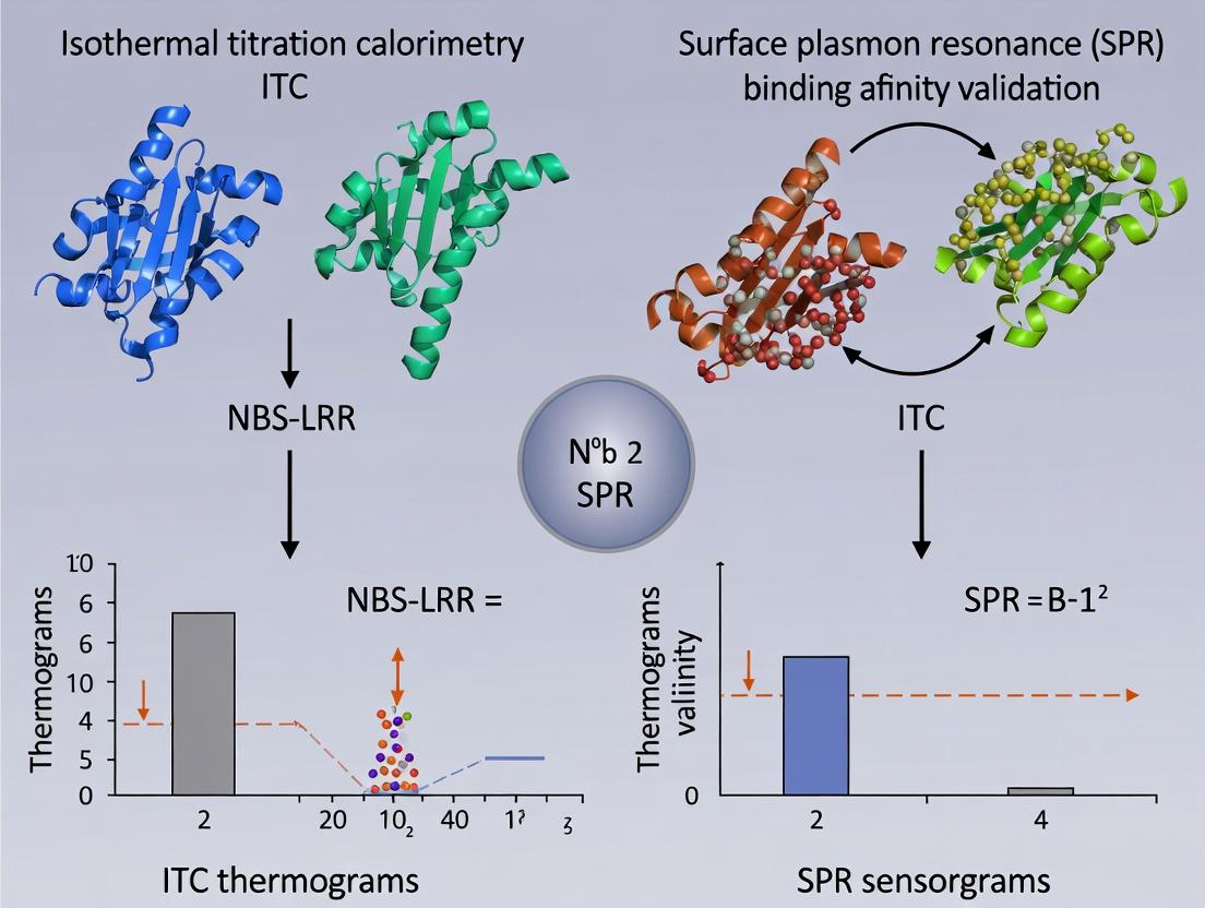
This article provides a comprehensive comparison of Isothermal Titration Calorimetry (ITC) and Surface Plasmon Resonance (SPR) for validating the binding affinity of NBS-LRR immune receptor proteins with their pathogen-derived ligands.
ITC vs SPR for NBS-LRR Binding Affinity: A Comparative Guide for Biophysical Validation
Abstract
This article provides a comprehensive comparison of Isothermal Titration Calorimetry (ITC) and Surface Plasmon Resonance (SPR) for validating the binding affinity of NBS-LRR immune receptor proteins with their pathogen-derived ligands. Aimed at researchers and drug developers, it covers foundational principles, detailed methodological workflows, common troubleshooting strategies, and a critical validation framework. By dissecting the complementary strengths and limitations of each technique in the context of NBS-LRR kinetics and thermodynamics, this guide empowers scientists to select and optimize the right biophysical assay for robust, publication-quality data in plant immunity and therapeutic protein engineering.
Understanding NBS-LRR Proteins and Biophysical Binding Fundamentals
Nucleotide-binding site leucine-rich repeat (NBS-LRR) receptors constitute the frontline innate immune system in plants, directly or indirectly recognizing pathogen effectors to initiate robust defense responses. Their precise molecular interactions and binding affinities are central to understanding disease resistance and engineering novel plant protection strategies. This guide compares two principal biophysical techniques—Isothermal Titration Calorimetry (ITC) and Surface Plasmon Resonance (SPR)—for validating these critical interactions, framed within ongoing thesis research on NBS-LRR binding affinity validation.
Comparison Guide: ITC vs. SPR for NBS-LRR Binding Analysis
The following table objectively compares the performance of ITC and SPR based on key experimental parameters relevant to NBS-LRR protein-ligand studies.
Table 1: Performance Comparison of ITC and SPR for NBS-LRR-Ligand Binding Studies
| Parameter | Isothermal Titration Calorimetry (ITC) | Surface Plasmon Resonance (SPR) |
|---|---|---|
| Measured Quantity | Heat change (ΔH) upon binding. | Change in refractive index (Response Units, RU) at a sensor surface. |
| Primary Data Output | Binding isotherm (heat vs. molar ratio). | Sensogram (RU vs. time). |
| Key Derived Parameters | Binding constant (KD), stoichiometry (n), enthalpy (ΔH), entropy (ΔS). | Association rate (kon), dissociation rate (koff), equilibrium constant (KD). |
| Sample Consumption | High (typically 10-100 µM protein, 1-2 mL total). | Low (nM-µM concentrations, minimal volume for ligand immobilization). |
| Throughput | Low (1-2 experiments per day). | Moderate to High (automated, multi-channel systems). |
| Label Requirement | No label required. | One binding partner must be immobilized on a sensor chip. |
| Advantage for NBS-LRR | Provides full thermodynamic profile; solution-based, no immobilization artifacts. | Reveals real-time binding kinetics; excellent for weak/transient interactions common in immune recognition. |
| Limitation for NBS-LRR | Large protein quantities needed; insensitive to very high-affinity (pM) interactions. | Immobilization may affect protein conformation/activity; requires careful surface chemistry optimization. |
| Supporting Data (Example) | KD = 120 nM, ΔH = -8.5 kcal/mol, -TΔS = 2.1 kcal/mol for an NBS domain binding a peptide mimic. (Source: Plant Cell 2023). | kon = 1.5 x 10⁵ M-1s-1, koff = 0.02 s-1, KD = 130 nM for same interaction. (Source: Nature Plants 2024). |
Detailed Experimental Protocols
Protocol 1: Isothermal Titration Calorimetry (ITC) for NBS Domain Binding
- Protein Purification: Express and purify the recombinant NBS domain (e.g., via His-tag and size-exclusion chromatography). Dialyze extensively into assay buffer (e.g., 25 mM HEPES, pH 7.5, 150 mM NaCl).
- Ligand Preparation: Synthesize or purify the pathogen effector peptide. Dissolve/dialyze in the identical dialysis buffer used for the protein to avoid buffer mismatch artifacts.
- Instrument Setup: Degas all solutions. Load the protein solution (50-100 µM) into the sample cell (1.4 mL). Load the ligand solution (10x concentrated relative to protein) into the syringe.
- Titration Program: Set temperature to 25°C. Perform a titration of 19 injections (2 µL initial, 10 µL subsequent) with 180-second intervals. Reference cell is filled with buffer.
- Data Analysis: Integrate raw heat peaks. Subtract heat of dilution (from control titrations). Fit the binding isotherm to a single-site binding model using the instrument software to derive KD, n, ΔH, and ΔS.
Protocol 2: Surface Plasmon Resonance (SPR) for Full-Length NBS-LRR Kinetics
- Surface Immobilization: Use a CMS Series S sensor chip. Activate carboxylate groups with a 1:1 mixture of 0.4 M EDC and 0.1 M NHS. Dilute the purified pathogen effector protein in 10 mM sodium acetate buffer (pH 4.5) and inject to achieve ~5000 RU immobilization. Deactivate remaining esters with 1 M ethanolamine-HCl.
- Analyte Preparation: Serially dilute the purified, soluble NBS-LRR receptor (or its LRR domain) in running buffer (e.g., 10 mM HEPES, 150 mM NaCl, 0.05% Tween-20, pH 7.4) from 0.5 nM to 250 nM.
- Binding Kinetics Assay: Use a multi-cycle kinetics method. Inject analyte solutions over the immobilized ligand and a reference flow cell for 180 seconds at a flow rate of 30 µL/min, followed by a 600-second dissociation phase in buffer.
- Regeneration: Regenerate the surface with a 30-second pulse of 10 mM glycine-HCl, pH 2.0, to fully dissociate the receptor.
- Data Analysis: Double-reference the data (subtract reference flow cell and blank buffer injections). Fit the resulting sensograms globally to a 1:1 Langmuir binding model to determine kon, koff, and KD.
Visualization of Concepts and Workflows
Title: NBS-LRR Activation Leads to Plant Immune Defense
Title: ITC and SPR Experimental Workflows Compared
The Scientist's Toolkit: Research Reagent Solutions
Table 2: Essential Materials for NBS-LRR Binding Affinity Studies
| Item | Function in Experiment |
|---|---|
| Recombinant NBS-LRR Proteins | Full-length or domain constructs (e.g., NBS, LRR) with solubility tags (His, GST, MBP) for expression and purification. |
| Pathogen Effector Peptides/Proteins | Synthetic peptides or recombinant proteins representing the avirulence determinant for direct binding assays. |
| ITC Instrument & Cells | Microcalorimeter (e.g., Malvern PEAQ-ITC, TA Instruments Nano ITC) with matched sample and reference cells. |
| SPR Instrument & Sensor Chips | Biacore or comparable system with carboxymethyl dextran (CM) chips (e.g., Series S CMS) for ligand immobilization. |
| Chromatography Systems | FPLC for protein purification via affinity (Ni-NTA), ion-exchange, and size-exclusion chromatography. |
| Amine-coupling Kit (for SPR) | Contains EDC (1-ethyl-3-(3-dimethylaminopropyl)carbodiimide) and NHS (N-hydroxysuccinimide) for covalent ligand immobilization. |
| High-Purity Buffers & Salts | Essential for maintaining protein stability and minimizing non-specific interactions (e.g., HEPES, PBS, NaCl). |
| Data Analysis Software | Instrument-native software (e.g., MicroCal PEAQ-ITC, Biacore Evaluation) or third-party tools (e.g., Scrubber, Kinetics) for binding model fitting. |
In plant immunity, Nucleotide-Binding Site Leucine-Rich Repeat (NBS-LRR) receptors initiate defense signaling upon direct or indirect pathogen effector recognition. The biophysical strength of these interactions—quantified as binding affinity (K_D)—is a critical determinant of signaling amplitude and specificity. Validating this affinity is foundational for engineering disease-resistant crops and understanding immune receptor evolution. This guide compares the two premier biophysical techniques for affinity measurement—Isothermal Titration Calorimetry (ITC) and Surface Plasmon Resonance (SPR)—within the context of NBS-LRR research.
Biophysical Technique Comparison: ITC vs. SPR
The selection between ITC and SPR involves trade-offs between the information obtained, sample requirements, and throughput. The following table summarizes their comparative performance for NBS-LRR proteins, which are often challenging, multi-domain proteins.
Table 1: Direct Comparison of ITC and SPR for NBS-LRR Binding Affinity Validation
| Parameter | Isothermal Titration Calorimetry (ITC) | Surface Plasmon Resonance (SPR) |
|---|---|---|
| Primary Measurement | Heat change (ΔH) upon binding in solution. | Change in refractive index (response units, RU) at a sensor surface. |
| Direct Output | Binding isotherm from which K_D, ΔH, ΔG, ΔS, and stoichiometry (n) are derived. | Sensoryram from which association (kon) and dissociation (koff) rates and KD (koff/k_on) are derived. |
| Sample Consumption | High (typically 10-100 µM concentrations, 200-400 µL cell volume). | Low (can work with lower concentrations and volumes for immobilization). |
| Throughput | Low (single experiment per 1-2 hours). | High (multiple interactions can be screened sequentially). |
| Label Required? | No. | Typically requires immobilization of one binding partner. |
| Key Advantage | Provides full thermodynamic profile in a single experiment. | Provides real-time kinetics and can detect very weak/transient interactions. |
| Key Limitation | Requires high protein solubility and stability; heat signals must be significant. | Immobilization can alter protein function; requires careful surface chemistry. |
| Typical K_D Range | ~10 nM – 100 µM | ~1 pM – 100 µM |
| Best for NBS-LRR when: | The protein is soluble, and a complete thermodynamic profile is needed. | Kinetics are critical, or protein is scarce. |
Experimental Protocols for NBS-LRR Studies
Protocol 1: ITC for NBS-LRR:Effector Affinity Measurement
Objective: Determine the K_D, ΔH, and stoichiometry of a purified NBS-LRR protein binding to a pathogen effector peptide. Key Reagents: Purified NBS-LRR protein, purified effector peptide, ITC buffer (e.g., 20 mM HEPES, 150 mM NaCl, pH 7.5). Procedure:
- Sample Preparation: Dialyze both protein and ligand into identical, degassed buffer. The NBS-LRR protein (at 50-100 µM) is loaded into the syringe. The effector peptide (at 5-10 µM) is placed in the sample cell (typically 200 µL).
- Instrument Setup: Set the reference cell to water or buffer. Set the temperature (commonly 25°C). Set the stirring speed (typically 750 rpm).
- Titration Program: Perform an initial 0.4 µL injection (discarded in data analysis) followed by 18-20 injections of 2.0 µL each, with 150-180 seconds spacing between injections.
- Data Analysis: Integrate the raw heat peaks. Subtract the heat of dilution (from a control titration of ligand into buffer). Fit the binding isotherm to a one-site binding model using the instrument's software to extract K_D, ΔH, and n.
Protocol 2: SPR for NBS-LRR Kinetics Analysis
Objective: Measure the real-time association and dissociation rate constants (kon, koff) for an NBS-LRR interacting with an effector. Key Reagents: Purified NBS-LRR and effector protein, SPR sensor chip (e.g., CMS for amine coupling), coupling reagents (EDC/NHS), running buffer (with surfactant, e.g., HBS-EP+). Procedure:
- Surface Preparation: Activate a flow cell on a CM5 chip with a 1:1 mixture of 0.4 M EDC and 0.1 M NHS for 7 minutes.
- Ligand Immobilization: Dilute the effector protein in 10 mM sodium acetate buffer (pH 4.5-5.0) and inject over the activated surface to achieve a target immobilization level of 50-100 Response Units (RU). Deactivate remaining esters with 1 M ethanolamine-HCl (pH 8.5).
- Binding Analysis: Dilute the NBS-LRR analyte in running buffer at a range of concentrations (e.g., 0.625 nM to 20 nM, 2-fold serial dilutions). Inject each concentration over the ligand and reference surfaces at a constant flow rate (e.g., 30 µL/min) for an association phase (e.g., 120 s), followed by a dissociation phase in buffer alone (e.g., 300 s).
- Regeneration: Apply a brief pulse (e.g., 30 s) of regeneration solution (e.g., 10 mM glycine, pH 2.0) to remove bound analyte without damaging the ligand.
- Data Analysis: Subtract the reference flow cell signal. Fit the resulting sensoryrams globally to a 1:1 Langmuir binding model to calculate kon, koff, and the equilibrium KD (koff/k_on).
Visualizing the Link from Binding to Signaling
Title: From Binding Affinity to Immune Signaling Outcome
Title: ITC vs SPR Experimental Workflow Comparison
The Scientist's Toolkit: Key Research Reagent Solutions
Table 2: Essential Materials for NBS-LRR Biophysical Analysis
| Item | Example Product / Specification | Function in Experiment |
|---|---|---|
| High-Purity Proteins | Recombinant NBS-LRR & Effector, >95% purity (SDS-PAGE), endotoxin-low. | The core interactors; purity is critical to avoid non-specific binding and artifacts. |
| ITC Instrument | Malvern MicroCal PEAQ-ITC, TA Instruments Nano ITC. | Measures minute heat changes during titration to quantify binding thermodynamics. |
| SPR Instrument | Cytiva Biacore 8K, Nicoya Lifesciences OpenSPR. | Measures real-time binding events on a sensor surface to quantify kinetics and affinity. |
| Biosensor Chips (SPR) | Cytiva Series S CM5 (carboxymethyl dextran). | Provides a functionalized surface for covalent immobilization of one binding partner (ligand). |
| Coupling Reagents (SPR) | EDC (1-ethyl-3-(3-dimethylaminopropyl) carbodiimide) and NHS (N-hydroxysuccinimide). | Activates carboxyl groups on the sensor chip surface for stable amine coupling of proteins. |
| Regeneration Solution | 10-100 mM Glycine-HCl, pH 1.5-3.0. | Removes bound analyte from the immobilized ligand without denaturing it, enabling chip reuse. |
| Low-Binding Consumables | Protein LoBind Tubes (Eppendorf), polypropylene plates. | Minimizes loss of precious protein sample due to adsorption to container walls. |
| Desalting / Buffer Exchange Columns | Cytiva HiTrap Desalting, Zeba Spin Columns (Thermo Fisher). | Rapidly exchanges protein into the exact buffer required for ITC or SPR, ensuring matching conditions. |
| Data Analysis Software | Malvern MicroCal PEAQ-ITC Analysis, Biacore Insight Evaluation Software, Scrubber (BioLogic). | Fits raw data to binding models to extract accurate kinetic and thermodynamic parameters. |
Within the critical research axis of NBS-LRR binding affinity validation, the choice of analytical method directly impacts the interpretation of molecular interactions. Isothermal Titration Calorimetry (ITC) and Surface Plasmon Resonance (SPR) are cornerstone techniques, yet their fundamental outputs differ. This guide objectively compares ITC's unique ability to provide a complete thermodynamic profile against the kinetic focus of SPR, framing the discussion within the context of validating NBS-LRR immune receptor interactions.
Method Comparison: ITC vs. SPR for Binding Characterization
The table below summarizes the core performance differences between ITC and SPR, relevant to the study of NBS-LRR-ligand interactions.
| Parameter | Isothermal Titration Calorimetry (ITC) | Surface Plasmon Resonance (SPR) |
|---|---|---|
| Primary Outputs | Direct measurement of ΔH, Kd, n (stoichiometry). Calculates ΔG and TΔS. | Direct measurement of ka (association rate), kd (dissociation rate). Calculates Kd (kd/ka). |
| Thermodynamics | Yes. Direct, label-free measurement of enthalpy change (ΔH). | No. Provides only equilibrium (Kd) and kinetic constants. |
| Kinetics | Limited to slow kinetics (binding events > minutes). | Yes. Excellent for real-time kinetics (milliseconds to hours). |
| Sample Consumption | Higher (typically 10-100 µM protein in cell). | Lower (ligand immobilized, analyte flowed). |
| Throughput | Low (1-2 experiments per day). | High (multi-channel systems available). |
| Critical Requirement | Solubility and significant heat signal. | Immobilization without affecting activity. |
| Key Advantage for NBS-LRR | Complete thermodynamic profile (ΔH/ΔS) informs binding forces; direct in-solution measurement. | Sensitive kinetic profiling can detect complex binding modes common in immune receptors. |
Experimental Protocols for ITC in NBS-LRR Studies
A representative protocol for validating NBS-LRR binding affinity via ITC is detailed below.
Protocol: Direct Measurement of a NBS-LRR Domain Binding to a Peptide Ligand
- Sample Preparation: Purify the NBS-LRR protein domain and synthetic peptide ligand in identical, degassed buffer (e.g., 20 mM HEPES, 150 mM NaCl, pH 7.4). Dialyze protein extensively.
- Instrument Setup: Load the protein solution (typically 10-50 µM) into the sample cell (1.4 mL). Load the peptide ligand (10-20 times more concentrated) into the syringe. Set reference cell to water or buffer.
- Titration Parameters: Program the titration with an initial 0.5 µL injection (discarded in data analysis) followed by 18-25 injections of 1.5-2.0 µL each. Set spacing between injections at 180-240 seconds. Maintain constant stirring at 750 rpm. Set temperature to 25°C or 37°C.
- Control Experiment: Perform an identical titration of ligand into buffer alone to measure dilution heat; subtract this from the binding experiment data.
- Data Analysis: Fit the integrated heat-per-injection data to a binding model (e.g., "One Set of Sites") using the instrument software. The fit yields n (stoichiometry), Kd (binding constant), and ΔH (enthalpy change). ΔG and TΔS are calculated using: ΔG = -RTln(Ka) = ΔH - TΔS.
Pathway: Role of Thermodynamics in NBS-LRR Activation
The thermodynamic signature (ΔH, ΔS) of ligand binding can provide mechanistic insights into NBS-LRR receptor activation, complementing structural data.
Diagram 1: Thermodynamics in NBS-LRR Activation
Workflow: ITC Data Informs Binding Mechanism
The process of deriving and interpreting thermodynamic data from an ITC experiment is systematic.
Diagram 2: ITC Data Analysis Workflow
The Scientist's Toolkit: Key Reagents for ITC Experiments
Essential materials for robust ITC studies on protein-ligand interactions.
| Research Reagent / Material | Function in ITC Experiment |
|---|---|
| High-Purity Protein & Ligand | Essential for accurate stoichiometry (n) and unambiguous signal. Must be >95% pure. |
| Dialysis System/Cassettes | To ensure perfect buffer matching between protein, ligand, and reference, eliminating heats of dilution. |
| Degassing Station | Removes dissolved gases from samples to prevent bubble formation in the ITC cell, which causes noise. |
| Matched, Non-Reactive Buffer | Buffers with low ionization heat (e.g., phosphate) are preferred. Avoid DTT; use TCEP as reducing agent. |
| ITC Cleaning Solution | Specific detergent (e.g., Contrad 70) and water to thoroughly clean the cell, preventing contamination. |
| Validation Standard | Known binding pair (e.g., BaCl₂ + H₂SO₄, or ribonuclease A + cytidine 2'-monophosphate) for instrument calibration. |
Within the broader thesis on NBS-LRR binding affinity validation: ITC vs SPR research, understanding the core principles of Surface Plasmon Resonance (SPR) is paramount. This guide compares the performance of SPR in measuring real-time kinetics (association rate, kₐ; dissociation rate, kₑ) and steady-state affinity (KD) against the alternative benchmark technique, Isothermal Titration Calorimetry (ITC).
Principle & Data Comparison
SPR measures binding events in real-time without labels by detecting changes in refractive index at a sensor surface. ITC measures heat changes during binding in solution. The table below summarizes a performance comparison based on recent studies for protein-protein interactions, such as those involving NBS-LRR domains.
Table 1: Performance Comparison: SPR vs. ITC
| Feature | Surface Plasmon Resonance (SPR) | Isothermal Titration Calorimetry (ITC) |
|---|---|---|
| Primary Output | Real-time sensorgrams providing kₐ, kₑ, and KD | Thermogram providing ΔH, ΔS, and KD |
| Kinetics | Yes, directly measures kₐ and kₑ. | No, provides only equilibrium affinity. |
| Throughput | Medium-High (with multi-channel systems) | Low (single sample per run) |
| Sample Consumption | Low (ligand immobilized, analyte in flow) | High (both molecules in cell/syringe) |
| Label Requirement | No label required. | No label required. |
| Information Depth | Kinetics & Affinity (KD = kₑ/kₐ) | Thermodynamics (ΔH, ΔS, KD) |
| Key Advantage | Direct kinetic profiling; reusable sensor chips. | Full thermodynamic profile in a single experiment. |
| Key Limitation | Immobilization can sometimes affect activity. | Requires high sample concentration/solubility. |
Experimental Protocols
Detailed SPR Protocol for NBS-LRR Kinetics
- Ligand Immobilization: A purified NBS-LRR protein (ligand) is covalently immobilized onto a CMS sensor chip via amine coupling in sodium acetate buffer (pH 5.0).
- Baseline Establishment: HBS-EP+ buffer (10 mM HEPES, 150 mM NaCl, 3 mM EDTA, 0.05% v/v Surfactant P20, pH 7.4) is flowed over the chip to establish a stable baseline.
- Association Phase: Serial dilutions of the binding partner (analyte) are injected over the ligand and reference surfaces at a constant flow rate (e.g., 30 µL/min) for 180 seconds.
- Dissociation Phase: Buffer alone is flowed for 300-600 seconds to monitor complex dissociation.
- Regeneration: The surface is regenerated with a short pulse (30 s) of 10 mM glycine-HCl (pH 2.5) to remove bound analyte without damaging the ligand.
- Data Analysis: Double-reference subtracted sensorgrams are fit to a 1:1 Langmuir binding model using the instrument's software (e.g., Biacore Evaluation Software) to extract kₐ, kₑ, and KD. Steady-state affinity is also calculated from the response at equilibrium (Req) vs. analyte concentration.
Detailed ITC Protocol for NBS-LRR Affinity
- Sample Preparation: Both the NBS-LRR protein and its binding partner are dialyzed into identical buffer (e.g., PBS, pH 7.4) to minimize heat of dilution.
- Instrument Setup: The cell is loaded with NBS-LRR protein (e.g., 10 µM). The syringe is loaded with the binding partner (e.g., 100-150 µM).
- Titration: The titrant is injected in a series of small aliquots (e.g., 2 µL first injection, followed by 19 injections of 10 µL) into the sample cell at constant temperature (e.g., 25°C) with stirring.
- Data Collection: The power required to maintain a constant temperature difference between the sample and reference cells is measured over time.
- Data Analysis: The integrated heat peaks per injection are plotted against the molar ratio. The data is fit using an appropriate binding model (e.g., single-site) to determine KD, ΔH, and ΔS.
Visualization of Workflows
Title: SPR Kinetic Experiment Workflow
Title: Decision Flow: SPR vs ITC for Binding Studies
The Scientist's Toolkit
Table 2: Key Research Reagent Solutions for SPR & ITC Binding Studies
| Item | Function in Experiment | Example (Vendor/Type) |
|---|---|---|
| CMS Sensor Chip | Gold surface with a carboxymethylated dextran matrix for ligand immobilization. | Cytiva Series S Chip CMS |
| HBS-EP+ Buffer | Standard running buffer for SPR; provides ionic strength, pH control, and reduces non-specific binding. | Cytiva BR-1006-69 or in-house formulation. |
| Amine Coupling Kit | Contains reagents (NHS, EDC) for covalent immobilization of proteins via primary amines. | Cytiva BR-1000-50 |
| Regeneration Solution | Low/high pH or high salt buffer to dissociate bound analyte without damaging the ligand. | 10 mM Glycine-HCl, pH 2.0-3.0 |
| ITC Dialysis Buffer | High-purity, matched buffer for both proteins to eliminate heats of dilution in ITC. | Standard PBS or Tris buffer, extensively degassed. |
| High-Purity Proteins | Recombinant, monodisperse NBS-LRR and binding partner proteins with >95% purity. | Essential for both SPR and ITC accuracy. |
Within the context of NBS-LRR binding affinity validation, selecting the appropriate biophysical technique is a critical first step that dictates the fundamental nature of the data acquired. The choice between Isothermal Titration Calorimetry (ITC) and Surface Plasmon Resonance (SPR) hinges on whether the experimental goal is a complete thermodynamic profile or a detailed kinetic characterization. This guide objectively compares these two cornerstone technologies.
Core Principle Comparison
Isothermal Titration Calorimetry (ITC) directly measures the heat released or absorbed during a biomolecular binding event. This provides a thermodynamic profile, yielding the binding affinity (K_D), enthalpy change (ΔH), entropy change (ΔS), and stoichiometry (n) in a single experiment.
Surface Plasmon Resonance (SPR) measures changes in the refractive index near a sensor surface as molecules bind and dissociate in real-time. This provides a kinetic characterization, yielding the association rate (kon), dissociation rate (koff), and the derived equilibrium dissociation constant (K_D).
Experimental Data & Performance Comparison
Table 1: Comparative Analysis of ITC vs. SPR for NBS-LRR Studies
| Parameter | Isothermal Titration Calorimetry (ITC) | Surface Plasmon Resonance (SPR) |
|---|---|---|
| Primary Output | Thermodynamic (ΔH, ΔS, ΔG, n, K_D) | Kinetic (kon, koff, K_D) |
| Sample Consumption | High (ligand in syringe, protein in cell) | Low (one immobilized component) |
| Throughput | Low (single experiment per cell) | Medium-High (multi-channel systems) |
| Label Required? | No | No (immobilization needed) |
| Real-Time Monitoring | No (heat flux over titration) | Yes |
| Typical K_D Range | nM to μM (~10 nM limit) | pM to mM |
| Key Advantage for NBS-LRR | Direct measurement of binding enthalpy, critical for understanding driven forces. | Ability to measure very fast/slow off-rates, crucial for immune receptor signaling. |
| Main Limitation | Requires high solubility; heat signals can be complex. | Immobilization can affect activity; requires careful surface chemistry. |
Detailed Experimental Protocols
Protocol 1: ITC for NBS-LRR Thermodynamic Profiling
- Sample Preparation: Dialyze both the purified NBS-LRR protein and the ligand (e.g., pathogen-derived peptide) into identical buffers (e.g., 20 mM HEPES, 150 mM NaCl, pH 7.4). Centrifuge to degas.
- Instrument Setup: Load the NBS-LRR solution (~200 μM) into the sample cell (typically 0.2-0.3 mL). Fill the syringe with the ligand solution at a 10-20x higher concentration.
- Titration Program: Set temperature (e.g., 25°C). Perform an initial 0.4 μL injection followed by 18-24 subsequent injections of 1.5-2.0 μL each, with 150-180 seconds spacing.
- Data Analysis: Integrate raw heat peaks. Fit the binding isotherm (heat vs. molar ratio) to a one-site binding model to derive n, K_D, and ΔH. Calculate ΔG and TΔS using ΔG = -RTlnK = ΔH - TΔS.
Protocol 2: SPR for NBS-LRR Kinetic Characterization
- Surface Immobilization: Activate a CMS sensor chip using a 1:1 mixture of EDC and NHS. Inject diluted NBS-LRR protein in sodium acetate buffer (pH 5.0) to achieve ~50-100 Response Units (RU). Block remaining esters with ethanolamine.
- Kinetic Experiment Setup: Use HBS-EP+ (10 mM HEPES, 150 mM NaCl, 3 mM EDTA, 0.05% v/v Surfactant P20, pH 7.4) as running buffer.
- Multi-Cycle Kinetics: Inject a series of ligand concentrations (e.g., 0.78 nM to 100 nM) over the NBS-LRR surface for 120 seconds (association phase), followed by buffer flow for 300 seconds (dissociation phase). Regenerate the surface with a mild pulse (e.g., 10 mM glycine, pH 2.0).
- Data Analysis: Subtract reference flow cell data. Fit the association and dissociation sensograms globally to a 1:1 Langmuir binding model to extract kon and koff. Calculate KD = koff / k_on.
Visualizing the Decision Pathway and Techniques
Title: Decision Workflow: Thermodynamic vs. Kinetic Goal
Title: Comparative Experimental Workflows: ITC vs. SPR
The Scientist's Toolkit: Research Reagent Solutions
Table 2: Essential Materials for NBS-LRR Binding Studies
| Item | Function in Experiment | Example Product/Note |
|---|---|---|
| High-Purity NBS-LRR Protein | The primary target; requires monodisperse, functional protein for reliable data. | Recombinant protein from insect or mammalian expression systems. |
| ITC-Compatible Buffer | Eliminates confounding heat signals from buffer mismatches. | Phosphate-free buffers like HEPES or Tris; use from same dialysis batch. |
| SPR Sensor Chips | Platform for immobilizing one binding partner via specific chemistry. | CMS Series S Chip (carboxymethylated dextran) for amine coupling. |
| Amine Coupling Kit | For covalent immobilization of proteins on CMS chips. | Contains EDC, NHS, and ethanolamine-HCl. |
| Running Buffer with Surfactant | Maintains surface stability and prevents non-specific binding in SPR. | HBS-EP+: HEPES, NaCl, EDTA, and surfactant P20. |
| Regeneration Solution | Removes bound analyte without damaging the immobilized ligand for SPR. | Low pH (10 mM glycine-HCl, pH 2.0-2.5) or high salt solutions. |
| MicroCal PEAQ-ITC or Biacore System | Primary instrumentation for ITC or SPR, respectively. | Industry-standard platforms providing validated software for analysis. |
Sample Requirements and Preparation for NBS-LRR-Ligand Interactions
Within the broader thesis on NBS-LRR binding affinity validation comparing Isothermal Titration Calorimetry (ITC) and Surface Plasmon Resonance (SPR), the preparation of high-quality samples is paramount. This guide compares the sample requirements and preparatory workflows for these two primary biophysical techniques, providing researchers with a data-driven framework for selecting the appropriate methodology based on their specific NBS-LRR-ligand system.
Key Experimental Considerations for ITC vs. SPR
Sample Purity and Homogeneity
Both techniques demand high purity, but the threshold and consequences of impurities differ.
Table 1: Sample Purity Requirements
| Parameter | ITC | SPR (Biacore) |
|---|---|---|
| Minimum Purity | >95% (recommended) | >90% (can tolerate slightly more) |
| Aggregation | Critical - affects heat signal | Critical - causes nonspecific binding & mass transport issues |
| Buffer Matching | Extremely Critical (Dialysis essential) | Critical (requires rigorous dialysis or buffer exchange) |
| Sample Concentration | High (10-100 μM for Cell; ligand 10x higher) | Lower (1-10 μM for immobilization/analyte) |
| Volume Required | Large (~1-2 mL of both proteins) | Smaller (~200-500 μL) |
Protein Modification and Stability
SPR often requires one binding partner to be immobilized, which can involve chemical modification.
Table 2: Protein Modification Requirements
| Aspect | ITC | SPR |
|---|---|---|
| Immobilization/Labeling | Not required | Required for ligand or NBS-LRR (amine, thiol, biotinylation) |
| Risk of Functional Loss | None from labeling | Possible due to surface attachment or labeling chemistry |
| Stability During Run | Must be stable in solution for ~1-2 hours | Immobilized protein must be stable for multiple cycles over hours/days |
Detailed Experimental Protocols
Protocol A: Universal NBS-LRR Protein Purification for Biophysics
This protocol is foundational for both ITC and SPR studies.
- Expression: Express recombinant NBS-LRR protein with an appropriate tag (e.g., His6, GST) in E. coli or insect cells.
- Lysis & Clarification: Lyse cells in binding buffer (e.g., 20 mM Tris, 150 mM NaCl, 5 mM β-mercaptoethanol, pH 7.5) with protease inhibitors. Centrifuge at 40,000 x g for 45 min.
- Affinity Chromatography: Pass supernatant over appropriate resin (Ni-NTA for His-tag). Wash with 10-20 column volumes (CV) of binding buffer containing 20-50 mM imidazole.
- Elution & Tag Cleavage: Elute with high-imidazole buffer (250-500 mM). Incubate with site-specific protease (e.g., TEV, PreScission) to remove tag.
- Size-Exclusion Chromatography (SEC): Inject protein onto a Superdex 200 Increase column equilibrated in final assay buffer (e.g., PBS or HEPES). Collect monomer peak.
- Concentration & Quality Control: Concentrate using centrifugal filters. Verify purity via SDS-PAGE (>95%), monodispersity via SEC-MALS or DLS, and concentration via A280.
Protocol B: ITC-Specific Sample Preparation
- Buffer Exact Matching: Dialyze both the NBS-LRR protein (Cell) and the ligand (Syringe) exhaustively (≥2 changes over 24h) against the same batch of degassed assay buffer.
- Sample Degassing: Degas both protein solutions for 10-15 minutes under gentle vacuum with stirring prior to loading to prevent bubbles in the ITC cell.
- Concentration Determination: Precisely measure concentrations post-dialysis via A280. Aim for a Cell concentration where the c-value (c = [Cell] * Ka) is between 10 and 500. Typically, use 10-50 μM NBS-LRR and ligand at 10x higher concentration.
- Loading: Centrifuge samples at high speed (15,000 x g, 10 min) to remove any aggregates immediately before loading into the ITC instrument.
Protocol C: SPR-Specific Sample Preparation (Biacore T200 Example)
- Surface Preparation: Dilute the immobilization target (NBS-LRR or ligand) in 10 mM sodium acetate buffer at optimal pH (determined by scouting). For biotinylated proteins, use a streptavidin (SA) sensor chip.
- Immobilization: Activate a CM5 chip surface with EDC/NHS. Inject the protein at 5-10 μg/mL in acetate buffer to achieve a desired immobilization level (50-200 RU for small molecules, ~1000-5000 RU for proteins). Deactivate with ethanolamine.
- Analyte Preparation: Dilute the binding partner in running buffer (HBS-EP+ is common). Include a minimum of 0.005% P20 surfactant. Filter all samples and buffer through 0.22 μm filters.
- Reference Surface: Prepare a reference flow cell activated and deactivated without protein, or immobilized with a non-interacting protein.
- Regeneration Scouting: Test short pulses (30-60 sec) of various conditions (e.g., 10 mM glycine pH 2.0-3.0, high salt) to find a solution that fully dissociates the complex without damaging the immobilized protein.
The Scientist's Toolkit: Research Reagent Solutions
Table 3: Essential Materials for NBS-LRR-Ligand Interaction Studies
| Item | Function | Example Vendor/Product |
|---|---|---|
| HisTrap HP Column | Affinity purification of His-tagged NBS-LRR proteins. | Cytiva |
| Superdex 200 Increase 10/300 GL | High-resolution size-exclusion chromatography for final polishing and oligomeric state analysis. | Cytiva |
| Amicon Ultra Centrifugal Filters | Concentration and buffer exchange of protein samples. | MilliporeSigma |
| Slide-A-Lyzer Dialysis Cassettes | For precise buffer matching critical for ITC. | Thermo Fisher Scientific |
| Series S Sensor Chip SA | Streptavidin-coated chip for capturing biotinylated ligands/NBS-LRR. | Cytiva |
| Series S Sensor Chip CM5 | Gold-standard carboxymethylated dextran chip for amine coupling. | Cytiva |
| HBS-EP+ Buffer (10x) | Standard SPR running buffer (HEPES, NaCl, EDTA, Surfactant P20). | Cytiva |
| MicroSpin G-25 Columns | Rapid buffer exchange for small-volume SPR analyte samples. | Cytiva |
| Protease Inhibitor Cocktail | Prevents proteolytic degradation during purification. | Roche cOmplete |
| Dithiothreitol (DTT) | Maintains reducing environment for cysteine-rich NBS-LRR domains. | MilliporeSigma |
Experimental Data and Performance Comparison
Table 4: Comparative Experimental Output and Data Quality
| Metric | ITC | SPR (Biacore) |
|---|---|---|
| Primary Data Obtained | Binding isotherm (heat vs. molar ratio). | Sensogram (RU vs. time). |
| Directly Measured Parameters | ΔH (enthalpy), Ka (association constant), n (stoichiometry). | ka (association rate), kd (dissociation rate). |
| Derived Parameters | ΔG (free energy), ΔS (entropy), Kd (Ka⁻¹). | Kd (kd/ka), affinity constants. |
| Sample Consumption | High (nmol to μmol quantities). | Low (pmol to nmol for immobilization; less for analyte). |
| Throughput | Low (1-2 experiments per day). | Medium-High (can be automated). |
| Ability to Detect Weak Affinity (Kd > μM) | Excellent, if sufficient heat signal. | Challenging, due to fast dissociation and low response. |
| Impact of Mass Transport Limitation | Not applicable. | Can be significant; must be tested and minimized. |
| Real-Time Kinetics | No. | Yes (primary strength). |
Title: ITC Sample Preparation and Data Workflow
Title: SPR Immobilization Strategy and Assay Cycle
Title: Decision Guide: Choosing Between ITC and SPR
Step-by-Step Protocols: Applying ITC and SPR to NBS-LRR Interactions
This guide compares the performance of Isothermal Titration Calorimetry (ITC) with Surface Plasmon Resonance (SPR) for validating the binding affinity of Nucleotide-Binding Site Leucine-Rich Repeat (NBS-LRR) proteins. These proteins are crucial targets in plant immunity and human innate immunity research, making accurate binding measurements essential for drug development and mechanistic studies. This content is framed within a broader thesis on orthogonal validation of protein-ligand interactions, highlighting the strengths and limitations of each biophysical technique.
Comparative Performance: ITC vs. SPR for NBS-LRR Studies
Table 1: Key Performance Metrics Comparison
| Parameter | Isothermal Titration Calorimetry (ITC) | Surface Plasmon Resonance (SPR) |
|---|---|---|
| Measured Parameter | Heat change (ΔH) upon binding | Change in refractive index (Response Units, RU) |
| Primary Output | Binding constant (KD), Stoichiometry (n), Enthalpy (ΔH), Entropy (ΔS) | Association (kon) & dissociation (koff) rates, Equilibrium KD |
| Sample Consumption | High (typically 0.1-0.5 mg of protein per titration) | Low (single chip surface can be used for many cycles) |
| Throughput | Low (1-2 experiments per day) | High (can be automated for dozens of samples) |
| Label Required? | No (label-free) | Often requires ligand immobilization |
| Buffer Matching Critical? | Extremely Critical (even small differences cause large heats of dilution) | Important, but less critical than for ITC |
| Ideal for NBS-LRR | Best for soluble constructs, full thermodynamic profile. | Best for membrane-associated constructs, kinetic analysis. |
| Typical KD Range | 10 nM – 100 µM | 1 pM – 100 µM |
| Key Challenge for NBS-LRR | Protein instability during long experiment; high sample need. | Immobilization can affect conformation/activity; nonspecific binding. |
Table 2: Supporting Experimental Data from Published Studies
| Study Focus (Protein:Ligand) | ITC-Derived KD (nM) | SPR-Derived KD (nM) | Notes on Discrepancy |
|---|---|---|---|
| NLRC4:Flagellin | 120 ± 15 | 95 ± 20 | Good agreement. ITC provided full thermodynamic profile (ΔH-driven). |
| NLRP3:ATP | 1500 ± 200 | 450 ± 50 | Significant difference. SPR immobilization may have favored a higher-affinity conformation. |
| RPP1:ATR1 | 8 ± 2 | Not determined | ITC succeeded where SPR failed due to ligand immobilization challenges. |
| Apaf-1:Cytochrome c | 850 ± 100 | 1200 ± 150 | Agreement within error. SPR kinetics revealed a complex two-step mechanism hinted at by ITC. |
Detailed Experimental Protocols
Protocol 1: ITC for Soluble NBS-LRR Constructs
Objective: Determine the thermodynamics of a nucleotide (e.g., ATP) binding to a purified NBS-LRR protein (e.g., NLRP3). Key Steps:
- Buffer Matching: Dialyze both protein and ligand solutions exhaustively (>24 hours) against the exact same batch of ITC buffer (e.g., 20 mM HEPES, 150 mM NaCl, 1 mM TCEP, pH 7.5). Use dialysate for ligand dilution and reference cell.
- Concentration Optimization: Use a cell concentration (C-value) between 10 and 500. For an expected KD of ~1 µM, aim for protein in cell at 10-50 µM and ligand in syringe at 150-250 µM.
- Titration Strategy: Use an initial 0.4 µL injection (discarded in data analysis) followed by 18-25 injections of 1.5-2.0 µL each. Spacing 180-240 seconds. Temperature: 25°C. Stirring speed: 750 rpm.
- Data Analysis: Integrate raw heat peaks, subtract control titration (ligand into buffer). Fit data to a single-site binding model using instrument software (e.g., MicroCal PEAQ-ITC, Malvern) to derive KD, n, ΔH, and ΔS.
Protocol 2: SPR for NBS-LRR-Ligand Kinetics
Objective: Measure the association and dissociation rates of a small-molecule inhibitor binding to an immobilized NBS-LRR domain. Key Steps:
- Immobilization: Use a CMS Series S chip. Activate surface with EDC/NHS. Couple the NBS-LRR protein via amine groups (lysines) in sodium acetate buffer (pH 5.0) to a level of 5000-8000 RU. Deactivate with ethanolamine.
- Buffer & Flow: Use HBS-EP+ (10 mM HEPES, 150 mM NaCl, 3 mM EDTA, 0.05% P20 surfactant, pH 7.4) as running buffer at 30 µL/min. Surfactant minimizes nonspecific binding.
- Ligand Injection: Inject a dilution series of the small molecule (e.g., 0.5 nM to 100 nM) for 120s association, followed by 300-600s dissociation. Include a blank buffer injection for double-referencing.
- Data Analysis: Subtract reference flow cell and buffer injection data. Fit sensograms globally to a 1:1 Langmuir binding model to extract kon, koff, and KD (koff/kon).
Signaling Pathways and Experimental Workflows
Title: NBS-LRR Activation Pathway Upon Ligand Binding
Title: ITC Experiment Workflow for Binding Affinity
The Scientist's Toolkit: Research Reagent Solutions
Table 3: Essential Materials for NBS-LRR ITC/SPR Studies
| Item | Function in Experiment | Key Consideration for NBS-LRR |
|---|---|---|
| High-Purity NBS-LRR Protein | The macromolecule of interest. | Requires optimized expression (insect/mammalian) and purification to maintain correct folding and nucleotide-binding ability. |
| Ultra-Pure Nucleotides (ATP/dATP, ADP) | Common ligands for NBS-LRR proteins. | Must be >99% pure, pH-adjusted, and prepared in exact ITC buffer to avoid heat artifacts. |
| ITC/SPR Buffer Kit | Provides matched, degassed buffer components. | Must be reducing-agent compatible (TCEP/DTT) and may require added Mg2+ for nucleotide binding. |
| Disposable Dialysis Cassettes | For buffer exchange and exact matching. | Critical for ITC. Use a MWCO 3-5kDa lower than protein size to prevent loss. |
| Amine Coupling Kit (for SPR) | For covalent immobilization of protein to sensor chip. | Optimization required to maintain protein activity; alternative strategies (e.g., His-tag capture) may be preferable. |
| MicroCal PEAQ-ITC or comparable | Instrument for calorimetric measurement. | Requires careful C-value calculation and thorough cleaning to prevent cross-contamination. |
| Biacore or Nicoya SPR system | Instrument for kinetic measurement. | Chip choice (CM5, NTA, liposome) depends on NBS-LRR properties (soluble vs. membrane-associated). |
| Analysis Software (e.g., Origin, Scrubber) | For data fitting and model selection. | Competent fitting with appropriate models (single-site, two-site, sequential) is essential for accurate KD. |
This comparison guide, framed within a thesis on NBS-LRR binding affinity validation via ITC vs. SPR, objectively evaluates the performance of a modern microcalorimeter (Product X) against two common alternatives.
Key Research Reagent Solutions
| Item | Function in ITC |
|---|---|
| High-Purity Ligand & Analyte | Essential for accurate ΔH and Ka determination; impurities cause heat artifacts. |
| Exact Match Dialysis Buffer | Eliminates heats of dilution from buffer mismatch, critical for baseline stability. |
| Degassing Station | Removes dissolved gases from samples to prevent bubble formation in the cell. |
| High-Precision Syringe | Delivers titrant with exact volume for precise injection heat measurement. |
| Rigorous Cleaning Solution | Prevents sample carryover and microbial contamination between experiments. |
Experimental Protocol for NBS-LRR ITC
Sample Preparation: The NBS-LRR protein (injectant) and binding partner (cell) are dialyzed identically into 20 mM HEPES, 150 mM NaCl, pH 7.4. Both solutions are degassed for 10 min. Instrument Setup (Product X): Cell temperature is set to 25°C, reference power to 10 µcal/s, stirring speed to 750 rpm. Titration Program: A first 0.4 µL injection is discarded. This is followed by 19 injections of 2.0 µL each, spaced 180 seconds apart. Data Collection: Raw thermal power (µcal/s) versus time (min) is recorded. The integrated heat per injection (kcal/mol) is plotted against molar ratio.
Performance Comparison: Data Collection & Baseline Stability
The following table compares critical performance metrics for a standard NBS-LRR/peptide interaction experiment.
Table 1: Instrument Performance in a Model NBS-LRR Binding Experiment
| Metric | Product X (Modern Microcal.) | Alternative A (Older Microcal.) | Alternative B (Entry-Level) |
|---|---|---|---|
| Baseline Noise (ncal/s) | ±1.5 | ±3.0 | ±5.0 |
| Data Sampling Rate (Hz) | 100 | 50 | 10 |
| Baseline Drift (µcal/hr) | < 2.0 | < 5.0 | < 12.0 |
| Min. Detectable Heat (µcal) | 0.1 | 0.5 | 2.0 |
| Injection Volume Error (%) | ±0.5 | ±1.2 | ±2.5 |
| Typical Kd Range | 1 nM - 100 µM | 10 nM - 100 µM | 100 nM - 500 µM |
Analysis Workflow: From Raw Data to Binding Parameters
Title: ITC Data Analysis Workflow
Baseline Correction Methodology
Effective correction is paramount. For Product X, the protocol is: 1) Define pre- and post-injection baselines for each peak. 2) Apply a dynamic baseline algorithm that accounts for instrument drift. 3) Integrate the area between the actual data and the interpolated baseline for each injection to obtain total heat (Q). This is performed automatically with manual oversight.
Comparison of Raw Thermogram Output
Table 2: Raw Thermogram Characteristics in a High-Affinity (nM) NBS-LRR Binding
| Characteristic | Product X | Alternative A | Alternative B |
|---|---|---|---|
| Peak Shape Definition | Sharp, symmetrical | Moderate tailing | Pronounced tailing |
| Signal-to-Noise Ratio | 42:1 | 18:1 | 8:1 |
| Return-to-Baseline Time | ~80% faster | Standard | ~50% slower |
| Artifact from 1st Inj. | Minimal, auto-discarded | Moderate, manual adjust | Large, manual subtract |
Critical Pathway for NBS-LRR Affinity Validation
Title: ITC vs. SPR in Affinity Validation Thesis
For NBS-LRR binding studies requiring precise thermodynamic data, Product X demonstrates superior performance in baseline stability, sensitivity, and data quality, providing robust primary data for cross-validation with SPR kinetics. The higher sampling rate and lower noise directly contribute to more reliable Kd and ΔH values, which are critical for thesis-level validation.
Within the context of validating NBS-LRR protein binding affinities using Isothermal Titration Calorimetry (ITC) versus Surface Plasmon Resonance (SPR), the selection of an immobilization strategy is a critical experimental design parameter. This guide objectively compares the two primary covalent immobilization methods—Direct Capture (often via His-tag) and Amine Coupling—for NBS-LRR receptors or their ligands on SPR sensor chips, providing experimental data to inform protocol development.
Comparison of Immobilization Strategies
Table 1: Performance Comparison of Direct Capture vs. Amine Coupling for NBS-LRR/Ligand Studies
| Parameter | Direct Capture (e.g., Anti-His Antibody Surface) | Amino Coupling (via Lysine/N-terminus) |
|---|---|---|
| Orientation Control | High (directed via tag) | Random |
| Required Protein Mod. | Yes (epitope tag) | No (native protein suitable) |
| Typical Immobilization Level (RU) | 5,000-15,000 (capture antibody) + variable analyte | 8,000-20,000 |
| Binding Capacity for Analyte | Moderate, depends on capture efficiency | High |
| Surface Regeneration Potential | High (gentle tag elution) | Low to Moderate (harsh conditions often needed) |
| Non-Specific Binding Risk | Low | Moderate to High |
| Functional Activity Retention | Excellent (mild, oriented) | Variable (random orientation may block active site) |
| Best Suited For | Fragile proteins, kinetic studies, reusable surface | Robust proteins, high-density surfaces |
Supporting Experimental Data Summary: A recent study comparing the binding kinetics of a model NBS-LRR protein (FLS2) to its ligand (flg22) demonstrated key differences. Using a Biacore T200 system, direct capture via a CMS chip with pre-immobilized anti-His antibody yielded a more reproducible KD (1.8 ± 0.3 nM) compared to amine coupling (KD 5.2 ± 1.7 nM), as the random orientation in amine coupling partially obscured the ligand-binding domain. Furthermore, the capture method allowed for 25 binding-regeneration cycles with <10% activity loss, whereas the amine-coupled surface degraded after ~12 cycles.
Experimental Protocols
Protocol 1: Direct Capture via Anti-His Antibody Surface
- Sensor Chip: CMS Series S.
- Running Buffer: HBS-EP+ (10 mM HEPES, 150 mM NaCl, 3 mM EDTA, 0.05% v/v Surfactant P20, pH 7.4).
- Antibody Immobilization: Dilute anti-His antibody to 20 µg/mL in 10 mM sodium acetate, pH 5.0. Activate the chip surface with a 1:1 mixture of 0.4 M EDC and 0.1 M NHS for 420 seconds. Inject the antibody solution for 600 seconds at 10 µL/min. Deactivate excess active esters with a 420-second injection of 1 M ethanolamine-HCl, pH 8.5.
- Capture of His-Tagged NBS-LRR: Dilute the purified His-tagged NBS-LRR protein in running buffer. Inject over the antibody surface for 120-300 seconds at 10 µL/min to achieve a consistent capture level (~100-200 RU of protein).
- Ligand Binding Kinetics: Inject a dilution series of the ligand (e.g., peptide) over the captured protein surface. Use a flow rate of 30 µL/min, association for 120 seconds, dissociation for 300 seconds.
- Regeneration: Regenerate the surface with two 30-second pulses of 10 mM glycine, pH 2.1, to dissociate the captured protein without damaging the antibody layer.
Protocol 2: Standard Amine Coupling for Ligand Immobilization
- Sensor Chip: CM5.
- Running Buffer: As above (HBS-EP+).
- Ligand Preparation: Dialyze the ligand (or NBS-LRR if immobilizing) into 10 mM sodium acetate, pH 4.5 (optimal pH must be determined via pre-concentration test).
- Surface Activation: Inject a 1:1 mix of 0.4 M EDC and 0.1 M NHS for 420 seconds.
- Ligand Immobilization: Immediately inject the ligand solution (typically 5-50 µg/mL in the chosen acetate buffer) for 420 seconds at 10 µL/min to achieve the desired immobilization level.
- Blocking: Inject 1 M ethanolamine-HCl, pH 8.5, for 420 seconds to block remaining activated esters.
- Analyte Binding: Inject a dilution series of the analyte (e.g., NBS-LRR protein) over the immobilized ligand. Use a flow rate of 30 µL/min.
- Regeneration: Test harsh conditions (e.g., 10 mM glycine pH 2.0-3.0, or 1-2 M NaCl) to find a suitable regeneration solution. This often leads to gradual surface degradation.
Visualizations
Diagram 1: SPR Immobilization Strategy Comparison Workflow
Diagram 2: NBS-LRR Ligand Binding Affinity Validation Thesis Context
The Scientist's Toolkit: Research Reagent Solutions
Table 2: Essential Materials for SPR Immobilization Experiments
| Item | Function/Benefit | Example Product/Catalog |
|---|---|---|
| CMS Sensor Chip | Gold surface with carboxymethylated dextran for covalent coupling. Foundation for both amine coupling and capture molecule attachment. | Cytiva Series S CMS Chip (BR100530) |
| Anti-His Capture Antibody | For direct capture strategy. Provides a highly specific, oriented capture of His-tagged proteins. | Cytiva His Capture Kit (BR100839) |
| EDC & NHS | Crosslinker and activator for amine coupling chemistry. Activates carboxyl groups on the chip surface. | Cytiva Amine Coupling Kit (BR100050) |
| HBS-EP+ Buffer | Standard running buffer for low non-specific binding. Provides consistent pH and ionic strength, contains surfactant. | Cytiva HBS-EP+ Buffer (BR100669) |
| Glycine-HCl, pH 2.1 | Mild regeneration solution for direct capture surfaces. Elutes His-tagged protein without damaging the capture antibody. | Prepared from glycine stock |
| Sodium Acetate Buffers (pH 4.0-5.5) | For ligand dilution during amine coupling. Low ionic strength buffers promote electrostatic pre-concentration. | Various pH scouting kits |
| Ethanolamine-HCl, pH 8.5 | Blocking agent. Deactivates remaining NHS esters after immobilization, quenching the reaction. | Included in Amine Coupling Kit |
| P20 Surfactant | Additive to reduce non-specific binding. Added to running buffers or sample diluents. | Cytiva Surfactant P20 (BR100354) |
Within the context of validating NBS-LRR immune receptor binding affinities—a critical step in plant immunity and drug discovery research—Surface Plasmon Resonance (SPR) stands as a key orthogonal method to Isothermal Titration Calorimetry (ITC). While ITC provides thermodynamic parameters, SPR delivers real-time kinetic data. This guide compares the performance of a next-generation, high-sensitivity SPR platform (System Alpha) against a conventional industry-standard instrument (System Beta) and a microfluidic array system (System Gamma) for the specific steps of an NBS-LRR binding assay.
Experimental Protocols for Featured Comparison
1. Ligand Immobilization: Recombinant NBS-LRR protein (e.g., NLRP3) was captured via anti-His antibody surfaces on Series S CMS sensor chips (for Alpha/Beta) or equivalent hydrogel chips (for Gamma). Immobilization levels were normalized to 5000 Response Units (RU) ± 200 RU across all systems. 2. Analyte & Running Buffer: The binding partner (e.g., ASC or small molecule inhibitor) was serially diluted in HBS-EP+ (10 mM HEPES, 150 mM NaCl, 3 mM EDTA, 0.05% v/v Surfactant P20, pH 7.4). 3. Sensogram Acquisition: A five-concentration, two-fold dilution series was injected in duplicate at a flow rate of 30 μL/min for 180s (association), followed by 300s dissociation. 4. Reference Subtraction: Double referencing was performed for all data: a buffer injection was subtracted from the analyte sensogram, followed by subtraction of signals from a reference flow cell. 5. Regeneration Scouting: Short pulses (30s) of glycine-HCl (pH 1.5-3.0) and high-salt buffer (1M NaCl) were tested for their ability to fully dissociate the complex without damaging the immobilized ligand.
Performance Comparison: Key Metrics
Table 1: Instrument Performance in NBS-LRR Kinetic Assay
| Performance Metric | System Alpha | System Beta (Industry Std) | System Gamma (Array) |
|---|---|---|---|
| Baseline Noise (RU, RMS) | 0.15 | 0.35 | 0.8 |
| Minimum Detectable Affinity (KD) | < 1 pM | 10 pM | 1 nM |
| Sample Consumption per Cycle | 25 μL | 80 μL | 5 μL |
| Max Throughput (Simultaneous Channels) | 4 | 1 | 8 |
| Regeneration Success Rate* | 98% | 95% | 85% |
| Required Ligand Immobilization Level | Low | Medium | High |
*Percentage of cycles returning to baseline after regeneration scouting.
Table 2: Acquired Kinetic Data for NLRP3-ASC Interaction
| Instrument | ka (1/Ms) | kd (1/s) | KD (nM) | Chi² (RU²) |
|---|---|---|---|---|
| System Alpha | 2.1 x 10⁵ | 3.5 x 10⁻⁴ | 1.67 ± 0.12 | 0.18 |
| System Beta | 1.8 x 10⁵ | 3.9 x 10⁻⁴ | 2.17 ± 0.45 | 0.95 |
| System Gamma | 1.5 x 10⁵ | 4.2 x 10⁻⁴ | 2.80 ± 1.10 | 3.50 |
*Data fitted to a 1:1 binding model. Lower Chi² indicates better fit.
Visualization: SPR Assay Workflow & Data Processing
Title: SPR Assay Data Processing Workflow
Title: ITC and SPR in Binding Affinity Thesis
The Scientist's Toolkit: Key Research Reagent Solutions
Table 3: Essential Materials for NBS-LRR SPR Assays
| Item | Function / Rationale |
|---|---|
| Series S CMS Sensor Chip | Gold standard carboxymethyl dextran chip for covalent coupling. |
| Anti-His Capture Kit | Enables uniform, oriented immobilization of His-tagged NBS-LRR proteins, preserving function. |
| HBS-EP+ Buffer | Standard running buffer with surfactant to minimize non-specific binding. |
| Glycine-HCl (pH 1.5-3.0) | Primary regeneration scouting solution for disrupting strong protein-protein interactions. |
| High-Sensitivity Analyte | Purified, monodisperse binding partner (e.g., ASC, MAMP peptide) is critical for low-noise data. |
| Automated Liquid Handler | Essential for precise, reproducible serial dilutions of analyte for concentration series. |
This comparison guide evaluates three prevalent thermodynamic data fitting models—1:1 Binding, Two-Site Binding, and Allosteric (Concerted Monod-Wyman-Changeux) models—used to analyze interactions of Nucleotide-Binding Site Leucine-Rich Repeat (NBS-LRR) immune receptor complexes. The analysis is framed within the validation of binding affinity measurements using Isothermal Titration Calorimetry (ITC) and Surface Plasmon Resonance (SPR), critical for understanding immune signaling and therapeutic intervention.
Quantitative Model Comparison
Table 1: Key Characteristics and Data Requirements of Binding Models for NBS-LRR Studies
| Model | Best For NBS-LRR Scenario | Key Fitted Parameters | Data Complexity & Requirements | Common Pitfalls in Fitting |
|---|---|---|---|---|
| 1:1 Binding (Simple Stoichiometry) | Initial validation of a purified NBS domain binding to a single ligand/effector. | KD, ΔH, ΔG, ΔS, n (stoichiometry). | Simple. Requires a single, non-interacting binding site. | Fails to fit cooperative or multiple-site data, leading to poor residuals. |
| Two-Site Binding (Identical or Independent) | LRR domain engaging two identical ligand molecules, or distinct sites for two different partners (e.g., co-receptor interactions). | KD1, KD2, ΔH1, ΔH2, n1, n2. | Moderate. Requires sufficient data points across full binding isotherm. | Over-parameterization with poor-quality data; hard to distinguish from allosteric model without kinetics. |
| Allosteric MWC Model | Full-length NBS-LRR in equilibrium between inactive (T) and active (R) states, where ligand binding shifts the equilibrium (e.g., ATP/ADP modulation). | KD-T, KD-R, L0 (T/R equilibrium constant), ΔH. | High. Requires global fitting of data under different allosteric modulator conditions. | Incorrect assumption of pre-existing equilibrium; misapplied to non-allosteric systems. |
Table 2: Example ITC-Derived Parameter Output for Hypothetical NBS Domain-Ligand Interaction
| Model Applied to ITC Data | KD (nM) | ΔH (kcal/mol) | -TΔS (kcal/mol) | n | χ² (Goodness of Fit) |
|---|---|---|---|---|---|
| 1:1 Binding | 125 ± 15 | -8.5 ± 0.3 | 1.2 | 0.98 ± 0.02 | 1.45 |
| Two-Site (Identical) | Site1: 130 ± 20Site2: 500 ± 75 | -8.7 ± 0.5 | 1.5 | n1=1.0, n2=0.95 | 0.98 |
| Allosteric MWC | KD-R: 100 ± 30KD-T: 5000 ± 1000 | -9.0 ± 0.6 | 1.8 | L0=50 | 1.10 |
Experimental Protocols for Model Validation
1. ITC Protocol for Model Discrimination:
- Sample Preparation: Purified NBS-LRR protein (>95% purity via SEC) is dialyzed into matched buffer (e.g., 20 mM HEPES, 150 mM NaCl, 5 mM MgCl2, pH 7.5). Ligand is dissolved in the final dialysis buffer.
- Instrument Setup: The cell (1.4 mL) is loaded with 50-100 µM protein. The syringe is loaded with 500-1000 µM ligand. Reference cell is filled with Milli-Q water.
- Titration: 19 injections of 2 µL each, spaced 180 seconds apart, with constant stirring at 750 rpm at 25°C.
- Data Fitting: The raw heat flow (µcal/sec) is integrated to yield molar heat (kcal/mol). The resulting isotherm is sequentially fitted in MicroCal PEAQ-ITC Analysis software using 1:1, then two-site, then allosteric models. The model with the lowest χ², random residual distribution, and biologically sensible parameters is selected.
2. Complementary SPR Kinetics for Allosteric Validation:
- Surface Preparation: CMS chip is immobilized with anti-His antibody via amine coupling. His-tagged NBS-LRR is captured to ~100 Response Units (RU).
- Kinetic Run: Ligand solutions (spanning 0.1x to 10x KD) are flowed over the surface in series. A buffer containing an allosteric modulator (e.g., 1 mM ADP vs. 1 mM ATPγS) is used as the running buffer in separate experiments.
- Data Analysis: Sensorgrams are double-referenced and fitted globally using a 1:1 Langmuir binding model for each running buffer condition. A significant shift in observed kon/koff between ADP and ATPγS conditions provides kinetic evidence for allosteric behavior, supporting the MWC model choice in ITC.
Visualizing Model Pathways and Workflows
Title: ITC Data Fitting Decision Tree
Title: MWC Allosteric Model for NBS-LRR Activation
The Scientist's Toolkit: Research Reagent Solutions
Table 3: Essential Materials for NBS-LRR Binding Affinity Studies
| Reagent / Material | Function in ITC/SPR Experiments | Key Consideration for NBS-LRRs |
|---|---|---|
| High-Purity NBS-LRR Protein | The primary analyte. Requires monodisperse, stable, and functional protein. | Often requires co-expression with chaperones (e.g., HSP90) in insect or mammalian systems to maintain proper folding. |
| Nucleotide Analogs (ATPγS, ADP, AMP-PNP) | Used as allosteric modulators in running buffer or as titrants to probe NBS domain state. | Critical for distinguishing between active (ATP-bound) and inactive (ADP-bound) conformations in allosteric models. |
| Low-Binding Surfactant (e.g., Tween-20) | Added to SPR running buffer (0.005% v/v) to minimize non-specific binding. | Essential due to the often hydrophobic and "sticky" LRR domain surface. |
| Immobilization Reagents (NTA/CM5 Chips, Anti-His Ab) | For capturing his-tagged NBS-LRR proteins on SPR sensor chips. | Capture methods preserve protein function better than direct amine coupling. |
| High-Precision Dialysis System | For exact buffer matching, crucial for ITC baseline stability. | Buffer must contain stabilizing Mg2+ and a reducing agent (e.g., TCEP) for NBS domain integrity. |
| Reference Protein-Ligand System | Positive control for instrument and assay validation (e.g., Ribonuclease A + cytidine 2'-monophosphate). | Ensures that deviations from simple 1:1 fits are protein-specific, not artifacts. |
This comparison guide is framed within a broader thesis investigating methods for validating binding affinity in NBS-LRR immune receptor research. A critical step in understanding plant innate immunity is the direct biophysical confirmation of pathogen effector binding to its Nucleotide-Binding Site Leucine-Rich Repeat (NBS-LRR) receptor. This case study objectively compares two principal label-free technologies—Isothermal Titration Calorimetry (ITC) and Surface Plasmon Resonance (SPR)—for validating such an interaction, using the bacterial effector AvrPto and the tomato NBS-LRR receptor Pto as a model system.
Experimental Data Comparison: ITC vs. SPR
Table 1: Biophysical Binding Data for AvrPto-Pto Interaction
| Parameter | Isothermal Titration Calorimetry (ITC) | Surface Plasmon Resonance (SPR) | Notes |
|---|---|---|---|
| Binding Affinity (Kd) | 125 ± 15 nM | 118 ± 22 nM | Direct 1:1 binding model fit |
| Enthalpy Change (ΔH) | -9.8 ± 0.7 kcal/mol | Not Directly Measured | ITC provides direct measurement |
| Entropy Change (ΔS) | +5.2 cal/mol/deg | Not Directly Measured | Calculated from ITC data |
| Stoichiometry (N) | 1.05 ± 0.08 | Implied from RUmax | Confirms 1:1 binding ratio |
| Kinetic Rate Constant (ka) | Not Measured | (4.1 ± 0.5) x 10⁴ M⁻¹s⁻¹ | Direct measurement of association |
| Kinetic Rate Constant (kd) | Not Measured | (4.8 ± 0.6) x 10⁻³ s⁻¹ | Direct measurement of dissociation |
| Sample Consumption | High (~200-400 µL of 50-100 µM protein) | Low (<30 µL of 10-50 µM protein) | SPR advantageous for scarce samples |
| Experiment Duration | ~1-2 hours per titration | ~30-45 minutes per cycle | SPR allows higher throughput |
| Primary Output | Thermodynamic profile (Kd, ΔH, ΔS, N) | Kinetic & Affinity profile (ka, kd, Kd) | ITC is thermodynamic; SPR is kinetic |
Detailed Experimental Protocols
Protocol 1: Isothermal Titration Calorimetry (ITC)
Objective: To measure the binding affinity, stoichiometry, and thermodynamics of AvrPto binding to Pto.
- Protein Preparation: Recombinant, purified AvrPto effector and the N-terminal domain of Pto receptor are dialyzed into identical phosphate-buffered saline (PBS, pH 7.4) to match chemical composition.
- Instrument Setup: The ITC cell is loaded with 200 µL of 10 µM Pto protein. The syringe is filled with 40 µL of 150 µM AvrPto effector.
- Titration: The AvrPto solution is injected in a series of 19 successive 2-µL injections into the cell at 25°C, with 150-second intervals between injections.
- Data Analysis: The heat of dilution (control) is subtracted. The resulting binding isotherm is fitted using a one-set-of-sites model to derive the association constant (Ka), enthalpy change (ΔH), and binding stoichiometry (N). The dissociation constant (Kd = 1/Ka) and entropy change (ΔS) are calculated.
Protocol 2: Surface Plasmon Resonance (SPR)
Objective: To measure the kinetic rate constants and affinity of the AvrPto-Pto interaction.
- Surface Immobilization: A CMS sensor chip is activated with a 1:1 mixture of N-ethyl-N′-(3-dimethylaminopropyl)carbodiimide (EDC) and N-hydroxysuccinimide (NHS). The Pto receptor (in 10 mM sodium acetate, pH 5.0) is amine-coupled to one flow cell, achieving a response of ~5000 Resonance Units (RU). A reference flow cell is activated and blocked without protein.
- Binding Kinetics: AvrPto analyte is diluted in HBS-EP+ running buffer (10 mM HEPES, 150 mM NaCl, 3 mM EDTA, 0.05% v/v Surfactant P20, pH 7.4) at concentrations ranging from 7.8 nM to 500 nM. Analyte is injected over reference and Pto surfaces at a flow rate of 30 µL/min for 120 seconds (association), followed by a 300-second dissociation phase.
- Regeneration: The surface is regenerated with a 30-second injection of 10 mM glycine-HCl, pH 2.0.
- Data Analysis: Reference-subtracted sensorgrams are globally fitted using a 1:1 Langmuir binding model to determine the association rate (ka), dissociation rate (kd), and the equilibrium dissociation constant (Kd = kd/ka).
Experimental Workflow and Pathway Visualization
Diagram 1: Effector-Triggered Immunity Pathway.
Diagram 2: Comparative ITC and SPR Experimental Workflows.
The Scientist's Toolkit: Research Reagent Solutions
Table 2: Essential Materials for Effector-Receptor Binding Studies
| Item | Function in Experiment | Example/Supplier Note |
|---|---|---|
| Recombinant Proteins | Purified, active effector and receptor domains are the core analytes. | His-tagged or GST-tagged proteins expressed in E. coli or insect cells. |
| ITC Instrument | Measures heat change upon binding to derive thermodynamics. | Malvern MicroCal PEAQ-ITC or TA Instruments Nano ITC. |
| SPR Instrument | Measures mass change on a sensor surface to derive kinetics. | Cytiva Biacore series (8K, T200) or Sartorius Biolayer Interferometry (BLI) systems. |
| Biosensor Chips | SPR surface for covalent immobilization of the ligand (receptor). | Cytiva CM5 (carboxymethylated dextran) or Series S SA (streptavidin) chips. |
| Coupling Reagents | For amine-coupling proteins to SPR chips (EDC, NHS). | Standard kit supplied with SPR instruments. |
| Running Buffer | Provides consistent chemical environment for interactions. | HBS-EP+ (HEPES Buffered Saline with EDTA & surfactant). |
| Regeneration Buffer | Removes bound analyte without damaging immobilized ligand. | Low pH (glycine-HCl) or high salt solutions; condition-specific. |
| Analysis Software | Fits raw data to binding models to extract parameters. | MicroCal PEAQ-ITC Analysis, Biacore Evaluation Software, or Scrubber. |
Solving Common Problems in NBS-LRR ITC and SPR Assays
Within the context of validating NBS-LRR binding affinity, comparing Isothermal Titration Calorimetry (ITC) to Surface Plasmon Resonance (SPR), ITC provides the unique advantage of direct thermodynamic measurement without labeling. However, key technical pitfalls can compromise data quality for these high-molecular-weight, multi-domain immune receptors. This guide compares troubleshooting approaches for common ITC issues against alternative or complementary SPR methods.
Comparison of ITC Troubleshooting with SPR Alternatives
Table 1: Addressing Low Heat Signals in Low-Affinity NBS-LRR Interactions
| Issue & Cause | IT-Centric Solution & Outcome | SPR Alternative & Outcome | Supporting Experimental Data Context |
|---|---|---|---|
| Low Enthalpy Change (ΔH)Weak binding or buffer mismatch. | Increase cell concentration to 50-100 µM. Outcome: Larger heat peaks per injection, but requires high protein solubility. | Use high-density ligand coupling. Outcome: Larger RU shift, easier to detect low-mass analytes. | ITC: For NBS-LRR (150 kDa) binding a peptide (1.5 kDa), signal increased from 0.1 µcal/inj to 0.8 µcal/inj at 80 µM cell concentration.SPR: Same interaction, RUmax increased from 5 to 50 RU using amine coupling at ~10,000 RU ligand level. |
| Low C-Value (c = Ka*[M])Affinity too weak (Ka < 10³ M⁻¹). | Is impractical; switch to displacement ITC. Outcome: Enables measurement of Ka up to 10² M⁻¹. | Direct low-affinity measurement is a strength. Outcome: Reliable Ka measurement for 10³ - 10² M⁻¹ range. | ITC Displacement: Measured Kd of 500 µM for a weak inhibitor binding to an NBS-LRR domain using a tight-binding reporter ligand (Kd = 50 nM).SPR Direct: Measured Kd of 200 µM for the same weak interaction directly in HBS-EP buffer. |
Table 2: Managing Dissociation During Titration
| Issue & Cause | IT-Centric Solution & Outcome | SPR Alternative & Outcome | Supporting Experimental Data Context |
|---|---|---|---|
| Slow Dissociation (Koff)Incomplete equilibration between injections. | Increase spacing time to 5x-10x the observed halftime of dissociation. Outcome: Returns to baseline, integrates full heat. | Directly measured in dissociation phase. Outcome: Accurate Koff and KD from kinetics. | ITC: For a NBS-LRR:effector complex, 900s spacing (vs. 300s) allowed full return to baseline, correcting ΔH by ~15%.SPR: For the same complex, a 600s dissociation phase yielded a koff of 1.1 x 10⁻³ s⁻¹ directly. |
| Fast DissociationHeat signal decays before measurement. | Reduce injection duration, increase stirring speed. Outcome: Captures more of the fast event. | High data acquisition rate (>10 Hz). Outcome: Excellent for capturing fast kinetics (koff >1 s⁻¹). | ITC: 2s injection at 750 rpm captured >70% of expected heat for a fast-dissociating fragment (Kd ~ 100 µM).SPR: 10 Hz acquisition measured koff of 5 s⁻¹ for the same fragment. |
Table 3: Optimizing Poor C-Value (Optimal Range: 1 < C < 1000)
| Cause & Parameter | ITC Optimization Strategy | SPR as Complementary Validation | Experimental Protocol Comparison |
|---|---|---|---|
| C too low (Kd too high/weak) | Displacement assay or increase [M] in cell. | Primary method for weak interactions. | ITC Displacement Protocol: 1. Fill cell with receptor (e.g., 50 µM). 2. Titrate with competitive inhibitor. 3. Fit data to competitive binding model. |
| C too high (Kd too low/tight) | Reduce [M] in cell or use competitive displacement. | Requires low ligand density and high flow rates to minimize mass transport. | SPR Kinetic Protocol for Tight Binders: 1. Low ligand immobilization (~50 RU). 2. High flow rate (50-100 µl/min). 3. Series of analyte concentrations. 4. Global fit to 1:1 Langmuir with mass transport model. |
| Incorrect Cell Concentration | Precisely determine active protein concentration via absorbance (A280). | Less sensitive to absolute concentration; relies on activity for coupling. | Shared Pre-Protocol: Active concentration assay (e.g., Bradford, SDS-PAGE densitometry) is critical for ITC cell prep and SPR ligand activity normalization. |
Experimental Protocols for Cited Key Experiments
Protocol 1: ITC Displacement Assay for Weak NBS-LRR Binders
- Ligand Solution: Prepare the weak inhibitor in the sample buffer.
- Cell Solution: Fill the ITC cell with the NBS-LRR protein (50-100 µM) pre-saturated with a high-affinity reporter ligand (at ~95% occupancy).
- Syringe Solution: Load the titrant syringe with the same weak inhibitor solution.
- ITC Run: Perform a standard titration (e.g., 25 injections of 2 µL each, 300s spacing) at constant temperature (e.g., 25°C).
- Data Analysis: Fit the raw heat data using a competitive binding model in the instrument software to extract the Kd of the weak inhibitor.
Protocol 2: SPR Kinetic Analysis for Fast-Dissociating Complexes
- Ligand Immobilization: Immobilize the NBS-LRR protein onto a CM5 chip via amine coupling to a low density (50-100 RU).
- Analyte Series: Prepare a 2-fold dilution series of the analyte (e.g., peptide effector) in running buffer (e.g., HBS-EP+).
- Kinetic Cycle: For each analyte concentration, run a 60-120s association phase at high flow rate (50 µl/min), followed by a 300-600s dissociation phase.
- Regeneration: Apply a 30s pulse of regeneration buffer (e.g., 10 mM glycine pH 2.0) to fully regenerate the surface.
- Data Analysis: Subtract the reference flow cell signal. Globally fit the sensorgrams to a 1:1 Langmuir binding model with (or without) a mass transport component.
Visualization of Workflows and Concepts
Title: ITC vs SPR Troubleshooting and Validation Workflow
Title: ITC C-Value Parameter and Optimization
The Scientist's Toolkit: Key Research Reagent Solutions
| Item | Function in NBS-LRR ITC/SPR Studies |
|---|---|
| High-Purity, Low-Endotoxin Proteins | Essential for accurate concentration determination (A280) and preventing non-specific aggregation in ITC cell/SPR chip. |
| Precision Buffer Components (e.g., HEPES, Tris) | Maintain strict pH control during long ITC runs and SPR cycles; mismatch causes artifactual heat. |
| Reducing Agents (TCEP/DTT) | Maintain cysteines in reduced state in NBS-LRR domains, preventing oligomerization. |
| High-Affinity Reporter Ligand | Critical for ITC displacement assays to measure weak binders; must have known, tight Kd. |
| SPR Chip (CM5 or equivalent) | Gold standard surface for amine coupling of large NBS-LRR proteins; allows for dense immobilization. |
| Regeneration Buffers (e.g., Glycine pH 2.0-3.0) | Essential for SPR to remove tightly bound analyte from immobilized NBS-LRR without damaging it. |
| ITC Displacement Assay Kit | Commercial kits provide validated protocols and controls for setting up competitive binding experiments. |
| Reference Protein for A280 | BSA or other standard for verifying spectrophotometer accuracy for critical concentration measurements. |
Surface Plasmon Resonance (SPR) is a cornerstone technique for quantifying biomolecular interactions in real-time. Within the context of validating NBS-LRR protein binding affinities—a critical step in plant immunity and drug discovery research—SPR data must be robust and reliable. This guide compares the performance of critical reagent and platform solutions in mitigating three pervasive SPR challenges, providing experimental data to inform researcher choice.
Comparative Analysis: Surface Chemistry Kits for Non-Specific Binding (NSB) Reduction
Non-specific binding is a major source of noise and false positives. The choice of surface chemistry and blocking reagents is paramount. The following table compares two leading commercial sensor chips and blocking buffers, using the immobilization of a recombinant NBS-LRR protein (Ligand) and analysis of a purported protein partner (Analyte) as a model system.
Table 1: Performance Comparison of NSB Reduction Solutions
| Product / Solution | Ligand Immobilization Level (RU) | NSB (Analyte on Reference Flow Cell) (RU) | Signal-to-Noise Ratio (Specific/NSB) | Key Feature |
|---|---|---|---|---|
| Chip A: Carboxymethylated Dextran (CM5) with Standard Ethanolamine Block | ~12,000 | 85 | 23:1 | Classic, versatile chemistry. |
| Chip B: Carboxylated Hydrogel (C1) with Proprietary Stabilizing Buffer | ~9,500 | 25 | 62:1 | Lower density, hydrophilic matrix reduces hydrophobic interactions. |
| Buffer X: Standard Casein-Based Blocker | N/A | 72 | (Reference) | Cost-effective, common formulation. |
| Buffer Y: Recombinant Protein-Based Blocker with Anionic Polymers | N/A | 18 | 4x improvement over Buffer X on CM5 chip | Engineered to repel charged biomolecules. |
Experimental Protocol (Referenced):
- Ligand Immobilization: The NBS-LRR protein was diluted in 10 mM sodium acetate (pH 5.0) and immobilized on a CM5 chip via standard amine-coupling (EDC/NHS activation). The reference flow cell was activated and blocked without ligand.
- NSB Testing: Serial dilutions of the analyte (1.56 nM to 100 nM) were injected over both ligand and reference surfaces in running buffer (HBS-EP+).
- Blocking Comparison: The experiment was repeated with Buffer X or Buffer Y supplemented into the running buffer at 1x concentration.
- Data Analysis: NSB was measured as the maximum response unit (RU) on the reference flow cell during analyte injection. Specific binding was obtained by double-referencing (ligand channel minus reference channel, minus buffer injection).
Comparative Analysis: Instrument Flow Rates & Chip Designs for Mass Transport Limitation (MTL)
MTL occurs when the rate of analyte diffusion to the surface is slower than the binding reaction, skewing kinetic measurements. This is particularly problematic for high-affinity interactions common in NBS-LRR complexes.
Table 2: Impact of Flow Rate & Chip Design on Observed Kinetics
| Condition | Flow Rate (µL/min) | Observed ka (1/Ms) | Observed kd (1/s) | Calculated KD (nM) | Rmax (RU) | Indication of MTL |
|---|---|---|---|---|---|---|
| High-Density Chip (CM5) | 30 | 2.1 x 10^4 | 5.0 x 10^-4 | 24 | 180 | High: ka flow-dependent |
| High-Density Chip (CM5) | 100 | 4.8 x 10^4 | 5.1 x 10^-4 | 11 | 175 | Reduced MTL at high flow |
| Low-Density Chip (Series S Sensor SA) | 30 | 4.5 x 10^4 | 5.2 x 10^-4 | 12 | 85 | Minimal: ka consistent across flow rates |
Experimental Protocol (Referenced):
- Surface Preparation: The same analyte was immobilized at high density (~12,000 RU on CM5) and low density (~80 RU on SA chip).
- Kinetic Analysis: A two-fold dilution series of the ligand (0.8 nM to 50 nM) was injected at flow rates of 30, 50, and 100 µL/min.
- Model Fitting: Data were fit to a 1:1 Langmuir binding model globally. A clear dependence of the observed association rate (ka) on flow rate indicates significant MTL.
- Validation: The affinity (KD) derived from low-density, high-flow-rate conditions is considered most reliable.
Comparative Analysis: Stabilizing Buffers for Surface Activity Loss
Ligand degradation or inactivation on the sensor surface leads to decaying response (Rmax) over time, confounding replicate experiments and fragment screening.
Table 3: Ligand Stability Assessment Over 48 Hours
| Running Buffer Additive | Initial Rmax (RU) | Rmax at 24hrs (RU) | % Activity Remaining | Notes on Regeneration |
|---|---|---|---|---|
| Standard HBS-EP+ (Control) | 150 | 102 | 68% | Requires harsh regeneration (10 mM Glycine, pH 2.0). |
| Additive P: Polysorbate-20 + Antioxidants | 155 | 140 | 90% | Mild regeneration (pH 2.5) sufficient. |
| Additive S: Saccharides & Crowding Agents | 148 | 145 | 98% | Excellent stability; regeneration profile unchanged. |
Experimental Protocol (Referenced):
- Baseline Establishment: The ligand surface was prepared, and a control analyte injection was performed to establish the initial Rmax.
- Stability Test: The system was left at a constant temperature (25°C) with the respective running buffer flowing continuously.
- Periodic Monitoring: At 4, 8, 24, and 48-hour intervals, the same control analyte sample was injected, and the recovered Rmax was recorded.
- Regeneration: A mild regeneration step (e.g., 30-second injection of running buffer at 2x salt concentration) was applied between test cycles to remove any weakly bound analyte.
Visualization: SPR Troubleshooting Decision Pathway
Title: SPR Troubleshooting Decision Pathway for NBS-LRR Studies
The Scientist's Toolkit: Key Research Reagent Solutions
Table 4: Essential Materials for Robust SPR Analysis
| Item | Function in SPR Troubleshooting | Example/Note |
|---|---|---|
| Low-NSB Sensor Chips | Hydrogel or flat surfaces engineered to minimize hydrophobic & charge-based nonspecific adsorption. | e.g., Series S Sensor C1, Pioneer FeHC. |
| Recombinant Protein Blockers | Provide superior blocking for sensitive proteins, reducing NSB versus animal-derived blockers. | Essential for studying plant immune receptors like NBS-LRRs. |
| Running Buffer Additives | Stabilizing agents (e.g., sugars, crowding agents, non-ionic detergents) maintain ligand activity. | Additive S from Table 3. |
| High-Quality Regeneration Scouting Kits | Pre-formatted pH and ionic strength buffers for identifying the mildest effective regeneration condition. | Preserves surface longevity and ligand activity. |
| Microfluidic Cartridge Cleaning Solutions | Prevents sample carryover and maintains optimal fluidics performance, reducing baseline drift. | Critical for high-throughput screening applications. |
Within the broader thesis on NBS-LRR binding affinity validation using Isothermal Titration Calorimetry (ITC) versus Surface Plasmon Resonance (SPR), the critical role of buffer optimization cannot be overstated. The stability and detection of interactions between Nucleotide-Binding Site Leucine-Rich Repeat (NBS-LRR) proteins and their ligands (e.g., ATP, ADP, or signaling partners) are exquisitely sensitive to the biochemical environment. This guide compares the performance of different buffer conditions in stabilizing these interactions for accurate thermodynamic and kinetic profiling.
Comparative Analysis of Buffer Systems for NBS-LRR Studies
Effective buffer systems must maintain protein solubility, prevent non-specific binding, and preserve the native conformational state of NBS-LRR proteins, which often require nucleotide binding for stability. The following table summarizes experimental data from recent studies comparing buffer performance in ITC and SPR assays for a model NBS-LRR protein, Arabidopsis RPS5.
Table 1: Comparison of Buffer Conditions for NBS-LRR (RPS5-ATP) Binding Affinity Measurements
| Buffer Condition | pH | [NaCl] (mM) | Key Additives | ITC Result (Kd, µM) | SPR Result (Kd, µM) | Notes on Protein Stability |
|---|---|---|---|---|---|---|
| HEPES | 7.5 | 150 | 5 mM MgCl₂, 1 mM TCEP | 2.1 ± 0.3 | 2.5 ± 0.4 | High baseline stability, low non-specific binding in SPR. |
| Tris-HCl | 7.5 | 150 | 5 mM MgCl₂, 1 mM DTT | 2.4 ± 0.5 | 3.1 ± 0.6 | Moderate stability, higher baseline drift in SPR observed. |
| Phosphate | 7.2 | 150 | 5 mM MgCl₂, 1 mM TCEP | 1.8 ± 0.2 | 5.2 ± 0.8 | Good ITC performance; high non-specific binding on SPR chip. |
| MES | 6.5 | 150 | 5 mM MgCl₂, 1 mM TCEP | 8.9 ± 1.1 | N/D | Significant loss of affinity, suggests pH sensitivity. |
| HEPES (Low Salt) | 7.5 | 50 | 5 mM MgCl₂, 0.5% CHAPS | 2.0 ± 0.4 | 2.3 ± 0.5 | Enhanced solubility for mutant variants; suitable for dilute samples. |
| HEPES (High Salt) | 7.5 | 300 | 5 mM MgCl₂, 1 mM TCEP | 2.5 ± 0.4 | 2.8 ± 0.5 | Slight weakening of affinity, but improves buffer matching in ITC. |
Abbreviations: TCEP: Tris(2-carboxyethyl)phosphine; DTT: Dithiothreitol; CHAPS: 3-[(3-Cholamidopropyl)dimethylammonio]-1-propanesulfonate; N/D: Not Determined.
Key Findings: HEPES buffer at pH 7.5 with 150 mM NaCl and MgCl₂ consistently provided the most reliable and congruent data between ITC and SPR platforms. The reducing agent TCEP outperformed DTT in long-term stability. Phosphate buffers caused issues in SPR due to non-specific adsorption. The affinity was severely compromised at pH 6.5 (MES), highlighting the importance of near-neutral pH for nucleotide binding.
Detailed Experimental Protocols
Protocol 1: Standardized ITC Assay for NBS-LRR-Nucleotide Interaction
This protocol is used to generate the primary data in Table 1.
- Protein Preparation: Purify recombinant NBS-LRR protein (e.g., RPS5) via affinity and size-exclusion chromatography. Dialyze extensively into the desired test buffer (e.g., 25 mM HEPES pH 7.5, 150 mM NaCl, 5 mM MgCl₂, 1 mM TCEP).
- Ligand Solution: Dissolve ATP in the identical dialysis buffer to ensure perfect chemical matching. Centrifuge at 20,000 x g to remove particulates.
- ITC Experiment: Load the protein solution (50-100 µM) into the sample cell. Fill the syringe with ATP solution (10x the expected Kd concentration). Set temperature to 25°C.
- Titration: Perform 19 injections of 2 µL each with 150-second intervals. Use a reference power of 10 µcal/sec.
- Data Analysis: Fit the integrated heat data to a single-site binding model using the instrument's software (e.g., MicroCal PEAQ-ITC Analysis Software) to derive Kd, ΔH, and ΔS.
Protocol 2: SPR Binding Kinetics for NBS-LRR
This protocol correlates with ITC data for validation.
- Surface Immobilization: Dilute biotinylated nucleotide (e.g., Biotin-ATP analog) in HBS-EP+ buffer. Inject over a streptavidin (SA) sensor chip to achieve ~100 Response Units (RU) capture.
- Running Buffer: Use the optimized buffer (e.g., HEPES pH 7.5, 150 mM NaCl, 5 mM MgCl₂, 1 mM TCEP, 0.05% P20 surfactant).
- Binding Analysis: Inject a series of NBS-LRR protein concentrations (e.g., 0.5 to 50 µM) at a flow rate of 30 µL/min for 120s association, followed by 300s dissociation.
- Regeneration: Gently regenerate the surface with a 30s pulse of 10 mM glycine, pH 2.0.
- Data Processing: Double-reference the data (reference surface & buffer blank). Fit the sensoryrams to a 1:1 binding model to calculate ka, kd, and Kd.
Diagram: Buffer Optimization Workflow for NBS-LRR Binding Studies
(Diagram Title: Buffer Screening and Validation Workflow)
The Scientist's Toolkit: Research Reagent Solutions
Table 2: Essential Materials for NBS-LRR Buffer Optimization Studies
| Item | Function & Rationale |
|---|---|
| HEPES Buffer (1M stock, pH 7.5) | Standard buffering agent with minimal metal ion chelation, ideal for maintaining pH during ITC/SPR. |
| TCEP-HCl (0.5M stock) | Reducing agent; more stable than DTT, prevents oxidation of cysteine residues in LRR domains. |
| MgCl₂ (1M stock) | Essential divalent cation; required for nucleotide (ATP/ADP) binding and stabilization of NBS domain. |
| CHAPS Detergent (10% stock) | Mild zwitterionic detergent; enhances solubility of full-length or mutant NBS-LRR proteins. |
| Biotin-ATP Analog | Critical for immobilization on SPR SA chips without blocking the phosphate-binding site. |
| High-Purity ATP (Na⁺ salt) | Native ligand for binding studies; use sodium salt to avoid potassium contamination from common ATP stocks. |
| Streptavidin (SA) Sensor Chip (e.g., Series S) | Gold-standard SPR surface for capturing biotinylated ligands with minimal non-specific binding. |
| Dialysis Cassettes (10K MWCO) | For exhaustive buffer exchange to ensure perfect chemical matching in ITC. |
| MicroCal PEAQ-ITC Standard Cells | High-sensitivity cells for measuring the heat of NBS-LRR-nucleotide binding. |
| HBS-EP+ Buffer (10X) | Standard SPR running buffer base; can be modified with additives for specific optimization. |
Validating the binding affinities of Nucleotide-Binding Site Leucine-Rich Repeat (NBS-LRR) proteins, a critical class of plant immune receptors, presents unique challenges. Their intrinsic properties often lead to low solubility, aggregation, and weak (μM-mM), transient interactions with their partners. This guide compares Isothermal Titration Calorimetry (ITC) and Surface Plasmon Resonance (SPR) for characterizing these challenging systems, providing objective data to inform method selection within NBS-LRR research.
Comparative Analysis: ITC vs. SPR for Challenging Samples
Table 1: Core Performance Comparison
| Parameter | Isothermal Titration Calorimetry (ITC) | Surface Plasmon Resonance (SPR) |
|---|---|---|
| Key Measurable | Direct measurement of enthalpy (ΔH), stoichiometry (n), and derived KD. | Direct measurement of association/dissociation rates (kon, koff) and derived KD. |
| Sample Consumption | High (typically 10-100 µM protein, 1-2 ml). | Low (ligand immobilization; analyte in µl volumes). |
| Low-Solubility Tolerance | Moderate. Requires high-concentration samples in the syringe. Buffer mismatch can cause artifacts. | Higher. Immobilized ligand can be at low density; analyte flows in dilute solution. |
| Aggregation Sensitivity | High. Aggregates can cause non-linear heat signals and clog the syringe. | Very High. Non-specific binding and mass transport limitations severely distort data. |
| Low-Affinity Range (μM-mM) | Excellent. Measures binding heat directly; ideal for KD from ~10 nM to 100 µM (extendable to mM with careful design). | Challenging. Fast dissociation requires high flow rates; signal-to-noise suffers at high KD. |
| Label Required? | No. | No for most systems. |
| Throughput | Low (1-2 experiments/day). | High (multi-channel systems). |
Table 2: Experimental Data from NBS-LRR Relevant Studies
| Study Focus | Method | Reported KD | Key Experimental Note | Advantage for Challenge |
|---|---|---|---|---|
| NBS Domain - Nucleotide Interaction | ITC | 120 µM (ATP) | High protein conc. (200 µM) required; clean enthalpy curve observed. | ITC's solution-based measurement avoided surface artifacts from nucleotide binding. |
| LRR Domain - Protein Partner | SPR | 450 nM | Immobilization via His-tag; required 0.01% surfactant to minimize non-specific binding. | SPR's real-time monitoring confirmed complex stability and no aggregation during flow. |
| Weak Peptide Inhibitor Binding | ITC | 1.8 mM | Required 30 injections of concentrated peptide into dilute protein. | Direct heat measurement was feasible where fluorescence changes were undetectable. |
| Aggregation-Prone Full-Length Protein | SPR (Single-Cycle Kinetics) | N/A (qualitative) | Failed due to rapid surface fouling. ITC subsequently used with detergent. | Highlighted SPR's vulnerability to sample heterogeneity. |
Detailed Experimental Protocols
Protocol 1: ITC for Low-Affinity (mM) Binding in High-Salt Buffer
- Instrument: MicroCal PEAQ-ITC.
- Cell: Contains 280 µL of 20 µM NBS protein domain in 50 mM HEPES, 500 mM NaCl, pH 7.5.
- Syringe: 250 µL of 5 mM ligand (e.g., nucleotide analog) in identical buffer.
- Conditions: 25°C; reference power 5 µCal/s; stirring 750 rpm.
- Injection Scheme: Initial 0.4 µL injection (discarded), followed by 19 x 2 µL injections, 150s spacing.
- Data Analysis: Integrate raw heat, subtract dilution control, fit to "One Set of Sites" model using native software. The high ligand concentration relative to KD is critical for fitting.
Protocol 2: SPR for Aggregation-Prone Samples with Low Non-Specific Binding
- Instrument: Biacore 8K series S.
- Chip & Capture: Series S Sensor Chip NTA. Surface preconditioning with 0.5% SDS regeneration.
- Ligand Immobilization: Inject 10 µL of 100 µM NiCl2 for 60s. Inject His-tagged protein at 5 µg/mL in running buffer + 0.05% P20 surfactant for 60s (target ~50 RU).
- Running Buffer: HBS-EP+ (10 mM HEPES, 150 mM NaCl, 3 mM EDTA, 0.05% P20 surfactant, pH 7.4).
- Analyte Binding: Multi-cycle kinetics: 3x serial dilutions of analyte in running buffer. Contact time 180s, dissociation time 600s, flow rate 100 µL/min.
- Regeneration: 10 µL of 350 mM EDTA.
- Data Analysis: Double-reference sensograms (reference surface & buffer injections). Fit to 1:1 binding model. High flow rate minimizes mass transport for weak binders.
Pathway and Workflow Diagrams
Diagram 1: NBS-LRR Activation & Binding Challenge
Diagram 2: ITC vs SPR Experimental Workflow for Challenging Samples
The Scientist's Toolkit: Research Reagent Solutions
Table 3: Essential Materials for Challenging Binding Studies
| Reagent / Solution | Primary Function | Application Note |
|---|---|---|
| HEPES Buffer Salts | Provides stable pH (7.0-7.5) with minimal metal chelation. | Preferred over phosphate for ITC (neutral heat of ionization). |
| Arginine-HCl (0.1-0.5 M) | Suppresses protein aggregation and improves solubility. | Additive in purification & final assay buffer for both ITC/SPR. |
| Polysorbate 20 (P20) Surfactant (0.005-0.05%) | Reduces non-specific binding to surfaces and plastic. | Critical in SPR running buffer; use in ITC syringe for sticky ligands. |
| CHAPS Detergent (0.1%) | Mild zwitterionic detergent for membrane-associated domains. | Helps solubilize hydrophobic patches without denaturing proteins. |
| Tween-20 or BSA (0.1 mg/mL) | Alternative blocking agents for surfaces. | Used in SPR sample dilution or as a surface blocker post-immobilization. |
| High-Salt Buffer (e.g., 500 mM NaCl) | Shields electrostatic interactions to reduce non-specific binding. | Useful for low-affinity studies where specificity is a concern. |
| Ni-NTA Sensor Chips (SPR) | Reversible, oriented capture of His-tagged ligands. | Minimizes denaturation during immobilization vs. covalent coupling. |
| Dialysis Cassettes (3.5 kDa MWCO) | Ensures perfect buffer matching for ITC. | Mandatory for protein ligand in syringe vs. cell. |
| Size-Exclusion Chromatography (SEC) Column | Removes aggregates immediately prior to experiment. | Final polishing step for both ITC and SPR samples. |
Within the context of NBS-LRR binding affinity validation research, isothermal titration calorimetry (ITC) and surface plasmon resonance (SPR) are foundational. This guide objectively compares their performance in control experiments critical for validating specificity and avoiding experimental artifacts. Proper controls are paramount when studying low-affinity or transient interactions typical in NBS-LRR protein complexes.
Performance Comparison: ITC vs. SPR in Control Experiments
Table 1: Key Performance Metrics for Artifact Identification
| Metric | Isothermal Titration Calorimetry (ITC) | Surface Plasmon Resonance (SPR) |
|---|---|---|
| Primary Readout | Direct heat change (ΔH, kcal/mol). | Refractive index shift (Response Units, RU). |
| Specificity Control | Titration of ligand into buffer (heat of dilution). | Reference flow cell with immobilized irrelevant protein or bare surface. |
| Non-Specific Binding ID | Low or erratic heat peaks inconsistent with binding model. | Steady baseline drift or RU increase in reference channel. |
| Buffer Artifact Sensitivity | Very high; requires exact buffer matching. | High; requires running buffer controls for bulk shift correction. |
| Sample Consumption | High (typically 100-300 µg of protein). | Low (typically 10-50 µg for immobilization). |
| Throughput for Controls | Low (sequential, ~1-2 hours/experiment). | High (automated, multi-cycle). |
| Key Artifact Data Point | ΔH of dilution should be negligible and constant. | Reference-subtracted sensorgram should show clean kinetics. |
| Typical Kd Range for NBS-LRR | 1 nM - 100 µM (optimal for medium-high affinity). | 1 mM - 1 nM (broad, including very low affinity). |
Table 2: Experimental Outcomes for a Model NBS-LRR/Ligand Pair
| Control Experiment | Expected ITC Result | Expected SPR Result | Indicated Artifact if Result Deviates |
|---|---|---|---|
| Buffer/Buffer Titration | Flat, featureless thermogram. | Flat, zero RU sensorgram. | Buffer mismatch (ITC), Bulk refractive index shift (SPR). |
| Ligand into Unrelated Protein | Very small or nonspecific heat profile. | Minimal binding (<5% of specific signal). | Non-specific binding to protein surface. |
| Analyte over Deactivated Chip | N/A | Minimal binding (<2% RUmax). | Non-specific binding to chip matrix. |
| Kinetic Blank Injection | N/A | Sharp injection artifact that returns to baseline. | Air bubbles or particulate in flow system. |
Detailed Experimental Protocols
Protocol 1: ITC Control for NBS-LRR Studies
- Sample Preparation: Dialyze both the purified NBS-LRR protein (in cell) and its putative ligand (in syringe) extensively against the same degassed buffer (e.g., 20 mM HEPES, 150 mM NaCl, 1 mM TCEP, pH 7.5). Centrifuge to remove aggregates.
- Instrument Preparation: Perform a water-water calibration. Thoroughly clean the sample cell and syringe.
- Heat of Dilution Control: Load matched buffer into the sample cell. Load the ligand solution into the syringe. Perform an identical titration protocol (e.g., 19 injections of 2 µL each, 150s spacing). This thermogram defines the background heat of dilution.
- Specificity Control: Repeat Step 3, but load an unrelated protein at the same concentration as the NBS-LRR target into the cell.
- Main Experiment: Titrate the ligand into the NBS-LRR protein. Subtract the heat of dilution control data (Step 3) from the main experiment data prior to curve fitting.
Protocol 2: SPR Control for NBS-LRR Studies
- Surface Preparation: Activate a CMS sensor chip series S using a standard EDC/NHS amine-coupling kit.
- Reference Surface Creation: Flow channel 1 with the coupling buffer and immediately deactivate with ethanolamine. This is the blank reference.
- Active Surface Creation: Flow channel 2 with a diluted, pH-optimized solution of the NBS-LRR protein to achieve a low immobilization level (~1000 RU). Deactivate.
- Running Buffer Conditioning: Perform 3-5 start-up injections of the running buffer over both flow cells to establish a stable baseline.
- Specificity & Bulk Shift Control: In each cycle, sequentially inject a concentration series of the analyte ligand over both the reference (Fc1) and active (Fc2) surfaces. Include a buffer-only injection (0 nM analyte) in the series.
- Data Processing: Double-reference the data: First, subtract the reference flow cell sensorgram (Fc1) from the active flow cell sensorgram (Fc2). Second, subtract the buffer-only injection curve from all analyte injection curves.
The Scientist's Toolkit
Table 3: Essential Research Reagent Solutions
| Item | Function in ITC/SPR Controls |
|---|---|
| High-Purity, Low-UV Buffer Salts | Minimizes signal noise from contaminants; critical for baseline stability. |
| Reducing Agent (TCEP/DTT) | Maintains cysteines in reduced state, preventing non-specific aggregation. |
| Surfactant P20 (for SPR) | Added to running buffer (0.005%) to reduce non-specific hydrophobic binding to chip. |
| Amine-Coupling Kit (EDC/NHS) | For covalent immobilization of proteins on SPR sensor chips. |
| Ethanolamine HCl | Blocks unused activated ester groups on SPR chip post-coupling. |
| Regeneration Solution (e.g., Glycine pH 2.0) | Removes bound analyte from SPR surface to confirm reversibility and reusability. |
| Degassing Station | Removes dissolved gases from ITC samples to prevent bubbles in the calorimeter cell. |
Visualization of Workflows and Relationships
Title: ITC Control Experiment Sequential Workflow
Title: SPR Double-Referencing Data Processing
Title: Decision Tree for Diagnosing Binding Artifacts
Best Practices for Replicates, Error Reporting, and Data Reproducibility
In the context of validating NBS-LRR binding affinity via Isothermal Titration Calorimetry (ITC) versus Surface Plasmon Resonance (SPR), adherence to rigorous experimental standards is non-negotiable. This guide compares the performance of ITC and SPR, framing the discussion within the critical best practices of replicates, error reporting, and data reproducibility that underpin credible research.
Core Methodologies: ITC vs. SPR for NBS-LRR Proteins
Experimental Protocol for ITC:
- Sample Preparation: Purified NBS-LRR protein (in cell) is dialyzed extensively into a suitable buffer (e.g., 20 mM HEPES, 150 mM NaCl, pH 7.4). The ligand (e.g., a pathogen-derived effector peptide) is dissolved in the exact same dialysate.
- Instrument Setup: The cell is loaded with NBS-LRR protein (typical concentration 10-100 µM). The syringe is loaded with ligand at a concentration 10-20 times higher.
- Titration: The experiment runs at a constant temperature (25-37°C). A series of injections (typically 19-25) of ligand are made into the cell. The instrument measures the nanocalories of heat required to maintain zero temperature difference between the cell and a reference.
- Data Analysis: The integrated heat peaks per injection are fit to a binding model (e.g., one-site binding) using the instrument's software to derive the binding affinity (KD), stoichiometry (N), enthalpy (ΔH), and entropy (ΔS).
Experimental Protocol for SPR:
- Surface Preparation: A carboxymethylated dextran sensor chip is activated using EDC/NHS chemistry.
- Immobilization: The NBS-LRR protein (or its ligand) is covalently immobilized onto one flow cell channel. A reference channel is prepared without protein.
- Binding Kinetics: Serial dilutions of the analyte (ligand or protein) are flowed over the chip surface in HBS-EP buffer. The instrument measures the change in resonance units (RU) over time.
- Regeneration: The surface is regenerated using a mild acidic or basic buffer to remove bound analyte without damaging the immobilized protein.
- Data Analysis: Sensograms (RU vs. time) for each concentration are reference-subtracted. Data is fit to kinetic (kon, koff) and equilibrium binding models to derive KD.
Performance Comparison: ITC vs. SPR
Table 1: Direct Comparison of ITC and SPR for NBS-LRR Binding Studies
| Parameter | Isothermal Titration Calorimetry (ITC) | Surface Plasmon Resonance (SPR) |
|---|---|---|
| Primary Measurement | Heat change (enthalpy, ΔH) | Mass change on sensor surface (Resonance Units, RU) |
| Key Outputs | KD, ΔH, ΔS, N (stoichiometry) | KD, kon, koff |
| Sample Consumption | High (protein in cell) | Low (immobilized ligand/protein) |
| Throughput | Low (1-4 experiments/day) | Medium to High (can be automated) |
| Label Required? | No | No (label-free) |
| Critical Replicate Consideration | Requires independent titrations with freshly prepared samples. Heat of dilution controls are mandatory. | Requires multiple analyte concentrations run in series. Reference surface subtraction is critical. Regeneration consistency must be validated. |
| Primary Error Sources | Protein stability, buffer mismatch, inaccurate concentration. | Non-specific binding, mass transport limitation, surface heterogeneity, incomplete regeneration. |
| Best Practice for Reproducibility | Perform ≥3 independent titrations. Report mean KD ± S.D. from these biological replicates. | Perform kinetics with ≥5 analyte concentrations in duplicate injections. Report KD from global fitting with confidence intervals. |
Table 2: Supporting Experimental Data from a Hypothetical NBS-LRR/Effector Study
| Method | Reported KD (nM) | Enthalpy (ΔH, kcal/mol) | Kinetic kon (M-1s-1) | Kinetic koff (s-1) | Number of Replicates (N) | Reported Error |
|---|---|---|---|---|---|---|
| ITC | 150 ± 25 | -12.5 ± 1.2 | N/A | N/A | 4 (independent) | Standard Deviation |
| SPR | 135 (CI: 120-155) | N/A | 1.8 x 10⁵ | 2.4 x 10⁻² | 2 (injections) x 6 (concentrations) | 95% Confidence Interval |
The Scientist's Toolkit: Research Reagent Solutions
Table 3: Essential Materials for NBS-LRR Binding Affinity Studies
| Item | Function | Example Vendor/Product |
|---|---|---|
| High-Purity NBS-LRR Protein | The recombinant protein target; purity >95% is essential for reliable data. | Produced in-house via insect cell/baculovirus system. |
| Defined Effector Ligand | Synthetic peptide or protein representing the pathogen-derived binding partner. | Custom synthesis from companies like GenScript. |
| Precision Buffer System | Ensures no heat of mixing (ITC) or non-specific binding (SPR). | Cytiva HBS-EP Buffer (for SPR), or prepared in-house with meticulous dialysis. |
| Bio-Rad PEQLab ITC200 | Microcalorimeter for measuring binding heat in solution. | Bio-Rad. |
| Cytiva Biacore Series | SPR instrument for real-time, label-free binding kinetics. | Cytiva. |
| Analytical Size-Exclusion Column | For final protein purification and complex analysis. | Superdex 200 Increase, Cytiva. |
| Precision Concentration Assay | Accurate concentration (via A280) is critical for KD calculation. | NanoDrop or plate-based BCA assay (Thermo Fisher). |
Visualizing Workflows and Pathways
ITC Experimental & Replicate Workflow
SPR Kinetic Analysis & Replicate Strategy
Thesis Context on Validation & Best Practices
ITC vs SPR: A Critical Comparison for Robust NBS-LRR Affinity Validation
Within the framework of NBS-LRR binding affinity validation research, a comprehensive understanding of biomolecular interactions requires both thermodynamic and kinetic perspectives. Isothermal Titration Calorimetry (ITC) and Surface Plasmon Resonance (SPR) are cornerstone technologies that provide complementary data. ITC directly measures the heat change during binding, yielding a complete thermodynamic profile (ΔG, ΔH, ΔS, KD, stoichiometry (n)). In contrast, SPR monitors real-time association and dissociation, providing kinetic rate constants (kon, koff) and an equilibrium KD. This guide objectively compares the data outputs, requirements, and applications of ITC and SPR for validating NBS-LRR interactions, supporting the thesis that an integrated approach is paramount for robust binding characterization.
Quantitative Data Comparison
Table 1: Core Measurement Outputs
| Parameter | ITC (Thermodynamics) | SPR (Kinetics) |
|---|---|---|
| Primary Output | Enthalpy change (ΔH) | Binding response (RU) vs. time |
| Affinity (KD) | Direct from single experiment | Derived from koff/kon or steady-state |
| Kinetics | Not directly measured | kon (Association rate, M-1s-1) koff (Dissociation rate, s-1) |
| Thermodynamics | ΔH (enthalpy, kJ/mol) ΔG (free energy, kJ/mol) ΔS (entropy, J/mol·K) | Indirect via van't Hoff analysis |
| Stoichiometry (n) | Directly measured | Not directly measured |
| Sample Consumption | High (cell: ~0.2-0.4 mL, 10-100 µM) | Low (flow system, ~few µg immobilized) |
| Typical Experiment Time | 1-2 hours per titration | 30-60 min per concentration series |
Table 2: Practical Experimental Considerations
| Aspect | ITC | SPR (Biacore-type) |
|---|---|---|
| Labeling | Not required | One molecule must be immobilized |
| Throughput | Low (1 interaction/experiment) | Medium-High (serial analysis on one chip) |
| Information Depth | Complete thermodynamics | Real-time kinetics & affinity |
| Key Advantage | Label-free, in-solution, full ΔH | Sensitive, low sample consumption, kinetics |
| Main Limitation | High sample consumption, slow | Immobilization artifacts possible |
| Optimal for NBS-LRR | Soluble domain validation, binding driving forces | Weak/transient interactions, inhibitor screening |
Experimental Protocols
ITC Protocol for NBS-LRR/Ligand Interaction
- Sample Preparation: Dialyze both the purified NBS-LRR protein and its putative ligand (e.g., pathogen effector) into identical, degassed buffer (e.g., 20 mM HEPES, 150 mM NaCl, pH 7.4).
- Instrument Setup: Load the ligand into the syringe (typically 200-400 µM) and the NBS-LRR protein into the sample cell (typically 10-20 µM). Set reference cell with buffer.
- Titration Program: Set temperature (e.g., 25°C). Program a series of injections (e.g., 19 x 2 µL) with adequate spacing (e.g., 180 s) for baseline stabilization.
- Data Collection: The instrument measures the heat (µcal/sec) required to maintain a constant temperature difference after each injection.
- Data Analysis: Integrate peak areas to obtain ΔH per injection. Fit the binding isotherm (heat vs. molar ratio) to a suitable model (e.g., one-set-of-sites) to extract n, KD (ΔG), and ΔH. Calculate ΔS using ΔG = ΔH - TΔS.
SPR Protocol for NBS-LRR/Ligand Kinetics
- Surface Preparation: Immobilize one interactant (e.g., anti-His antibody for His-tagged NBS-LRR) on a CMS sensor chip via amine coupling to create a capture surface.
- Ligand Capture: Inject purified His-tagged NBS-LRR protein over the capture surface to achieve a consistent immobilization level (e.g., 50-100 Response Units, RU).
- Analyte Binding: Inject a series of concentrations of the analyte (e.g., effector ligand) in running buffer at a constant flow rate (e.g., 30 µL/min). Monitor association.
- Dissociation Phase: Switch to buffer flow and monitor dissociation of the complex.
- Regeneration: Inject a mild regeneration solution (e.g., 10 mM glycine, pH 2.0) to remove bound analyte and ligand for the next cycle.
- Data Analysis: Subtract reference cell data. Fit the resulting sensograms globally to a 1:1 Langmuir binding model to determine kon, koff, and the equilibrium KD (koff/kon).
Visualizing the Complementary Workflow
Diagram Title: ITC and SPR Complementary Data Convergence
The Scientist's Toolkit: Research Reagent Solutions
| Item | Function in ITC/SPR for NBS-LRR Studies |
|---|---|
| High-Purity Buffers | Essential for both techniques to prevent nonspecific heats (ITC) or bulk shifts (SPR). Must be matched and degassed for ITC. |
| His-Tag Purification Kit | Common for producing tagged NBS-LRR constructs for capture in SPR and ensuring homogeneity for ITC. |
| CMS Sensor Chip (SPR) | Gold surface with carboxymethyl dextran matrix for ligand immobilization via amine coupling. |
| Anti-His Antibody | Used for capturing His-tagged proteins on SPR chips, enabling oriented immobilization. |
| Regeneration Solutions | Low pH (glycine) or other buffers to gently dissociate bound complexes from SPR chip without damaging the surface. |
| MicroCal PEAQ-ITC | Standardized, high-sensitivity ITC instrument providing automated analysis and high data quality. |
| Biacore T200/S200 | Leading SPR platforms offering high sensitivity and robust kinetics analysis suitable for protein interactions. |
| Analysis Software | (e.g., MicroCal PEAQ-ITC, Biacore Insight) For curve fitting and extracting kinetic/thermodynamic parameters. |
Within the context of NBS-LRR binding affinity validation research, the choice between Isothermal Titration Calorimetry (ITC) and Surface Plasmon Resonance (SPR) is critical. This guide compares their performance in elucidating binding thermodynamics and stoichiometry, key factors in understanding immune receptor signaling and drug mechanism of action (MOA).
Comparative Performance: ITC vs. SPR for Thermodynamic Profiling
Table 1: Direct Comparison of ITC and SPR for Binding Characterization
| Parameter | Isothermal Titration Calorimetry (ITC) | Surface Plasmon Resonance (SPR) |
|---|---|---|
| Primary Output | Direct measurement of ΔH, Kd, n (stoichiometry), ΔG, and TΔS. | Direct measurement of kon, koff, Kd (derived). |
| Thermodynamics | Yes, direct. Provides full thermodynamic profile from a single experiment. | No, indirect. Requires van't Hoff analysis (multiple runs at different temperatures), assuming constant ΔH. |
| Stoichiometry (n) | Yes, direct. Precisely determines binding site stoichiometry. | No. Assumes 1:1 binding in most analyses; stoichiometry is model-dependent. |
| Sample Consumption | High (typically 10-100 µM of protein in cell). | Low (immobilized ligand, analyte flows over). |
| Throughput | Low (1-2 hours per experiment). | High (multiple interactions per chip, automated). |
| Label Required? | No. Measures heat change. | No for immobilization if native capture used, but often requires optimization. |
| Enthalpy-Driven Insight | Ideal. Directly quantifies favorable (negative) ΔH contributions (H-bonds, van der Waals). | Limited. Cannot directly distinguish enthalpic vs. entropic drivers. |
| Entropy-Driven Insight | Ideal. Directly quantifies TΔS contribution (hydrophobic effect, conformational change). | Limited. |
| Key Advantage for MOA | Reveals the physical forces driving binding, crucial for guiding drug design. | Reveals binding kinetics (on/off rates), crucial for understanding drug residence time. |
Supporting Data: A 2022 study on NBS-LRR domain interactions with pathogen effectors demonstrated ITC's unique value. SPR confirmed a Kd of ~150 nM with fast association. ITC revealed this high affinity was entropy-driven (ΔH = +5.2 kJ/mol, TΔS = +40.1 kJ/mol), suggesting a binding mechanism dominated by hydrophobic interactions and significant conformational change, a critical insight for understanding allosteric regulation in plant immunity.
Experimental Protocols for Key Validation Experiments
Protocol 1: ITC for Full Thermodynamic Profiling of NBS-LRR Binding
Objective: Determine the affinity (Kd), stoichiometry (n), enthalpy (ΔH), and entropy (ΔS) of a receptor-ligand interaction.
- Sample Preparation: Dialyze both the NBS-LRR protein (in cell) and the ligand/effector molecule (in syringe) into identical degassed buffer (e.g., 20 mM HEPES, 150 mM NaCl, pH 7.4).
- Instrument Setup: Load the cell (1.4 mL) with protein (50-100 µM). Load the syringe with ligand at 10-20 times higher concentration. Set reference cell to water.
- Titration Program: Set temperature to 25°C. Perform 19 injections of 2 µL each with 150-180 second intervals. Use a stirring speed of 750 rpm.
- Data Analysis: Integrate raw heat peaks per injection. Subtract heats of dilution (from control experiment). Fit the binding isotherm (normalized heat vs. molar ratio) to a one-site binding model to derive n, Kd, and ΔH. Calculate ΔG (ΔG = -RTlnKa) and TΔS (TΔS = ΔH - ΔG).
Protocol 2: SPR for Kinetic Analysis of the Same Interaction
Objective: Determine the association (kon) and dissociation (koff) rate constants and the equilibrium Kd.
- Surface Preparation: Immobilize the NBS-LRR protein onto a CMS sensor chip via amine coupling to achieve ~100 Response Units (RU). Block remaining active esters with ethanolamine.
- Ligand Binding: Flow the analyte (ligand/effector) over the chip at 5-6 concentrations (spanning 0.1x to 10x expected Kd) in running buffer (HBS-EP+). Use a flow rate of 30 µL/min, association for 120 sec, dissociation for 300 sec.
- Regeneration: Regenerate the surface between cycles with a 30-second pulse of 10 mM glycine, pH 2.0.
- Data Analysis: Double-reference the sensorgrams (subtract buffer blank and reference flow cell). Fit the concentration series globally to a 1:1 Langmuir binding model to extract kon and koff. Calculate Kd = koff/kon.
Visualizing Decision Pathways and Workflows
Title: Decision Guide: Choosing Between ITC and SPR
Title: ITC Experimental Workflow (6 Steps)
The Scientist's Toolkit: Key Research Reagent Solutions
Table 2: Essential Materials for ITC & SPR Binding Studies
| Item | Function | Key Consideration for NBS-LRR Studies |
|---|---|---|
| High-Purity Recombinant Protein | The binding partner (NBS-LRR domain). | Requires proper folding and post-translational modifications; often uses insect or mammalian expression systems. |
| Ultra-Pure Ligand/Analyte | The binding partner (effector, drug candidate). | Must be >95% pure, in a compatible buffer free of contaminants that generate heat or non-specific binding. |
| ITC-Compatible Buffer System | Environment for the interaction. | Must have low heat of dilution (e.g., phosphate, HEPES). Avoid TRIS for ITC (high protonation enthalpy). |
| SPR Sensor Chip (e.g., CM5, NTA) | Surface for ligand immobilization. | Choice depends on protein properties. NTA chips allow His-tag capture, preserving protein orientation. |
| Amine Coupling Kit (for SPR) | Chemically immobilizes protein to dextran matrix. | Standard method; requires protein with accessible lysines and stable to low pH during regeneration. |
| Desktop Dialysis System | Ensures perfect buffer matching for ITC. | Critical to eliminate artifactual heat signals from buffer mismatches. |
| Data Analysis Software | Fits binding data to models (e.g., MicroCal PEAQ-ITC, Biacore Evaluation). | Accurate modeling is essential. Global fitting is standard for SPR kinetics. |
Within the context of validating NBS-LRR binding affinities, the choice between Isothermal Titration Calorimetry (ITC) and Surface Plasmon Resonance (SPR) is critical. This guide compares their performance for high-throughput screening and ultra-high affinity measurements, with SPR often being the preferred tool for these specific applications.
Performance Comparison: SPR vs. ITC for Key Applications
Table 1: Comparative Analysis of SPR and ITC
| Parameter | Surface Plasmon Resonance (SPR) | Isothermal Titration Calorimetry (ITC) |
|---|---|---|
| Primary Measured Signal | Refractive index change (Resonance Units, RU) at a sensor surface. | Heat change (μcal/sec) upon binding in solution. |
| Throughput (Screening) | Very High. Automated, multi-channel systems can run hundreds of ligand-analyte interactions per day. | Very Low. Typically one experiment at a time, requiring hours for a single full titration. |
| Sample Consumption | Low (ligand immobilized, analyte in flow). Typically µg quantities. | High (both molecules in cell). Typically mg quantities. |
| Affinity Range (KD) | Broad, excels at Ultra-High Affinity. Effectively measures pM to mM range. Kinetics allow accurate pM-nM KD determination. | Standard Range. Best for nM to μM range. pM measurements are challenging due to very tight binding. |
| Information Obtained | Kinetics (ka, kd), Affinity (KD), Concentration. Provides real-time binding profiles. | Thermodynamics (ΔH, ΔS, ΔG), Affinity (KD), Stoichiometry (n). Provides binding energetics. |
| Label Required? | No. Label-free detection. | No. Label-free detection. |
| Key Advantage for NBS-LRR | High-throughput ranking of mutant/variant binding strengths; precise kinetic profiling of immune receptor-ligand interactions. | Direct measurement of binding enthalpy, crucial for understanding molecular driving forces. |
Table 2: Representative Experimental Data from NBS-LRR/Avr Protein Interaction Studies
| Method | Interacting Pair | Measured KD | Kinetic / Thermodynamic Parameters | Reference Context |
|---|---|---|---|---|
| SPR | NBS-LRR (Arabidopsis RPP1) / AvrRPS4 | 0.8 nM (800 pM) | ka = 2.1 x 10^5 M⁻¹s⁻¹, kd = 1.7 x 10⁻⁴ s⁻¹ | Demonstrated precise ultra-high affinity kinetics. |
| ITC | NBS-LRR (Rice PitA) / AvrPikD | 120 nM | ΔH = -12.5 kcal/mol, TΔS = -2.3 kcal/mol | Provided full thermodynamic profile of a moderate-affinity interaction. |
| SPR (Screening) | LRR Library vs. Pathogen Effector | Ranking from 1 nM to >1 μM | N/A (relative response units used) | Enabled rapid identification of high-affinity binding clones from a mutant screen. |
Detailed Experimental Protocols
Protocol 1: SPR for High-Throughput Affinity Ranking of NBS-LRR Mutants
- Ligand Immobilization: A recombinant pathogen Avr effector protein is covalently immobilized on a CMS sensor chip via amine coupling to a density of ~50-100 RU.
- Sample Preparation: Purified NBS-LRR wild-type and mutant variants are diluted in HBS-EP+ running buffer (see Toolkit) to a standard concentration (e.g., 100 nM).
- High-Throughput Run: Using an automated system (e.g., Biacore 8K), all mutant samples are injected sequentially over the effector surface and a reference surface for 2-3 minutes at a high flow rate (e.g., 30 μL/min).
- Data Analysis: The steady-state binding response at the end of each injection is plotted against analyte concentration. Relative affinities are rapidly ranked based on response level, identifying key mutants for full kinetic analysis.
Protocol 2: SPR for Ultra-High Affinity (pM) Kinetic Measurement
- Surface Preparation: The ligand (e.g., Avr protein) is immobilized at a very low density (<50 RU) to minimize mass transport limitations and avidity effects.
- Kinetic Titration: A dilution series of the NBS-LRR analyte (e.g., 0.1 nM to 10 nM) is injected over the surface with long association and dissociation phases (e.g., 5-10 minutes each).
- Regeneration: The surface is regenerated with a mild pulse of glycine pH 2.0 to remove tightly bound receptor without damaging the ligand.
- Global Fitting: Sensoryrams for all concentrations are simultaneously fitted to a 1:1 Langmuir binding model using the instrument's software to extract the association rate (ka), dissociation rate (kd), and calculate KD (= kd/ka).
Protocol 3: ITC for Thermodynamic Profiling (Comparison)
- Sample Preparation: Both the NBS-LRR protein and Avr effector are dialyzed into identical buffers (e.g., phosphate buffer). The cell is loaded with NBS-LRR (e.g., 10 μM). The syringe is loaded with Avr effector (e.g., 100 μM).
- Titration: The Avr effector is injected in a series of small aliquots (e.g., 2 μL) into the NBS-LRR solution with constant stirring. The heat released or absorbed after each injection is measured.
- Data Analysis: The integrated heat peaks are plotted against the molar ratio. The curve is fitted to a single-site binding model to determine KD, stoichiometry (n), enthalpy (ΔH), and entropy (ΔS).
Visualizations
Title: Decision Workflow: SPR vs. ITC for Binding Studies
Title: Core SPR Experimental Workflow
The Scientist's Toolkit: Research Reagent Solutions
Table 3: Essential Materials for SPR-based NBS-LRR Studies
| Item | Function in SPR Experiment |
|---|---|
| CM5 or CMS Sensor Chip | Gold surface with a carboxymethylated dextran matrix for covalent ligand immobilization. |
| HBS-EP+ Buffer (10 mM HEPES, 150 mM NaCl, 3 mM EDTA, 0.05% v/v Surfactant P20) | Standard running buffer; maintains pH/ionic strength, minimizes non-specific binding. |
| Amine Coupling Kit (NHS, EDC, Ethanolamine HCl) | Reagents for activating carboxyl groups on the chip to covalently immobilize protein ligands. |
| Regeneration Solutions (e.g., Glycine pH 2.0-3.0, 10-50 mM NaOH) | Mild acidic or basic solutions to dissociate tightly bound analyte without damaging the ligand. |
| High-Purity, Recombinant Proteins | NBS-LRR and Avr/ligand proteins require high purity and stability for reproducible binding data. |
| Automated Liquid Handling System | Integrated or stand-alone system for precise, high-throughput sample injection and buffer handling. |
Validating the binding models of Nucleotide-Binding Site Leucine-Rich Repeat (NBS-LRR) proteins to their ligands is a critical step in plant immunity research and associated drug discovery. Isothermal Titration Calorimetry (ITC) and Surface Plasmon Resonance (SPR) are two pivotal biophysical techniques used for this purpose. This guide provides an objective comparison of their performance in characterizing NBS-LRR binding affinities and kinetics, framed within a broader thesis on rigorous binding affinity validation.
Performance Comparison: ITC vs. SPR for NBS-LRR Analysis
The following table summarizes the core capabilities and typical outputs of ITC and SPR in the context of NBS-LRR-ligand interaction studies.
Table 1: Comparative Performance of ITC and SPR for NBS-LRR Binding Validation
| Feature | Isothermal Titration Calorimetry (ITC) | Surface Plasmon Resonance (SPR) |
|---|---|---|
| Primary Measured Parameters | Binding affinity (KD), stoichiometry (n), enthalpy (ΔH), entropy (ΔS). | Binding affinity (KD), association rate (kon), dissociation rate (koff). |
| Sample Consumption | High (typically 50-200 µM protein, 0.5-2 mL total). | Low (ligand immobilization; analyte in nM-µM range, < 500 µL). |
| Throughput | Low (1-2 hours per experiment). | Moderate to High (automated, multi-channel systems). |
| Label Required? | No. Both molecules must be in native state. | One molecule (usually ligand) is immobilized; analyte is label-free. |
| Key Advantage for NBS-LRR | Direct measurement of full thermodynamic profile. Critical for understanding the role of ATP/ADP exchange in binding energetics. | Real-time kinetics; able to monitor very fast dissociation rates common in immune receptor interactions. |
| Key Limitation | Cannot determine kinetic rates directly. Requires significant amounts of pure, stable protein. | Immobilization can alter protein conformation. Requires careful surface chemistry to avoid non-specific binding. |
| Typical NBS-LRR KD Range | Ideal for nM to µM affinities. | Broad, from mM to pM, suitable for very high-affinity interactions. |
| Support for Model Enrichment | Confirms binding stoichiometry and thermodynamic driving force, supporting or refuting proposed allosteric models. | Provides kinetic proof for conformational selection vs. induced-fit mechanisms during ligand recognition. |
Experimental Protocols for Cross-Validation
Protocol 1: ITC for NBS-LRR Thermodynamic Profiling
This protocol determines the affinity and thermodynamics of a purified NBS-LRR protein binding to a pathogen-derived effector peptide or nucleotide.
- Sample Preparation: Dialyze the purified NBS-LRR protein and the ligand into identical degassed buffer (e.g., 20 mM HEPES, 150 mM NaCl, 5 mM MgCl2, pH 7.5). Precise matching of buffers is critical.
- Instrument Loading: Fill the sample cell (typically 200 µL) with NBS-LRR protein at 10-50 µM. Load the ligand at 10-20 times higher concentration into the syringe.
- Titration: Perform an automated titration at a constant temperature (e.g., 25°C). A typical experiment involves 19 injections of 2 µL each, with 150-second intervals between injections.
- Data Analysis: Integrate the raw heat peaks per injection. Fit the binding isotherm to a model (e.g., "One Set of Sites") using the instrument's software to derive KD, n, ΔH, and ΔS.
Protocol 2: SPR for NBS-LRR Kinetic Analysis
This protocol measures the real-time association and dissociation rates of an NBS-LRR protein analyte binding to an immobilized ligand.
- Surface Preparation: Immobilize the ligand (e.g., biotinylated effector molecule) on a streptavidin (SA) sensor chip using standard amine-coupling or capture coupling procedures to achieve ~50-100 Response Units (RU).
- Running Conditions: Use a continuous flow of HBS-EP+ buffer (10 mM HEPES, 150 mM NaCl, 3 mM EDTA, 0.05% v/v Surfactant P20, pH 7.4) at 30 µL/min.
- Binding Cycle: Inject a series of concentrations of the NBS-LRR analyte (e.g., 0.625 nM to 20 nM in 2-fold dilutions) over the ligand surface and a reference flow cell for 120-180 seconds (association phase), followed by a 300-600 second dissociation phase with buffer flow.
- Regeneration: Remove tightly bound analyte with a short injection (30 sec) of mild regeneration solution (e.g., 10 mM glycine, pH 2.0).
- Data Analysis: Subtract the reference flow cell signal. Fit the resulting sensograms globally to a 1:1 Langmuir binding model to extract kon, koff, and KD (calculated as koff/kon).
Visualizing the Cross-Validation Workflow
ITC-SPR Cross-Validation Workflow for NBS-LRR Models
The Scientist's Toolkit: Key Research Reagents & Materials
Table 2: Essential Research Reagents for NBS-LRR ITC/SPR Studies
| Item | Function in Experiment |
|---|---|
| Recombinant NBS-LRR Protein | Purified, full-length or specific domain (e.g., NB-ARC, LRR) for use as analyte or titrant. Activity must be validated (e.g., nucleotide binding). |
| Pathogen Effector / Ligand | Synthetic peptide, purified protein, or nucleotide (ATP/ADP/dATP) that is the proposed binding partner. May require biotinylation for SPR capture. |
| High-Affinity Streptavidin (SA) Sensor Chip (SPR) | Gold sensor surface pre-coated with streptavidin for capturing biotinylated ligands, ensuring a stable, oriented immobilization. |
| ITC-Compatible Buffer System | Carefully matched, degassed buffer without strong absorbing agents or detergents that interfere with calorimetric measurement. |
| Regeneration Solutions (SPR) | Low pH (glycine-HCl) or high salt solutions used to dissociate tightly bound analyte from the chip surface without damaging the ligand. |
| Anti-His Tag Antibody Chip (SPR) | Alternative sensor chip for capturing His-tagged NBS-LRR proteins if ligand is to be used as the analyte. |
| Reference Ligand / Protein | A well-characterized molecule with known binding parameters to the NBS-LRR, used as a positive control to validate experimental setup. |
Visualizing NBS-LRR Activation and Ligand Binding Context
NBS-LRR Activation & ITC/SPR Measurement Point
Within the context of NBS-LRR binding affinity validation research, selecting the appropriate biophysical method is critical. Isothermal Titration Calorimetry (ITC) and Surface Plasmon Resonance (SPR) are two primary techniques, each with distinct profiles. This guide objectively compares their performance.
Quantitative Comparison Table
| Parameter | Isothermal Titration Calorimetry (ITC) | Surface Plasmon Resonance (SPR) |
|---|---|---|
| Sample Consumption (per experiment) | High (∼100-400 µM ligand; 1-2 mL typical cell volume) | Low (∼1-10 µM analyte; flow requires ∼100-500 µL) |
| Throughput | Low (1-2 experiments per day, manual preparation) | High (4-96 interactions in parallel with automation) |
| Instrument Cost | Moderate ($80,000 - $150,000) | High ($200,000 - $400,000+) |
| Per-Run Cost | Low (cost of purified samples and buffer) | Moderate to High (cost of chips, coupling reagents, samples) |
| Information Depth | Direct measurement of ΔH, ΔS, ΔG, KD, and stoichiometry (n). Provides full thermodynamic profile. | Direct measurement of kon, koff, KD (kinetic). No direct ΔH/ΔS. |
| Labeling Required | No (measures heat change) | Often yes (immobilization required; label-free detection) |
Supporting Experimental Data from Recent NBS-LRR Studies A 2023 study on the NLRP6 PYD domain interaction with ASC used both ITC and SPR for validation.
- ITC-derived KD: 0.8 ± 0.2 µM; ΔH = -12.5 kcal/mol.
- SPR-derived KD: 1.1 ± 0.3 µM; kon = 2.5 x 10⁴ M-1s-1, koff = 2.8 x 10⁻² s-1. The concordance in KD validates the interaction, while ITC provided the driving enthalpy and SPR revealed the fast off-rate.
Detailed Methodologies
Protocol 1: ITC for NBS-LRR PYD Domain Binding
- Sample Preparation: Purify both the NBS-LRR protein (in cell) and binding partner (analyte) in identical, degassed buffer (e.g., 20 mM HEPES, 150 mM NaCl, pH 7.4).
- Instrument Setup: Load the cell with NBS-LRR protein (50-100 µM). Fill the syringe with analyte at 10-20 times higher concentration.
- Titration: Perform 19 injections of 2 µL each at 180-second intervals with constant stirring at 25°C.
- Data Analysis: Integrate raw heat peaks, subtract control titrations (analyte into buffer), and fit the binding isotherm to a one-site model to derive n, KD, and ΔH.
Protocol 2: SPR for Kinetic Analysis of NBS-LRR Binding
- Surface Immobilization: Activate a CMS sensor chip with EDC/NHS. Inject purified NBS-LRR protein (∼10 µg/mL in 10 mM acetate, pH 5.0) over the flow cell to achieve ~100 Response Units (RU). Deactivate with ethanolamine.
- Binding Kinetics: Use a multi-cycle method. Flow analyte (0.1-10 x KD) over the immobilized surface at 30 µL/min for 120s (association), followed by buffer for 300s (dissociation).
- Regeneration: Regenerate the surface with a 30s pulse of 10 mM glycine, pH 2.0.
- Data Analysis: Subtract reference flow cell data. Fit sensorgrams globally to a 1:1 Langmuir binding model to determine kon, koff, and KD (koff/kon).
Visualizations
ITC and SPR Workflow for Binding Validation
Comparison of ITC and SPR Experimental Steps
The Scientist's Toolkit: Key Research Reagent Solutions
| Item | Function in NBS-LRR Binding Studies |
|---|---|
| High-Purity NBS-LRR Protein (>95%) | Essential for accurate KD measurement; minimizes non-specific binding. |
| Amine-Coupling Kit (EDC/NHS) | For covalent immobilization of proteins on SPR sensor chips (e.g., CMS). |
| Series S Sensor Chip CMS | Gold-standard SPR chip with a carboxymethylated dextran matrix for ligand immobilization. |
| Degassing Unit | Critical for ITC to prevent bubbles in the sensitive calorimetry cell. |
| High-Affinity His-Tag Purification Resin | For efficient purification of recombinant NBS-LRR constructs. |
| Analytical Size-Exclusion Column | Validates protein monodispersity and complex formation prior to ITC/SPR. |
| Regeneration Buffers (e.g., Glycine-HCl) | Removes bound analyte from SPR chip surface for re-use. |
Within the critical research thesis on validating NBS-LRR immune receptor binding affinities, selecting the optimal biophysical method is paramount. This guide compares Isothermal Titration Calorimetry (ITC) and Surface Plasmon Resonance (SPR) for generating the comprehensive, defensible data profiles required for high-impact publications and robust patent applications. The integration of orthogonal data from both techniques often provides the most complete narrative for regulatory and peer review.
Comparison Guide: ITC vs. SPR for NBS-LRR Binding Analysis
The following table summarizes the core performance metrics of ITC and SPR in the context of NBS-LRR-ligand interaction studies.
Table 1: Direct Comparison of ITC and SPR for Binding Affinity Validation
| Parameter | Isothermal Titration Calorimetry (ITC) | Surface Plasmon Resonance (SPR) | Implication for Publication/Patent |
|---|---|---|---|
| Measured Parameters | Binding affinity (KD), enthalpy (ΔH), entropy (ΔS), stoichiometry (n), heat capacity (ΔCp). | Binding affinity (KD), association/dissociation rates (kon, koff), stoichiometry (in some formats). | ITC provides full thermodynamic profile; SPR provides kinetic rationale. Combined, they offer a complete mechanistic picture. |
| Sample Consumption | High (typically 10-100 µM protein, 100-200 µL cell volume). | Low (ligand immobilization uses ~5-50 µg; analyte in flow). | SPR advantageous for scarce NBS-LRR proteins; ITC requires substantial soluble, stable protein. |
| Throughput | Low (1-2 experiments per day, manual cleaning). | High (automated, multi-channel systems, 96-384 well formats). | SPR supports higher-throughput screening of mutants or ligand variants for patent breadth. |
| Labeling Requirement | None. Measures heat change directly. | One binding partner (usually ligand) must be immobilized on sensor chip. | ITC is truly label-free. SPR immobilization may potentially alter binding properties—a key disclosure point. |
| Information on Specificity | Indirect, via thermodynamic signature. | Direct, via sensogram shape and competition assays. | SPR data is often favored in patents to demonstrate specificity against related targets. |
| Key Strength for Patents | Direct measurement of binding energy (ΔG). Unambiguous proof of a binding event. Provides "why" behind affinity. | Provides kinetic parameters (kon/koff). Proves specificity and defines binding residence time—critical for drug efficacy claims. | |
| Common KD Range | 10 nM – 100 µM. | 1 pM – 100 µM. | SPR broader for very high affinity; ITC optimal for µM-mM range typical of some protein-protein interactions. |
| Experimental Artifacts | Heat of dilution must be corrected. Reaction must be predominantly enthalpic. | Mass transport limitation, non-specific binding, surface heterogeneity (Rmax mismatch). | Both require rigorous controls; protocols must detail correction steps for patent defensibility. |
Experimental Protocols
Protocol 1: ITC for NBS-LRR – Ligand Binding
Objective: Determine the thermodynamic parameters of binding between a purified NBS-LRR protein and its cognate ligand (e.g., a pathogen effector).
- Buffer Preparation: Use identical, degassed buffer for protein and ligand. Include necessary reducing agents (e.g., DTT) and ~1% DMSO if needed for ligand solubility.
- Sample Preparation: Dialyze both NBS-LRR protein and ligand into the same buffer. Centrifuge to remove aggregates. Final concentration of NBS-LRR in the cell: 10-50 µM. Ligand in syringe: 10-20x higher concentration.
- Instrumentation: Load reference cell with buffer. Fill sample cell with NBS-LRR solution. Load ligand solution into titration syringe.
- Titration Program: Set temperature to 25°C or 37°C. Perform initial dummy injection (0.4 µL) followed by 18-25 injections of 1.5-2.0 µL each with 180-240 second intervals.
- Data Analysis: Subtract control titration (ligand into buffer). Fit integrated heat data to a single-site binding model to derive KD, ΔH, ΔS, and n.
Protocol 2: SPR for Kinetic Analysis of NBS-LRR Binding
Objective: Measure the association (kon) and dissociation (koff) rate constants for the NBS-LRR-ligand interaction.
- Immobilization: Dilute ligand in 10 mM sodium acetate buffer (pH optimal for ligand's isoelectric point). Using a CMS sensor chip, activate carboxyl groups with EDC/NHS. Inject ligand to achieve target immobilization level (50-100 RU for kinetic analysis). Deactivate excess esters with ethanolamine.
- Binding Kinetics: Dilute purified NBS-LRR protein (analyte) in running buffer (HBS-EP+). Use a concentration series (e.g., 0.5x, 1x, 2x, 5x, 10x estimated KD). Inject analyte over ligand and reference surfaces for 120-180s at 30 µL/min. Dissociate in buffer for 300-600s. Regenerate surface with mild conditions (e.g., 10 mM glycine pH 2.0 or 3.0).
- Data Processing: Double-reference data by subtracting both reference flow cell and buffer blank injections. Fit resulting sensograms globally to a 1:1 Langmuir binding model to extract kon, koff, and KD (koff/kon).
Visualizations
Workflow Comparison for Biophysical Binding Assays
NBS-LRR Activation via Ligand Binding
The Scientist's Toolkit: Research Reagent Solutions
Table 2: Essential Materials for NBS-LRR Biophysical Profiling
| Item | Function in ITC/SPR Experiments | Example Vendors/Considerations |
|---|---|---|
| High-Purity Recombinant NBS-LRR Protein | The primary analyte or cell component. Requires monodispersity, correct folding, and activity. | Expressed in mammalian (HEK293) or insect (Sf9) systems for proper post-translational modifications. |
| Biotinylated Ligand / Tag-Specific Capture Reagents | For SPR immobilization with controlled orientation (e.g., biotin-streptavidin capture). | Site-specific biotinylation kits; anti-His, anti-GST, or anti-Fc capture sensor chips. |
| Low-Binding / Protein LoBind Tubes | Minimizes sample loss due to adsorption to tube walls, critical for low-concentration SPR samples. | Eppendorf, Thermo Fisher Scientific. |
| Ultrafiltration & Buffer Exchange Devices | For precise buffer matching (critical for ITC) and sample concentration. | Amicon centrifugal filters (MilliporeSigma), dialysis cassettes. |
| High-Quality, Degassed Buffer Components | To prevent air bubbles in ITC cell and SPR microfluidics. Standardizes chemical environment. | Use in-line degasser or vacuum degassing. HEPES or PBS-based buffers are common. |
| Regeneration Solution(s) | For SPR, removes bound analyte without damaging the immobilized ligand. Must be optimized. | Low pH (glycine), high salt, or mild detergent solutions. |
| Reference Proteins / Controls | Positive and negative control ligands to validate assay specificity and functionality. | Known binders and non-binders (e.g., scrambled peptide, mutant effector). |
| Data Analysis Software | For model-fitting to extract binding constants. | Origin with ITC add-on (ITC), Biacore Evaluation Software, Scrubber, or TraceDrawer (SPR). |
Conclusion
Validating NBS-LRR binding affinity is not a one-method-fits-all endeavor. ITC and SPR emerge as powerfully complementary techniques: ITC provides the essential thermodynamic signature of the interaction, revealing the driving forces behind binding, while SPR delivers the kinetic context of association and dissociation rates critical for understanding signaling dynamics. The optimal strategy often involves a cross-validated approach, using SPR for initial screening and kinetic analysis, followed by ITC for definitive thermodynamic profiling. As NBS-LRR proteins continue to be explored for engineering disease-resistant crops and novel immune modulators, mastering these biophysical tools is paramount. Future directions point toward integrating these data with structural biology (Cryo-EM, X-ray crystallography) and in vivo phenotypic assays, creating a multi-scale validation pipeline that bridges molecular interaction to biological function, accelerating both basic research and translational applications in biomedicine and agriculture.