Mastering the ClinGen SVI Functional Assay Worksheet: A Complete Guide for Precision Medicine Research
This comprehensive guide provides researchers, scientists, and drug development professionals with an in-depth exploration of the ClinGen Sequence Variant Interpretation (SVI) Functional Assay Documentation Worksheet.
Mastering the ClinGen SVI Functional Assay Worksheet: A Complete Guide for Precision Medicine Research
Abstract
This comprehensive guide provides researchers, scientists, and drug development professionals with an in-depth exploration of the ClinGen Sequence Variant Interpretation (SVI) Functional Assay Documentation Worksheet. We cover the foundational framework and purpose of the worksheet, detailed methodologies for its application in variant classification, troubleshooting common challenges, and best practices for assay validation. This article synthesizes the latest standards and expert recommendations to empower precise and reproducible functional evidence generation, critical for advancing genomic medicine and therapeutic development.
Understanding the ClinGen SVI Worksheet: The Blueprint for Functional Evidence
Application Note: The ClinGen Sequence Variant Interpretation Working Group (SVI WG)
The Clinical Genome Resource (ClinGen) is an NIH-funded initiative dedicated to building a central resource defining the clinical relevance of genes and variants for use in precision medicine and research. The Sequence Variant Interpretation (SVI) Working Group is a core component of ClinGen, tasked with developing, refining, and standardizing the process for interpreting the pathogenicity of sequence variants, a critical bottleneck in genomic medicine.
Within the context of thesis research on the ClinGen SVI Functional Assay Documentation Worksheet, this application note details the mission and framework of the SVI WG. The Working Group’s primary mission is to create consensus recommendations for the consistent application of the ACMG/AMP (American College of Medical Genetics and Genomics/Association for Molecular Pathology) variant interpretation guidelines. A key output has been the development of the Functional Assay (PS3/BS3) Criterion Documentation Worksheet, which provides a structured framework for evaluating the clinical validity of functional studies, thereby increasing the rigor and reproducibility of evidence used in variant classification.
Quantitative Data on ClinGen Impact
Table 1: Key Quantitative Metrics of ClinGen's Growth and Reach (Representative Data)
| Metric | Value / Description | Source/Timeframe |
|---|---|---|
| Expert Curated Variant Pathogenicity Assertions | Over 72,000 variants | ClinGen Public Data, 2024 |
| Expert Curated Gene-Disease Validity Assertions | Over 1,400 gene-disease relationships | ClinGen Public Data, 2024 |
| Number of Active Clinical Domain Working Groups | 50+ | ClinGen Website, 2024 |
| SVI Recommendation Publications (e.g., in Genetics in Medicine) | 10+ major publications | PubMed, 2018-2024 |
Protocol: Utilizing the SVI Functional Assay Documentation Worksheet
This protocol outlines the methodology for applying the SVI WG's Functional Assay Documentation Worksheet to evaluate experimental evidence for variant pathogenicity, a core process in the thesis research.
Materials and Reagents (The Scientist's Toolkit)
Table 2: Research Reagent Solutions for Functional Assay Development & Validation
| Item | Function in Assay Context |
|---|---|
| Isogenic Cell Line Pairs | Engineered cell lines (e.g., via CRISPR-Cas9) differing only at the variant of interest; critical for controlling genetic background. |
| Validated Primary Antibodies | For immunoassays (Western blot, immunofluorescence) to assess protein expression, localization, or post-translational modifications. |
| Reporter Plasmid Systems | To measure pathway activity (e.g., luciferase-based reporters for transcription factor activity). |
| High-Fidelity Polymerase & Sanger Sequencing Kits | For verifying plasmid constructs and genotyping engineered cell lines. |
| Positive & Negative Control Plasmids/Variants | Well-characterized pathogenic and benign variants for assay calibration and establishing dynamic range. |
| Statistical Analysis Software (e.g., R, GraphPad Prism) | For robust data analysis, determining statistical significance, and calculating effect sizes. |
Methodology
- Assay Selection and Design: Select a functional assay that directly probes the molecular function of the gene product (e.g., enzyme activity, protein-protein interaction, electrophysiology). The assay should be mechanistically linked to the disease pathology.
- Development of Experimental and Control Materials: a. Create or source an appropriate biological model (e.g., isogenic cell lines, recombinant proteins) for the variant of interest (VOI) and corresponding wild-type (WT). b. Establish positive controls (known pathogenic variants) and negative controls (known benign variants). Include appropriate empty vector and/or mock transfection controls.
- Experimental Replication and Blinding: a. Perform a minimum of three independent biological replicates, each with technical replicates. b. Where feasible, implement blinding by coding sample identities (WT, VOI, controls) during data acquisition and initial analysis.
- Data Acquisition and Primary Analysis: Conduct the assay according to optimized laboratory protocols. Record raw data.
- Statistical Analysis and Effect Size Calculation: a. Perform appropriate statistical tests (e.g., t-test, ANOVA) to compare the VOI result to the WT and control variant results. b. Calculate the effect size (e.g., Cohen's d, percent activity relative to WT). The SVI worksheet emphasizes the importance of effect size over p-value alone for evidence strength calibration.
- Worksheet Completion and Evidence Calibration: a. Populate the SVI Functional Assay Documentation Worksheet with detailed experimental parameters, raw data, statistical results, and effect sizes. b. Use the decision tree within the worksheet to map the assay results (precision, recall, statistical effect) to the appropriate evidence level: Strong (PS3/BS3), Moderate, Supporting, or Stand-Alone.
- Independent Validation: Ideally, key findings should be validated in a secondary, orthogonal assay or by an independent laboratory.
Workflow for SVI Functional Assay Evidence Generation
SVI's Role in the Variant Interpretation Ecosystem
The Critical Role of Functional Assays in ACMG/AMP Variant Classification
Application Notes
Functional assays are essential for resolving variant pathogenicity within the ACMG/AMP framework, particularly for variants of uncertain significance (VUS). The ClinGen Sequence Variant Interpretation (SVI) Working Group has standardized the evaluation of functional data (PS3/BS3 criterion) to ensure consistency. High-throughput, well-validated assays that recapitulate the molecular mechanism of disease are given the greatest weight. Assays must demonstrate robust statistical analysis, clear separation between known pathogenic and benign controls, and reproducibility across laboratories. Data from such assays can provide Strong or Supporting evidence for either pathogenicity or benignity, directly impacting clinical classification and therapeutic decision-making.
Table 1: ClinGen SVI Recommendations for Functional Evidence Strength
| Evidence Level | Minimum Requirements (Quantitative) | Typical Assay Types |
|---|---|---|
| Strong (PS3) | Effect size >70-80% of pathogenic controls; Precision (CI) not overlapping benign range; N≥3 replicates. | Saturation genome editing, multiplexed functional assays, high-confidence clinical validity. |
| Supporting (PS3/BS3) | Moderate effect size (e.g., 50-70%); Statistically significant difference from controls; Clear separation. | Medium-throughput cell-based assays (luciferase, localization, flow cytometry). |
| Stand-Alone (BA1/BS1) | Functional result identical to common benign polymorphism; Large-scale population data. | Functional population cohort studies. |
| Non-Contributory | Insufficient precision; Overlap between variant and control distributions; Poor assay calibration. | Poorly calibrated or non-quantitative assays. |
Table 2: Example Functional Assay Performance Metrics for a Hypothetical Channelopathy Gene
| Variant | Normalized Current (% of WT) | 95% CI | N | Classification | ACMG Code Applied |
|---|---|---|---|---|---|
| WT Control | 100 | 95-105 | 10 | Benign | N/A |
| Known Pathogenic | 15 | 10-20 | 10 | Pathogenic | PS3 (Strong) |
| Known Benign | 98 | 92-104 | 10 | Benign | BS3 (Strong) |
| VUS 1 | 18 | 12-24 | 8 | Likely Pathogenic | PS3 (Strong) |
| VUS 2 | 85 | 78-92 | 8 | Likely Benign | BS3 (Supporting) |
Detailed Experimental Protocols
Protocol 1: Multiplexed Assay of Variant Effect (MAVE) via Saturation Genome Editing
Purpose: To functionally characterize all possible single-nucleotide variants in a gene ex vivo with high throughput and native genomic context. Workflow:
- Design & Cloning: Design sgRNA libraries targeting exons of interest. Clone into lentiviral backbone with a repair template cassette containing a random barcode region.
- Delivery & Editing: Transduce diploid human cell lines (e.g., HAP1) with lentiviral library and Cas9. Use HDR to introduce variant libraries.
- Selection & Sorting: Apply a selective pressure relevant to gene function (e.g., drug for a kinase, fluorescence for a transporter). Use FACS to separate cells into bins based on fitness/activity.
- Sequencing & Analysis: Extract genomic DNA from each bin. Amplify barcode regions and perform NGS. Calculate variant effect scores by comparing barcode counts in selected vs. unselected populations.
Protocol 2: Heterologous Cell-Based Electrophysiology for Ion Channel Variants
Purpose: To quantitatively assess the functional impact of ion channel gene variants on current amplitude and kinetics. Workflow:
- Site-Directed Mutagenesis & cRNA Preparation: Introduce variant into wild-type cDNA plasmid using PCR-based mutagenesis. Linearize plasmid and transcribe cRNA in vitro.
- Oocyte or Mammalian Cell Expression: Inject cRNA into Xenopus laevis oocytes or transfect mammalian cells (HEK293T/CHO) with expression plasmid.
- Two-Electrode Voltage Clamp (TEVC) or Patch Clamp: After 24-72 hours incubation, impale oocytes with recording and ground electrodes (TEVC) or form a gigaohm seal on mammalian cells (patch clamp). Hold cells at defined potentials and apply voltage-step protocols.
- Data Acquisition & Analysis: Record ionic currents. Analyze peak current amplitude, current-voltage relationships, activation/inactivation time constants, and voltage dependence. Normalize all data to wild-type controls from the same batch.
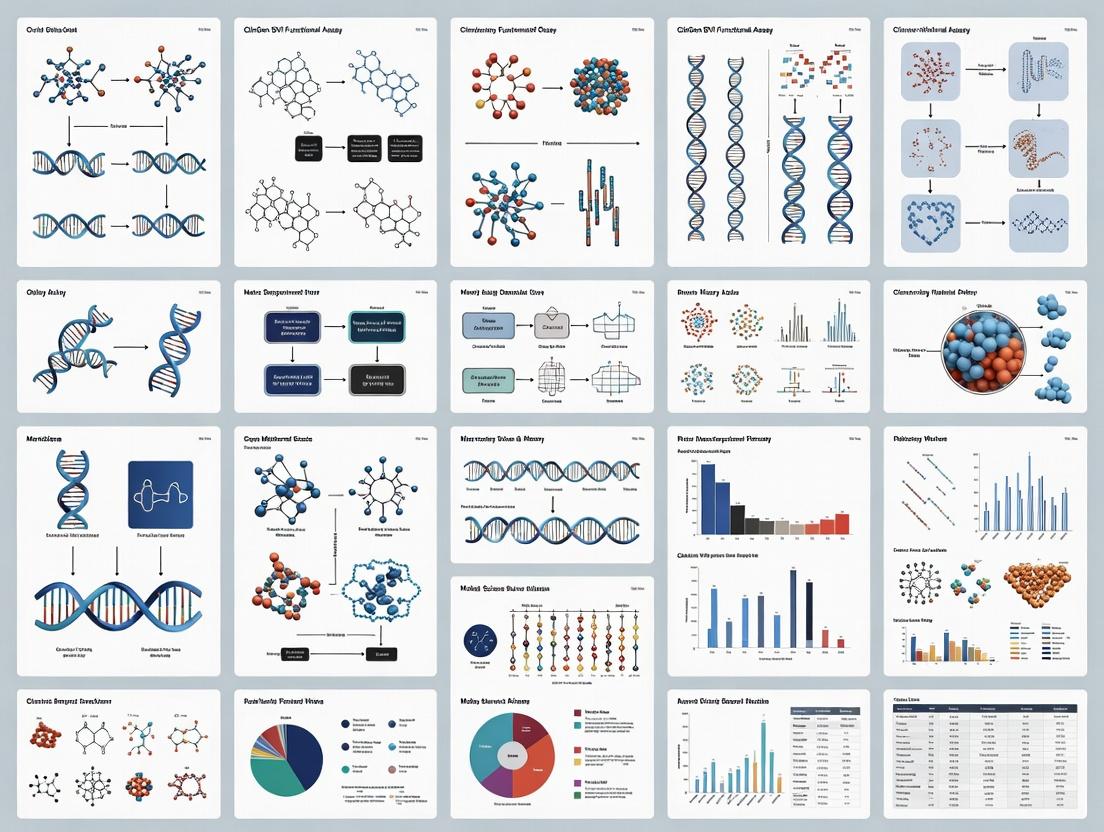
Diagrams
Title: Functional Assay Integration in Variant Classification Workflow
Title: Molecular Pathways and Assay Targets for Variant Functional Analysis
The Scientist's Toolkit
Table 3: Essential Research Reagent Solutions for Functional Assays
| Reagent/Material | Supplier Examples | Function in Variant Functional Analysis |
|---|---|---|
| Pre-Validated cDNA ORF Clones | DNASU, Addgene, Horizon Discovery | Wild-type expression backbone for site-directed mutagenesis to ensure consistent baseline activity. |
| High-Fidelity Site-Directed Mutagenesis Kits | Agilent, NEB, Thermo Fisher | Accurate introduction of specific nucleotide variants into expression constructs with low error rates. |
| ClinGen-Curated Control Variant Sets | Coriell Institute, ATCC | Essential known pathogenic and benign variants for assay calibration and validation (SVI requirement). |
| Reporter Cell Lines (Luciferase, GFP) | Horizon Discovery, ATCC | Engineered lines with integrated reporters for measuring pathway activity (e.g., p53, MAPK) upon variant expression. |
| Genome Editing Tools (CRISPR/Cas9) | Synthego, Integrated DNA Technologies | For creating isogenic cell lines or performing MAVEs in native genomic context. |
| Heterologous Expression Systems (Oocytes, HEK293) | Xenopus1, ECACC | Standardized cellular backgrounds for electrophysiology or protein interaction studies, minimizing confounding variables. |
| High-Content Imaging Systems | PerkinElmer, Thermo Fisher | Quantify protein localization, cell morphology, or fluorescent reporter changes in a high-throughput format. |
| Data Analysis Software (Patch Clamp, NGS) | Molecular Devices, Geneious, Custom Pipelines | Specialized software for rigorous quantitative analysis, ensuring statistical robustness for ACMG/AMP codes. |
Application Notes and Protocols
Within the ClinGen Sequence Variant Interpretation (SVI) framework, standardized documentation of functional assay data is critical for consistent variant pathogenicity assessment. This protocol details the purpose, scope, and key components of the Functional Assay Documentation Worksheet, a central tool for curating and evaluating evidence (PS3/BS3) according to the 2015 ACMG/AMP guidelines.
Purpose: The primary purpose of the worksheet is to provide a structured, transparent, and reproducible format for summarizing the experimental details, results, and interpretation of a functional study. It enables the standardization of evidence strength calibration across different genes, diseases, and assay types.
Scope: The worksheet scope encompasses all in vitro and in vivo functional assays used to characterize the impact of a genetic variant on molecular or cellular phenotypes relevant to disease mechanism. It is not intended for clinical diagnostic assays or computational predictions alone.
Key Components: The worksheet is organized into distinct modules capturing metadata, experimental design, results, and final classification.
Table 1: Core Quantitative Metrics for Functional Assay Calibration
| Metric | Description | Target Threshold (Typical) | Calculation |
|---|---|---|---|
| Effect Size | Magnitude of difference between variant and control. | >70-80% loss/gain for Strong | (Variant Activity / WT Activity) x 100% |
| Statistical Significance (p-value) | Probability that observed difference is due to chance. | p < 0.05 | Student's t-test, ANOVA |
| Number of Replicates (n) | Independent experimental repetitions. | n ≥ 3 | Reported per construct/line |
| Dynamic Range | Assay's ability to detect full spectrum of functional effects. | Must distinguish WT from known null. | (WT Signal - Null Control Signal) |
| Intra-assay Variability (CV) | Precision within a single experiment. | < 20% | (Standard Deviation / Mean) x 100% |
| Inter-assay Variability | Reproducibility across independent experiments. | < 25% | Comparison of experiment means |
Experimental Protocols for Key Assay Types
Protocol 1: Luciferase Reporter Assay for Transcriptional Activity
- Objective: Quantify the impact of a variant in a transcription factor on its ability to drive gene expression.
- Materials: See "The Scientist's Toolkit" below.
- Methodology:
- Cloning: Site-directed mutagenesis to introduce variant into expression plasmid for transcription factor.
- Cell Seeding: Seed HEK293T cells in 24-well plate at 1 x 10^5 cells/well.
- Transfection: Co-transfect 100 ng of transcription factor plasmid (WT or variant), 100 ng of firefly luciferase reporter plasmid (with responsive elements), and 10 ng of Renilla luciferase control plasmid (pRL-SV40) using lipid-based transfection reagent per manufacturer's protocol.
- Incubation: Incubate cells for 48 hours at 37°C, 5% CO2.
- Lysis & Measurement: Lyse cells with 100 µL Passive Lysis Buffer. Measure firefly and Renilla luciferase activity sequentially using a dual-luciferase reporter assay system on a luminometer.
- Analysis: Normalize firefly luminescence to Renilla luminescence for each well. Calculate mean ± SD of normalized relative light units (RLU) for at least three independent transfections. Express variant activity as a percentage of WT control. Perform unpaired t-test.
Protocol 2: Cell-Based Protein Localization Assay by Confocal Microscopy
- Objective: Determine if a variant alters the subcellular localization of a protein (e.g., nuclear, cytoplasmic, membrane).
- Materials: Expression plasmid with N- or C-terminal fluorescent tag (e.g., GFP), appropriate cell line, fluorescent microscope, image analysis software.
- Methodology:
- Cloning & Transfection: Introduce variant into tagged expression plasmid. Transfect cells on glass-bottom dishes.
- Fixation & Mounting: At 24h post-transfection, fix cells with 4% PFA for 15 min, permeabilize with 0.1% Triton X-100 if needed, and mount with DAPI-containing medium.
- Imaging: Acquire z-stack images using a confocal microscope with consistent settings (laser power, gain, exposure) across samples. Image ≥ 50 cells per construct.
- Quantification: Use software to define cellular compartments (nucleus via DAPI, cytoplasm). Calculate fluorescence intensity ratio (e.g., Nucleus/Cytoplasm or Membrane/Cytosol) for each cell. Compare distribution of ratios between WT and variant.
Visualizations
Title: Functional Assay Documentation and Classification Workflow
Title: Four Core Modules of the Documentation Worksheet
The Scientist's Toolkit: Essential Research Reagents
Table 2: Key Reagents for Functional Genomics Studies
| Reagent / Solution | Function / Application | Key Consideration |
|---|---|---|
| Site-Directed Mutagenesis Kit | Introduces specific nucleotide changes into plasmid DNA. | Efficiency and fidelity are critical for high-throughput variant generation. |
| Mammalian Expression Vectors | Plasmid for expressing gene of interest in cell models. | Choice of promoter, tag (e.g., GFP, HA), and selection marker matters. |
| Lipid-Based Transfection Reagent | Delivers nucleic acids into mammalian cells. | Optimize for cell type used; balance efficiency with cytotoxicity. |
| Dual-Luciferase Reporter Assay System | Quantifies transcriptional activity via firefly/Renilla luminescence. | Provides internal normalization (Renilla) for transfection efficiency. |
| Validated Antibodies | Detects endogenous or tagged proteins in WB, IF, IP. | Specificity and lot-to-lot consistency are paramount for reproducibility. |
| CRISPR/Cas9 Gene Editing Tools | Creates isogenic cell lines with endogenous variant knock-in. | Gold standard for physiological relevance; controls for genomic context. |
| qPCR Master Mix with ROX | Quantifies mRNA expression levels for target genes. | Requires validated primers and normalization to housekeeping genes. |
| Cell Viability Assay (e.g., MTT, ATP) | Assesses cytotoxicity linked to variant expression. | Important control to distinguish specific functional loss from cell death. |
Within the ClinGen Sequence Variant Interpretation (SVI) framework, the PP1/BS3 codes are critical for integrating functional assay data into clinical variant classifications. PP1 (Pathogenic Strong) supports pathogenicity based on well-established functional evidence, while BS3 (Benign Strong) supports benignity when functional studies show no deleterious effect. This document provides application notes and protocols for generating and interpreting functional assay data to satisfy these evidence codes, as part of a broader thesis on standardizing the SVI functional assay documentation worksheet.
Decoding PP1 and BS3: Criteria and Data Requirements
The application of PP1 and BS3 requires that assay results be mapped to specific clinical criteria defined by the ACMG/AMP guidelines. The following table summarizes the core quantitative and qualitative benchmarks.
Table 1: PP1/BS3 Evidence Criteria and Data Thresholds
| Evidence Code | Clinical Interpretation | Key Assay Criteria | Typical Quantitative Thresholds (Example: Enzyme Activity) | Required Data Robustness |
|---|---|---|---|---|
| PP1 | Strong Evidence for Pathogenicity | Assay detects a damaging effect on gene/protein function. | Activity < 10% of wild-type; Dominant-negative effect ≥ 125% of control; Significant loss-of-function in validated system. | Replication across independent experiments; Use of appropriate controls; Concordance with known pathogenic variants. |
| BS3 | Strong Evidence for Benignity | Assay shows no damaging effect on gene/protein function. | Activity ≥ 80% of wild-type; No significant difference from wild-type (p > 0.05). | Assay must be calibrated with known pathogenic variants; Sufficient statistical power to detect a meaningful effect. |
Core Experimental Protocols for Functional Validation
To generate data suitable for PP1/BS3 classification, robust and validated experimental protocols are essential. Below are detailed methodologies for key assay types commonly referenced in SVI documentation.
Protocol 1: Quantitative Protein Function Assay (e.g., Enzyme Kinetics)
- Objective: To measure and compare the specific activity of wild-type and variant proteins.
- Materials: Purified recombinant protein (WT and variant), specific substrate, reaction buffer, spectrophotometer/fluorimeter.
- Procedure:
- Express and purify WT and variant proteins using identical conditions (e.g., HEK293T system, affinity tag purification).
- Determine protein concentration via Bradford or BCA assay, confirmed by SDS-PAGE.
- In triplicate, incubate a fixed amount of protein (e.g., 10 nM) with a range of substrate concentrations (e.g., 0.1–10 x Km).
- Measure initial reaction velocity (V0) by tracking product formation over time.
- Fit data to the Michaelis-Menten equation to derive kinetic parameters (Kcat, Km).
- Data Analysis: Calculate relative activity (Variant Kcat / WT Kcat). For BS3, variant activity must be ≥80% of WT with no statistically significant difference. For PP1 supporting loss-of-function, variant activity is typically <10% of WT.
Protocol 2: Cellular Localization and Trafficking Assay
- Objective: To assess if a variant disrupts subcellular localization (e.g., for membrane receptors, ion channels).
- Materials: Expression plasmids (WT/variant), fluorescent tag (e.g., GFP), appropriate cell line, confocal microscope, organelle markers.
- Procedure:
- Transfect cells with GFP-tagged WT or variant constructs.
- At 24-48 hours post-transfection, fix cells and stain with organelle-specific dyes (e.g., ER tracker, DAPI for nucleus).
- Acquire high-resolution z-stack images via confocal microscopy.
- Perform co-localization analysis (e.g., Pearson's coefficient) using image analysis software (e.g., ImageJ/Fiji).
- Data Analysis: Significant mislocalization (e.g., retention in ER vs. plasma membrane) compared to WT and known pathogenic controls supports PP1. Normal WT-like localization supports BS3.
Pathway and Decision Logic Visualizations
Decision Logic for Applying PP1/BS3 Evidence Codes
Functional Assay Validation Workflow
The Scientist's Toolkit: Key Research Reagent Solutions
Table 2: Essential Materials for Functional Assay Development
| Reagent / Solution | Function in Assay | Key Considerations for SVI Documentation |
|---|---|---|
| Validated Reference Plasmid | Wild-type cDNA construct for benchmarking. | Must match reference transcript; source (e.g., Addgene, cDNA clone) must be documented. |
| Site-Directed Mutagenesis Kit | Introduction of the specific variant into the reference plasmid. | Protocol must include sequence verification of the final construct. |
| Calibration Controls | Known pathogenic and benign variant constructs. | Critical for assay calibration; required for both PP1 and BS3 application. |
| Cell Line with Relevant Background | Host for protein expression (e.g., null background, patient-derived). | Must be justified. Use of isogenic controls is ideal. |
| Activity-Specific Substrate/Reporter | Quantifies protein function (e.g., luminescent, fluorescent). | Should have a validated dynamic range and linear response. |
| High-Affinity Antibodies (if used) | For protein detection, localization, or immunoprecipitation. | Must specify clone, vendor, and dilution; validation data is preferred. |
| Statistical Analysis Software | For comparing variant to WT and control data. | Must apply appropriate tests (e.g., t-test, ANOVA); report p-values and n. |
Application Notes
Within ClinGen Sequence Variant Interpretation (SVI) functional assay documentation, the principles of calibration, scalability, and reproducibility are paramount for robust variant classification and data sharing.
Calibration ensures that assay outputs (e.g., readouts of protein function, splicing efficiency) are accurately mapped to a standardized, biologically-relevant scale, enabling definitive assignment of variant pathogenicity (e.g., benign, pathogenic). Calibrated assays use established positive and negative controls to define the dynamic range and decision thresholds.
Scalability addresses the need for assays to transition from low-throughput, proof-of-concept studies to higher-throughput formats capable of evaluating dozens to hundreds of variants efficiently. This is critical for ClinGen's goal of systematically interpreting large volumes of genetic variants.
Reproducibility demands that assay protocols are documented with sufficient detail and controls that independent laboratories can replicate the experimental process and achieve concordant results. This principle underpins the reliability of data submitted to the ClinGen SVI documentation worksheet.
Table 1: Key Metrics for Functional Assay Validation
| Metric | Target for Calibration | Target for Scalability | Target for Reproducibility |
|---|---|---|---|
| Dynamic Range (Signal-to-Noise) | ≥ 10-fold | Maintained in scaled format | Coefficient of Variation (CV) < 20% |
| Control Performance (Positive/Negative) | Z' factor > 0.5 | Automated scoring possible | Inter-assay CV < 25% |
| Throughput (Variants/Experiment) | Not primary | > 96 variants per run | Consistent across operators |
| Data Concordance (Inter-lab) | N/A | N/A | > 90% for control variants |
Table 2: ClinGen SVI Evidence Code Mapping for Functional Data
| Assay Performance Characteristic | Corresponding PS3/BS3 Evidence Strength Calibration |
|---|---|
| Excellent (Fully calibrated, high reproducibility) | Supports Strong (PS3) or Stand-alone (BS3) |
| Moderate (Good calibration, moderate reproducibility) | Supports Moderate |
| Insufficient/Uncalibrated | Supports Supporting or No Evidence |
Experimental Protocols
Protocol 1: Calibration of a Mammalian Cell-Based Transcriptional Reporter Assay
Objective: To establish calibrated thresholds for loss-of-function (LOF) and normal function for a tumor suppressor gene variant assay.
Reagent Preparation:
- Generate expression constructs for calibration controls: Wild-type (WT) cDNA, known pathogenic truncation variant (Positive LOF control), empty vector (Negative control).
- Seed HEK293T cells in 96-well plates at 20,000 cells/well in antibiotic-free medium.
Transfection & Assay:
- Transfert each calibration construct in triplicate using a standardized lipid-based method.
- Co-transfect with a constitutive Renilla luciferase plasmid for normalization.
- At 48 hours post-transfection, lyse cells and measure firefly and Renilla luciferase activity using a dual-luciferase assay kit.
Data Analysis & Threshold Setting:
- Calculate normalized activity: (Firefly / Renilla) for each well.
- Set the LOF Threshold as the mean normalized activity of the positive LOF control + 3 standard deviations.
- Set the Normal Function Threshold as the mean normalized activity of the WT control - 3 standard deviations.
- Variants falling below the LOF threshold are classified as LOF; those above the normal function threshold are classified as functional; variants in between require further scrutiny.
Protocol 2: Scalable Functional Evaluation Using Saturation Genome Editing
Objective: To assess the functional impact of all possible single-nucleotide variants in a critical exon at scale.
Library Design & Cloning:
- Design an oligo library containing all possible nucleotide substitutions for the target exon.
- Clone the library into the endogenous genomic locus in haploid human cells using CRISPR-Cas9 and a template delivery vector containing a resistance marker.
Selection & Sequencing:
- Apply a relevant phenotypic selection (e.g., drug resistance, cell survival, FACS sorting based on a fluorescent reporter).
- Culture pooled edited cells under selective and non-selective conditions for 14-21 population doublings.
- Harvest genomic DNA from pre-selection and post-selection pools. Amplify the target region via PCR and perform high-depth next-generation sequencing (NGS).
Scalable Data Analysis:
- Enrichment scores for each variant are calculated from the change in allele frequency between selection and non-selection pools using specialized pipelines (e.g.,
MAGeCK). - Scores are compared to pre-calibrated thresholds from known controls included in the library to classify variants as functional or LOF.
- Enrichment scores for each variant are calculated from the change in allele frequency between selection and non-selection pools using specialized pipelines (e.g.,
Mandatory Visualization
Assay Development Workflow & Core Principles
Generic Functional Assay Signaling & Analysis Pathway
The Scientist's Toolkit: Research Reagent Solutions
Table 3: Essential Materials for SVI Functional Assays
| Item | Function in Context | Key Consideration for Principles |
|---|---|---|
| Calibrated Control Plasmids (WT, Pathogenic, Benign) | Define assay dynamic range and classification thresholds. Critical for calibration. | Sequence-verified, from reputable repository (e.g., Addgene). |
| Reference Standard Cell Line (e.g., HEK293, HAP1) | Provides consistent, reproducible cellular context for assays. | Use low-passage, authenticated stocks; regular mycoplasma testing. |
| Dual-Luciferase Reporter Assay System | Quantifies transcriptional activity with internal normalization. Enables calibration. | High signal-to-noise ratio kits ensure robust dynamic range. |
| CRISPR-Cas9 Ribonucleoprotein (RNP) Complex | Enables scalable genome editing for saturation variant libraries. | High-purity Cas9 and sgRNAs ensure editing efficiency and reproducibility. |
| NGS Library Prep Kit (for scalable assays) | Prepares amplicons from variant pools for high-throughput sequencing. | Low-error polymerase and unique molecular identifiers (UMIs) reduce noise. |
| Validated Primary Antibody (for protein-based assays) | Detects protein expression, localization, or post-translational modifications. | Antibody validation for specific application (e.g., knockout cell line). |
Data Analysis Pipeline Software (e.g., MAGeCK, custom R/Python) |
Processes high-throughput data to calculate variant enrichment scores. | Documented, version-controlled code is essential for reproducibility. |
| ClinGen SVI Documentation Worksheet | Standardized framework for reporting assay details and results. | Ensures all core principles are addressed for community evaluation. |
Step-by-Step Guide: Completing the SVI Worksheet for Your Assay
Introduction Within the ClinGen Sequence Variant Interpretation (SVI) Functional Assay Documentation Worksheet research framework, defining the biological context is the critical first step for assay development and validation. This pre-worksheet checklist ensures that the proposed functional assay is grounded in a comprehensive understanding of the relevant biological system, directly informing the ACMG/AMP PP3/BS3 criteria for variant pathogenicity assessment. This document provides application notes and protocols to guide researchers through this foundational process.
Application Notes A well-defined biological context establishes the assay's relevance to human disease. It requires mapping the gene product's role within precise molecular pathways and physiological systems. Key considerations include:
- Gene-Disease Validity: Confirm the gene-disease relationship as defined by the relevant ClinGen Gene-Disease Validity curation.
- Molecular Mechanism: Distinguish between loss-of-function (LOF) or gain-of-function (GOF) mechanisms for the disease in question.
- Functional Domains: Identify critical protein domains (e.g., catalytic, DNA-binding) where variants are likely to be disruptive.
- System Complexity: Determine if the assay needs to capture simple biochemical activity, protein-protein interactions, or complex cellular phenotypes.
Table 1: Quantitative Parameters for Biological Context Definition
| Parameter | Description | Example Data Source | Relevance to Assay Design |
|---|---|---|---|
| Expression Specificity | Tissue/cell type enrichment of gene expression. | GTEx (Median TPM > 50), Human Protein Atlas. | Guides choice of cellular model system (endogenous vs. overexpression). |
| Protein Abundance | Estimated copies per cell. | PaxDb, quantitative proteomics studies. | Informs required sensitivity of the detection method. |
| Variant Distribution | Location of known pathogenic vs. benign variants. | gnomAD, ClinVar. | Identifies critical functional domains for targeting in assay. |
| Pathogenic Mechanism Ratio | % LOF vs. GOF for disease-associated variants. | ClinVar summaries, literature review. | Determines assay readout direction (rescue vs. toxicity). |
Protocol 1: Establishing the Molecular Pathway Context
Objective: To diagram the immediate molecular network and upstream/downstream effects of the gene product of interest.
Methodology:
- Literature Curation: Perform a systematic search using PubMed and OMIM for reviews on the gene's function. Prioritize recent studies (<5 years) and human genetic evidence.
- Database Mining:
- Extract known protein-protein interactions from BioGRID, STRINGdb (confidence score > 0.7).
- Map to canonical signaling pathways using KEGG Pathway and Reactome.
- Pathway Synthesis: Integrate data to create a simplified pathway model highlighting:
- Direct molecular inputs (e.g., activating signals, upstream regulators).
- The core function of the protein (e.g., kinase activity, transcriptional activation).
- Direct molecular outputs (e.g., phosphorylated substrates, target genes).
- Assay Anchor Point Identification: Select the most representative and measurable node or edge in this pathway as the primary target for the functional assay.
Visualization: Core Pathway Mapping for Assay Design
Protocol 2: Defining the Cellular and Physiological Context
Objective: To identify the appropriate cellular models and phenotypic endpoints that reflect the gene's role in human biology and disease.
Methodology:
- Disease Phenotype Analysis: Review Human Phenotype Ontology (HPO) terms associated with the gene-disease pair. Categorize phenotypes as cellular (e.g., "reduced cell proliferation"), tissue/organ (e.g., "cardiomyopathy"), or organismal.
- Cell Model Selection Criteria:
- Endogenous Expression: Verify model expresses the gene at relevant levels (via RNA-seq or protein atlas data).
- Genetic Tractability: Assess feasibility of CRISPR/Cas9 editing, RNAi, or transgene expression.
- Phenotypic Relevance: Determine if the cell type exhibits a relevant baseline phenotype or can be induced to do so (e.g., stress assay).
- Endpoint Correlation: Match a quantifiable cellular endpoint (e.g., reporter activity, viability, localization) to the key physiological phenotype.
Visualization: Biological Context Decision Workflow
The Scientist's Toolkit: Research Reagent Solutions
Table 2: Essential Materials for Biological Context Definition
| Item | Function in Context Definition | Example/Supplier |
|---|---|---|
| Gene-Specific Antibodies (Validated) | Confirm protein expression, localization, and stability in chosen cell models. | Cell Signaling Technology, Abcam. |
| CRISPR/Cas9 Knockout Kits | Create isogenic cell lines to establish baseline phenotype and validate assay specificity. | Synthego, Horizon Discovery. |
| Pathway-Specific Reporter Constructs | Quantify activity of the relevant pathway downstream of the protein of interest. | Luciferase-based reporters (Cignal, Qiagen). |
| Validated cDNA ORF Clones | Source of wild-type sequence for rescue experiments and benchmark for activity. | Mammalian expression-ready clones (GenScript, DNASU). |
| Pathogenic & Benign Control Variants | Critical for establishing the dynamic range and predictive value of the assay. | Sourced from ClinVar-curated variants or saturation mutagenesis studies. |
| Pathway Inhibitors/Activators | Pharmacological tools to modulate the pathway and test assay responsiveness. | Tocris Bioscience, Selleckchem. |
| Public Data Portals | Access expression, variant, and interaction data for hypothesis generation. | GTEx, gnomAD, BioGRID, ClinVar. |
This document provides a detailed guide for completing the Assay Description and Design Rationale section of the ClinGen Sequence Variant Interpretation (SVI) Functional Assay Documentation Worksheet. This section is critical for establishing the assay's biological relevance and technical robustness, enabling consistent interpretation of functional data for variant pathogenicity assessment in clinical genomics and therapeutic development.
This subsection requires a concise yet comprehensive overview of the experimental system, measured variable, and assay outcome. The goal is to allow an independent researcher to understand the assay's fundamental purpose.
- Core Components Table:
| Component | Description | Example Entry |
|---|---|---|
| Biological System | The cellular or biochemical environment (e.g., cell line, animal model, purified protein system). | HEK293T cells transiently expressing wild-type or variant MYH7 protein. |
| Measured Variable | The specific molecular or phenotypic readout. | ATPase activity normalized to total protein concentration. |
| Assay Outcome | The specific metric used for variant classification (e.g., percentage of wild-type activity, fold-change). | Percent residual ATPase activity (%WT). Variants with <30% WT activity are considered loss-of-function. |
- Key Validation Data Table:
| Validation Parameter | Typical Target | Example Data from Literature (MYH7 ATPase Assay) |
|---|---|---|
| Assay Dynamic Range | Signal linearity across relevant protein concentrations. | Linear (R² > 0.98) from 0.1 to 10 µg of lysate protein. |
| Inter-assay Precision (CV) | Coefficient of Variation for replicate experiments. | CV < 15% for n=3 independent experiments. |
| Signal-to-Noise Ratio | Distinction between positive/negative controls. | S/N > 10 for WT vs. vector-only control. |
| Z'-Factor | Statistical parameter for high-throughput assay quality. | Z' > 0.5 for 96-well plate format. |
Design Rationale: Establishing Biological Relevance
This subsection justifies why the assay is an appropriate model of the gene/product's function and how it connects to disease mechanisms. It must reference the disease context from the ClinGen Variant Curation Expert Panel's (VCEP) specified disease mechanism.
Experimental Protocol: Establishing a Relevant Cell System
- Objective: To generate cellular material expressing the protein of interest under controlled conditions.
- Methodology:
- Construct Design: Clone the cDNA of the gene of interest (e.g., MYH7) into a mammalian expression vector with a constitutive promoter (e.g., CMV) and an epitope tag (e.g., FLAG) for detection.
- Variant Introduction: Introduce patient-specific variants using site-directed mutagenesis. Validate all constructs by Sanger sequencing.
- Cell Culture & Transfection: Maintain appropriate cell lines (e.g., HEK293T) in recommended media. Transfect cells with wild-type, variant, and empty vector (negative control) plasmids using a validated method (e.g., polyethylenimine (PEI) transfection). Use a minimum of two biological replicates.
- Lysate Preparation: 48 hours post-transfection, harvest cells in lysis buffer (e.g., RIPA buffer with protease inhibitors). Clarify lysates by centrifugation. Determine protein concentration using a standardized assay (e.g., BCA assay).
- Quality Control: Analyze expression levels by Western blot using an antibody against the epitope tag or endogenous protein to confirm equal expression and stability of variant proteins.
Pathway Diagram: Connecting Assay to Disease Biology
Diagram Title: Biological Rationale Linking Gene Function to Disease Phenotype
Experimental Design and Controls
Detail the experimental layout, including controls essential for data normalization and interpretation.
- Standard Experimental Run Layout Table:
| Well/Group | Condition | Purpose | N (per experiment) |
|---|---|---|---|
| 1-3 | Vector Only (Mock) | Background/Noise Control | 3 |
| 4-9 | Wild-Type (WT) Reference | 100% Activity Baseline | 6 |
| 10-21 | Test Variants (e.g., V1, V2) | Variant Functional Assessment | 6 per variant |
| 22-24 | Known Pathogenic Variant | Positive Control for Loss-of-Function | 3 |
| 25-27 | Known Benign Polymorphism | Negative Control (WT-like function) | 3 |
- Experimental Workflow Diagram:
Diagram Title: Workflow for Functional Assay Execution and Analysis
The Scientist's Toolkit: Key Research Reagent Solutions
| Item | Function & Rationale |
|---|---|
| Mammalian Expression Vector (e.g., pcDNA3.1) | Backbone for cDNA expression; allows constitutive high-level protein production in transfected cells. |
| Site-Directed Mutagenesis Kit | Enables precise introduction of nucleotide variants into expression constructs to model patient alleles. |
| Transfection-Grade Plasmid Prep Kit | Produces pure, endotoxin-free plasmid DNA critical for high transfection efficiency and cell viability. |
| Polyethylenimine (PEI) Transfection Reagent | Cost-effective cationic polymer for high-efficiency transient transfection of adherent cell lines like HEK293. |
| Lysis Buffer (RIPA) with Protease Inhibitors | Efficiently extracts total protein while preserving function and preventing degradation. |
| Colorimetric/Fluorometric ATPase Assay Kit | Provides optimized reagents for specific, sensitive, and quantitative measurement of ATP hydrolysis activity. |
| Anti-FLAG/HA/Tag Antibody | Allows specific immunodetection of transfected protein independent of native protein, enabling expression normalization. |
| Statistical Analysis Software (e.g., GraphPad Prism) | For rigorous data analysis, including outlier testing, ANOVA, and post-hoc tests to compare variants to WT. |
Detailed Protocol: Kinetic ATPase Activity Assay
This protocol follows from the cell lysate preparation described in Section 2.
- Reagent Preparation: Thaw cell lysates on ice. Prepare ATP substrate solution per kit instructions. Prepare a phosphate standard curve (0, 2, 4, 6, 8, 10 nmol) in lysis buffer.
- Reaction Setup: In a clear 96-well plate, add 10 µg of normalized lysate (WT, variant, controls) to wells. Adjust volume to 30 µL with lysis buffer. Include substrate-only (blank) wells.
- Initiate Reaction: Add 20 µL of ATP substrate solution (final ATP concentration: e.g., 1 mM) to each well using a multichannel pipette. Mix immediately by gentle shaking.
- Incubation: Incubate the plate at 30°C (or physiological temperature) for 30 minutes. Optimization Note: Time and temperature should be established in pilot studies to ensure linear reaction kinetics.
- Stop Reaction & Develop Color: Add 50 µL of malachite green-based detection reagent to each well to stop the reaction and initiate color development. Incubate at room temperature for 20 minutes for stable color formation.
- Absorbance Measurement: Read the absorbance at 620 nm using a plate reader.
- Data Calculation: Subtract the average blank absorbance. Calculate the amount of inorganic phosphate (Pi) released for each sample using the standard curve linear equation. Express activity as nmol Pi released/min/µg protein. Normalize variant data to the mean of the WT controls from the same experiment run to calculate % Wild-Type Activity.
In the ClinGen Sequence Variant Interpretation (SVI) Functional Assay Documentation Worksheet framework, meticulous documentation of experimental details is paramount for the credible classification of genomic variants. This protocol provides standardized application notes for documenting controls, replicates, and statistical analyses, ensuring reproducibility and reliability of functional assay data submitted for clinical interpretation.
Essential Documentation Components & Quantitative Benchmarks
Table 1: Minimum Documentation Standards for SVI Functional Assays
| Component | Purpose in SVI Context | Minimum Recommended Standard | Justification |
|---|---|---|---|
| Positive Control | Establishes assay dynamic range; validates protocol. | Well-characterized pathogenic variant (ClinVar pathogenic assertion). | Calibrates expected signal for loss/gain-of-function. |
| Negative Control | Defines baseline/noise level; confirms assay specificity. | Wild-type sequence or confirmed benign variant. | Distinguifies true variant effect from background. |
| Technical Replicates | Measures intra-assay precision (pipetting, instrument noise). | n ≥ 3 per sample per run. | Quantifies random error within a single experiment. |
| Biological Replicates | Captures biological variability (different cell lines, donors, clones). | n ≥ 2 independent transfections/isolations. | Ensures observed effect is not an artifact of a single preparation. |
| Independent Experiments | Demonstrates reproducibility across time and reagent batches. | N ≥ 2 completely separate experiments. | Gold standard for assessing overall result robustness. |
| Statistical Test | Provides quantitative confidence in observed differences. | Test appropriate for data distribution (e.g., t-test, ANOVA, Mann-Whitney). | Required for SVI's evidence scoring (PS3/BS3). |
| Effect Size & Confidence Intervals | Quantifies magnitude and precision of the variant effect. | Report mean difference & 95% CI or standardized effect size (e.g., Cohen's d). | Critical for distinguishing clinical significance. |
Detailed Experimental Protocols
Protocol 2.1: Designing and Implementing Controls for a Luciferase Reporter Assay
- Objective: To measure the impact of a variant on transcriptional activity in a TP53 reporter assay.
- Materials: See "Research Reagent Solutions" below.
- Method:
- Plate Layout: Seed 293T cells in a 96-well plate at 10,000 cells/well.
- Transfection Complexes: Prepare separate lipofectamine complexes for:
- Test: Plasmid containing the variant of interest.
- Negative Control: Wild-type TP53 expression plasmid.
- Positive Control (Loss-of-Function): Known pathogenic TP53 truncation variant (e.g., R213*).
- Assay Control: Empty vector (to define baseline reporter activity).
- Transfection Control: Co-transfect a Renilla luciferase plasmid in all wells.
- Transfection: Apply complexes to designated wells (n=6 technical replicates per condition).
- Harvest & Read: At 48h post-transfection, lyse cells and measure firefly and Renilla luciferase activity sequentially.
- Normalization: Divide firefly luminescence by Renilla luminescence for each well to control for transfection efficiency.
- Analysis: Calculate normalized relative luminescence units (RLU) for each condition.
Protocol 2.2: Executing and Documenting Replicates for a Saturation Genome Editing (SGE) Fitness Assay
- Objective: To determine variant effect on cellular fitness via SGE in a haploid cell line.
- Method:
- Variant Library Design: Design and synthesize a library targeting the gene of interest with all possible single-nucleotide substitutions.
- Transduction & Selection: Transduce the library into HAP1 cells at low MOI to ensure single variant integration. Apply selection.
- Biological Replicates: Initiate two independent transductions on different days with freshly thawed cells and library aliquots.
- Passaging: Passage each replicate culture for ~14 population doublings.
- Timepoints: Harvest genomic DNA (gDNA) at Day 0 (post-selection baseline) and Day 14 (endpoint) from each replicate.
- Sequencing & Analysis: Amplify integrated regions from gDNA and perform high-throughput sequencing. Analyze variant frequency changes between timepoints.
Protocol 2.3: Statistical Analysis for a Cellular Localization Assay
- Objective: To quantify significant mislocalization of a protein variant compared to wild-type.
- Method:
- Imaging: Acquire high-resolution images of cells transfected with GFP-tagged wild-type and variant constructs (N=3 independent experiments, ≥30 cells per condition per experiment).
- Quantification: Use image analysis software to calculate a "nuclear-to-cytoplasmic (N:C) ratio" for each cell.
- Statistical Testing:
- Normality Test: Perform Shapiro-Wilk test on N:C ratio datasets.
- If data are normal: Conduct an unpaired, two-tailed Student's t-test between wild-type and variant groups (pooling cells from all replicates).
- If data are non-normal: Conduct a Mann-Whitney U test.
- Account for Experiment Effect: Perform a two-way ANOVA with "variant" and "experiment day" as factors if pooling is inappropriate.
- Reporting: Document p-value, test used, sample size (n cells, N experiments), and mean ± SD for each condition.
Visualizations
Diagram Title: SVI Assay Documentation Workflow
Diagram Title: Replication Hierarchy for Robust Evidence
The Scientist's Toolkit: Research Reagent Solutions
Table 2: Essential Materials for Functional Assay Documentation
| Item / Solution | Function in Documentation Context | Example (Non-endorsing) |
|---|---|---|
| Certified Reference Materials | Provide standardized positive/negative controls for assay validation. | NIST genomic DNA standards, ATCC cell lines with characterized variants. |
| Dual-Luciferase Reporter Assay System | Enables normalization via internal control (Renilla), critical for precise technical replicate analysis. | Promega Dual-Glo Luciferase Assay. |
| Validated Knockout Cell Lines | Serve as isogenic negative controls for complementation assays (e.g., CRISPR-Cas9 generated). | Horizon Discovery HAP1 or parental KO lines. |
| Fluorescent Protein-Tagged ORF Clones | Standardized vectors for consistent expression across experiments in localization assays. | Harvard Plasmids (Addgene) ORFeome collections. |
| High-Fidelity DNA Polymerase | Ensures accurate amplification for sequencing-based assays, minimizing errors in replicate analysis. | Q5 High-Fidelity DNA Polymerase. |
| Statistical Analysis Software | Performs appropriate statistical tests and generates confidence intervals for evidence scoring. | GraphPad Prism, R Stats package. |
| Electronic Lab Notebook (ELN) | Centralized, timestamped platform for recording replicates, controls, and raw data metadata. | Benchling, LabArchives. |
1. Introduction and Context Within the ClinGen Sequence Variant Interpretation (SVI) framework, the Functional Assay Documentation Worksheet provides a structured approach for evaluating the strength of functional data for variant pathogenicity classification. A critical component of this process is the interpretation of assay results through validated scoring scales and the definition of thresholds that distinguish between normal and abnormal function. This protocol details the methodologies for establishing and applying these scales, framed within the broader thesis research on standardizing functional evidence curation for the ClinGen SVI initiative.
2. Quantitative Data Summary: Common Scoring Scales
Table 1: Common Functional Assay Scoring Scales and Clinical Interpretations
| Scale Type | Typical Range | Normal/Control Mean ± SD | Pathogenic Threshold | Supporting Evidence Strength (PS3/BS3) | Common Assay Application |
|---|---|---|---|---|---|
| Residual Activity (%) | 0% - 150% | 100% ± 15% | < 20% (LoF) | Strong (PS3) | Enzymatic activity, transcriptional activation. |
| Fold-Change vs. WT | Variable | 1.0 ± 0.2 | < 0.3 (LoF) > 2.0 (GoF) | Moderate (PS3/BS3) | Reporter assays, protein-protein interaction. |
| Z-Score | -∞ to +∞ | 0 ± 1 | Z < -3.09 (p<0.001) | Strong (PS3) | High-throughput functional genomics. |
| Likelihood Ratio (LR) | >0 | N/A | LR ≥ 18.7 (Pathogenic) LR ≤ 0.053 (Benign) | Very Strong (PS3/BS3) | Integrated, calibrated population data. |
3. Experimental Protocols for Key Methodologies
Protocol 3.1: Establishing a Residual Activity Scale for an Enzymatic Assay Objective: To determine the percentage of wild-type (WT) enzymatic activity retained by a variant and define pathogenicity thresholds. Materials: See Scientist's Toolkit. Procedure:
- Expression Constructs: Clone WT and variant cDNA into identical expression vectors.
- Cell Transfection: Transfect a standardized amount (e.g., 1 µg) of each plasmid into triplicate samples of an appropriate cell line using a consistent method (e.g., lipid-based).
- Lysate Preparation: 48 hours post-transfection, harvest cells and prepare lysates in non-denaturing buffer. Determine total protein concentration.
- Enzymatic Reaction: Perform the enzyme-specific reaction using equal amounts of total protein from each lysate. Include a vector-only control. Measure product formation kinetically or at endpoint.
- Activity Normalization: Normalize the raw activity of each sample to its expression level (via Western blot densitometry).
- Scale Calibration: Set the mean normalized activity of the WT replicates to 100%. Calculate the residual activity for each variant replicate as a percentage of the WT mean.
- Threshold Definition: Using data from known benign (population variants) and known pathogenic (null) controls, establish thresholds. Typically, activity <20% of WT supports pathogenicity for loss-of-function (LoF) variants, while activity >70% may support benignity. The range of 20-70% is considered intermediate.
Protocol 3.2: High-Throughput Variant Functional Assessment using a Z-Score Scale Objective: To score a large set of variants in a single experiment and derive a statistical threshold for abnormality. Materials: Saturation mutagenesis library, deep sequencing capability, relevant phenotypic reporter (e.g., growth, fluorescence). Procedure:
- Library Construction & Transduction: Create a pooled variant library covering the gene of interest. Introduce the library into the assay system (e.g., mammalian cells, yeast) at high coverage (≥500x per variant).
- Selection/Reporter Readout: Apply the functional selection (e.g., drug for survival, FACS for fluorescence) or measure the reporter output.
- Deep Sequencing: Isolate genomic DNA or plasmid DNA from pre-selection (input) and post-selection/output (output) populations. Sequence amplified target regions to high depth.
- Variant Abundance Calculation: Calculate the frequency of each variant in the input and output pools.
- Enrichment Score: For each variant, compute a functional score (e.g., log2(output frequency / input frequency)).
- Z-Score Calculation: Compute the mean and standard deviation of the functional scores for all synonymous (assumed neutral) variants in the experiment. For each variant, calculate its Z-score: (Variant Score - Mean of Synonymous Scores) / SD of Synonymous Scores.
- Threshold Definition: A Z-score < -3.09 (corresponding to p<0.001 in a one-tailed test, assuming normality) is a commonly used stringent threshold for functional impairment, supporting pathogenicity.
4. Visualizations
Diagram 1: ClinGen SVI Functional Evidence Curation Workflow
Diagram 2: Threshold Definition Using Control Variants
5. The Scientist's Toolkit: Research Reagent Solutions
Table 2: Essential Materials for Functional Assay Development and Calibration
| Item | Function & Rationale |
|---|---|
| Validated WT and Mutant Expression Constructs | Isogenic backbone ensures measured differences are due to the variant, not expression context. Critical for calibration. |
| Clinically Annotated Control Variants | Known pathogenic and benign variants are required to empirically set assay thresholds and validate the scoring scale. |
| Standardized Cell Line (e.g., HEK293T, HeLa) | Consistent cellular background reduces experimental noise, enabling reliable detection of variant-specific effects. |
| Normalization Reporter Plasmid (e.g., Renilla luciferase) | Controls for transfection efficiency and cell viability in transient assays, allowing for activity normalization. |
| Precision Activity Assay Kit (e.g., luminescent, fluorescent) | Provides sensitive, quantitative readout with a wide dynamic range for accurate residual activity measurement. |
| High-Fidelity Taq Polymerase & Cloning Reagents | Ensures error-free generation of variant constructs to prevent introduction of confounding mutations. |
| Automated Liquid Handler / Plate Reader | Increases throughput and reproducibility by minimizing manual pipetting errors and enabling precise kinetic reads. |
| Statistical Analysis Software (e.g., R, Prism) | Essential for robust data analysis, including calculation of means, SDs, Z-scores, and statistical significance testing. |
1. Introduction and Context This application note details the systematic application of the ClinGen SVI (Sequence Variant Interpretation) functional assay documentation worksheet to a novel gene therapy target, the TDP1 gene, for the treatment of Spinocerebellar Ataxia with Axonal Neuropathy (SCAN1). This work is situated within a broader thesis on standardizing functional evidence curation for clinical variant interpretation, demonstrating how the worksheet framework ensures reproducibility and robust evidence scoring in a therapeutic development pipeline.
2. Target Overview and Pathogenic Mechanism SCAN1 (OMIM: 607250) is an autosomal recessive disorder caused by biallelic pathogenic variants in the TDP1 gene, encoding Tyrosyl-DNA Phosphodiesterase 1. The canonical pathogenic variant is the homozygous p.His493Arg missense mutation. TDP1 repairs topoisomerase I (TOP1)-mediated DNA damage. The p.His493Arg variant results in a catalytically deficient enzyme, leading to the accumulation of TOP1-DNA cleavage complexes (TOP1cc), stalled replication forks, and subsequent neuronal apoptosis.
3. Quantitative Data Summary Table 1: Key Biochemical and Cellular Phenotypes of TDP1 p.His493Arg
| Assay Parameter | Wild-Type TDP1 | p.His493Arg Mutant | Assay System | Source |
|---|---|---|---|---|
| Catalytic Activity (kcat, min⁻¹) | 28.4 ± 3.1 | 0.05 ± 0.01 | Recombinant Protein | Takashima et al., 2002 |
| TOP1cc Clearance (% of WT) | 100% | <5% | Patient Lymphoblastoid Cells | El-Khamisy et al., 2005 |
| DNA Damage (γH2AX foci per cell) | 2.1 ± 0.8 | 18.7 ± 4.2 | SCAN1 Patient Fibroblasts | Katyal et al., 2007 |
| Neuronal Survival (% vs Control) | 100% | 42% ± 7% | Tdp1⁻/⁻ Mouse Neurons + Mutant Vector | Hassan et al., 2017 |
4. Experimental Protocols
Protocol 4.1: In Vitro TDP1 Phosphodiesterase Activity Assay (Adapted from Interthal et al., 2005) Objective: Quantify the enzymatic cleavage of a 3'-phosphotyrosyl DNA substrate. Reagents: Recombinant WT or mutant TDP1 protein, 5'-Cy3-labeled oligonucleotide substrate with a 3'-phosphotyrosine moiety, reaction buffer (50 mM Tris-HCl pH 7.5, 80 mM KCl, 2 mM EDTA, 1 mM DTT, 40 μg/mL BSA). Procedure:
- Dilute substrate to 500 nM in reaction buffer.
- Initiate reaction by adding TDP1 enzyme to a final concentration of 50 nM.
- Incubate at 37°C for 0, 2.5, 5, 10, and 20 minutes.
- Terminate reactions with an equal volume of 95% formamide/20 mM EDTA.
- Heat-denature samples at 95°C for 5 minutes and separate products on a 20% denaturing polyacrylamide gel.
- Visualize and quantify using a fluorescence gel scanner. Calculate kcat.
Protocol 4.2: Cellular TOP1cc Clearance Assay (Adapted from El-Khamisy et al., 2005) Objective: Measure the persistence of TOP1-DNA covalent complexes in cells after camptothecin (CPT) challenge. Reagents: Patient-derived or isogenic cell lines, 10 mM Camptothecin stock (in DMSO), lysis buffer (6 M guanidine HCl, 10 mM Tris-HCl pH 8.0, 100 mM Na₂HPO₄/NaH₂PO₄, 5 mM imidazole), Ni-NTA magnetic beads. Procedure:
- Treat 1x10⁶ cells with 10 μM CPT for 30 minutes. Replace with drug-free medium for a 0-60 minute recovery period.
- Lyse cells in 1 mL lysis buffer. Sonicate briefly to shear DNA.
- Incubate lysates with Ni-NTA beads for 4 hours at RT to capture His-tagged TOP1cc.
- Wash beads 3x with lysis buffer + 0.1% Triton X-100, then 2x with wash buffer (8 M urea, 10 mM Tris-HCl pH 8.0, 100 mM Na₂HPO₄/NaH₂PO₄, 0.1% Triton X-100).
- Elute TOP1cc and subject to Western blot using anti-TOP1 antibody. Quantify band intensity.
5. Signaling Pathway and Experimental Workflow
6. The Scientist's Toolkit: Research Reagent Solutions Table 2: Essential Research Reagents for TDP1 Functional Analysis
| Reagent/Category | Specific Example/Product | Function in Assay |
|---|---|---|
| Recombinant Protein | Purified human WT and p.His493Arg TDP1 (e.g., Abcam ab165409) | Substrate for direct in vitro enzymatic activity assays. |
| Specialized Substrate | 3'-Phosphotyrosine DNA oligonucleotide (custom synthesis) | Specific chemical substrate to measure TDP1 phosphodiesterase activity. |
| Cell Line Models | SCAN1 patient-derived fibroblasts (Coriell Institute); Tdp1⁻/⁻ mouse embryonic stem cells. | Disease-relevant cellular context for TOP1cc clearance and DNA damage assays. |
| TOP1 Poison | Camptothecin (CPT) (Sigma-Aldrich C9911) | Induces TOP1-DNA covalent complexes to challenge the TDP1 repair pathway. |
| DNA Damage Marker | Anti-γH2AX (phospho S139) antibody (e.g., Millipore 05-636) | Immunofluorescence or flow cytometry readout for persistent DNA double-strand breaks. |
| Affinity Capture Beads | Nickel-Nitrilotriacetic Acid (Ni-NTA) Magnetic Beads (e.g., Qiagen 36113) | For isolating His-tagged TOP1cc complexes in cellular clearance assays. |
| Gene Delivery Vector | AAV9-TDP1 expression construct (for rescue studies) | Tool for functional complementation in patient cells or animal models. |
Overcoming Challenges: Expert Tips for Robust Functional Assay Documentation
Common Pitfalls in Experimental Design and How to Avoid Them
Within the ClinGen Sequence Variant Interpretation (SVI) framework, robust functional assay data are critical for accurate pathogenicity classification of genetic variants. This document outlines common experimental design flaws in generating such evidence and provides detailed protocols to avoid them, thereby enhancing data quality for the SVI documentation worksheet.
Pitfall 1: Inadequate Controls
Failure to include appropriate controls is the most frequent and critical flaw, leading to uninterpretable results.
Quantitative Impact of Inadequate Controls:
| Control Type | Purpose | Consequence if Omitted | Recommended Minimum in Assay |
|---|---|---|---|
| Positive Control (Wild-type) | Establishes normal functional signal/baseline. | Cannot assess variant impact severity. | Included in every run (n≥3). |
| Negative Control (Known Pathogenic) | Confirms assay can detect loss-of-function. | High risk of false negative results. | At least one well-characterized variant. |
| Empty Vector/Transfection Control | Measures background noise. | Overestimation of residual function. | Included in every experiment. |
| Technical Replicate Control | Assesses intra-assay variability. | Unreliable estimate of data precision. | Minimum n=3 independent replicates. |
Detailed Protocol: Implementing a Comprehensive Control Strategy for a Luciferase Reporter Assay Objective: To test the impact of a TP53 promoter variant on transcriptional activity.
- Plasmid Constructs:
- Experimental: Reporter plasmid with variant promoter sequence.
- Positive Control: Reporter plasmid with confirmed wild-type promoter.
- Negative Control 1: Reporter plasmid with a known pathogenic promoter variant.
- Negative Control 2: Empty reporter backbone plasmid (no promoter).
- Cell Seeding: Seed HEK293T cells in a 96-well plate at 20,000 cells/well in triplicate for each construct.
- Transfection: Use a standardized lipid-based method. Include an internal control Renilla luciferase plasmid (e.g., pRL-SV40) in all transfections to normalize for transfection efficiency.
- Harvest & Assay: 48h post-transfection, lyse cells and measure Firefly and Renilla luminescence using a dual-luciferase assay kit.
- Data Analysis: Normalize Firefly luminescence to Renilla for each well. Calculate mean and standard deviation for each construct group. Express experimental variant activity as a percentage of the wild-type positive control mean.
Pitfall 2: Insufficient Biological Replicates and Statistical Power
Underpowered experiments produce irreproducible results that cannot be reliably submitted to the ClinGen SVI worksheet.
Protocol: Power Analysis and Replication Design
- Pilot Experiment: Perform an initial experiment with the wild-type and one known loss-of-function variant (negative control) with n=4 replicates each.
- Calculate Effect Size & Variability: Determine the mean difference and pooled standard deviation (SD) between the control groups.
- Perform A Priori Power Analysis: Using statistical software (e.g., G*Power), set α=0.05, power (1-β)=0.80. Input the effect size (e.g., Cohen's d) and SD from the pilot.
- Determine Required N: The analysis outputs the required sample size per group. For typical functional assays with moderate variability, n=6-12 independent biological replicates (distinct transfections/passages) are often necessary. Biological replicates are not technical replicates (multiple wells from the same transfection).
Pitfall 3: Assay Conditions Not Reflecting Physiological Context
Assays performed in non-relevant cell lines or with non-physiological overexpression can yield misleading data.
Protocol: Designing a Physiologically Relevant Protein Interaction Assay (Co-Immunoprecipitation) Objective: Assess the impact of a BRCA1 missense variant on its interaction with BARD1.
- Cell Model Selection: Use a mammalian cell line endogenously expressing both proteins (e.g., MCF-10A) over a model lacking context (e.g., HEK293).
- Expression System: Employ CRISPR/Cas9-mediated gene editing to generate isogenic cell lines harboring the variant, avoiding transient overexpression artifacts. Confirm near-physiological expression levels via western blot.
- Endogenous Co-IP: a. Lyse 5x10^6 cells per isogenic line in 1 mL of mild non-denaturing lysis buffer. b. Pre-clear lysate with 20 μL Protein A/G beads for 30 min at 4°C. c. Incubate supernatant with 2 μg of anti-BRCA1 antibody or species-matched IgG (negative control) overnight at 4°C. d. Add 50 μL beads for 2h, then wash 4x with lysis buffer. e. Elute proteins in 2X Laemmli buffer, boil, and analyze by western blot for BRCA1 and co-precipitated BARD1.
- Quantification: Use densitometry to calculate the BARD1:BRCA1 ratio in the IP fraction for wild-type and variant lines, normalized to input levels.
The Scientist's Toolkit: Key Research Reagent Solutions
| Item | Function & Rationale |
|---|---|
| Isogenic Cell Line Pairs (Wild-type/Variant) | Gold standard for functional studies; eliminates genetic background noise, critical for SVI evidence. |
| Dual-Luciferase Reporter Assay System | Enables normalized measurement of promoter/enhancer activity; internal Renilla control corrects for confounding variables. |
| CRISPR/Cas9 Gene Editing Tools | Allows for precise knock-in of variants or creation of knockout controls in physiologically relevant models. |
| Validated, Knockdown-Confirmed Antibodies | Essential for protein-based assays (WB, IP); ensures specificity and reduces false positives/negatives. |
| Standardized Reference Plasmid (e.g., pGL4) | Provides consistent promoter/enhancer backbone for reporter assays, improving inter-laboratory comparability. |
| Quantitative Power Analysis Software (G*Power) | Statistically validates experimental design before execution, ensuring robust and publishable results. |
Visualizations
Title: Common Pitfalls, Consequences, and Solutions in Functional Assay Design
Title: Workflow for Statistical Power and Replicate Determination
Title: Essential Control and Replicate Elements for a Single Assay
Within the ClinGen Sequence Variant Interpretation (SVI) functional assay documentation framework, the unavailability of ideal positive and negative controls represents a significant methodological gap. This document provides application notes and protocols for researchers to design robust experimental strategies that maintain validity and interpretability under these constraints. The focus is on generating reliable evidence for variant pathogenicity classification in clinical genomics and drug development.
Strategic Framework for Control Gaps
When canonical controls are unavailable, a multi-layered approach is required to compensate and ensure assay reliability.
Table 1: Alternative Control Strategies & Their Applications
| Control Gap | Proposed Alternative Strategy | Key Validation Metrics | Applicable Assay Types |
|---|---|---|---|
| Missing Positive Control | Use orthogonal benchmarks (e.g., computational predictions, known functional residues). | Effect size correlation (R²), reproducibility (CV < 20%). | Enzymatic activity, protein-protein interaction. |
| Missing Negative Control | Use benign variant databases (gnomAD), saturation mutagenesis data. | Specificity score, signal-to-background ratio (> 3:1). | Cellular localization, transcriptional activation. |
| No Isogenic Cell Line | Use CRISPR correction on patient lines or independent siRNA/shRNA knockdown. | Rescue of phenotype (> 70%), concordance with orthogonal data. | Cell proliferation, apoptosis, reporter assays. |
| No Wild-Type Reference | Utilize comparative models (e.g., paralogous proteins, phylogenetic conservation). | Conservation score (GERP > 2), model confidence score. | Structural stability, ligand binding. |
Detailed Experimental Protocols
Protocol 1: Utilizing Orthogonal Benchmarking for Missing Controls
Objective: To validate a functional assay for a novel missense variant in the BRCA1 RING domain without a known pathogenic positive control variant.
- Computational Benchmarking:
- Collect in-silico predictions from 5+ tools (e.g., REVEL, PolyPhen-2, SIFT).
- Define a benchmark set of 20 variants with known functional outcomes from literature.
- Calculate correlation between assay output and computational pathogenicity scores.
- Assay Execution (Ubiquitination Assay):
- Transfert HEK293T cells with plasmids: BRCA1 variant + BARD1 WT + Ubiquitin-HA.
- Lyse cells after 48h in RIPA buffer with protease inhibitors.
- Immunoprecipitate BRCA1 using anti-FLAG magnetic beads.
- Elute and perform Western Blot probing with anti-HA antibody to detect ubiquitin conjugation.
- Quantify band intensity relative to total BRCA1 (anti-FLAG blot).
- Data Interpretation:
- Normalize activity of test variant to WT BRCA1 (set at 100%).
- Compare variant activity to the orthogonal benchmark set. Activity < 40% of WT, coupled with high REVEL score (>0.7), supports likely pathogenic classification.
Protocol 2: CRISPR-Based Rescue as an Isogenic Control
Objective: To generate a controlled system for assessing variants in a patient-derived cell line lacking a WT control.
- Design & Synthesis:
- Design sgRNAs flanking the variant using CRISPR design tools (e.g., CRISPick).
- Synthesize a single-stranded DNA donor template containing the WT sequence.
- Cell Line Engineering:
- Electroporate patient-derived fibroblasts with Cas9 RNP complex and donor template.
- Culture for 7 days, then single-cell sort into 96-well plates.
- Expand clones and sequence the target locus to identify isogenic WT-corrected clones.
- Functional Validation:
- Run the primary functional assay (e.g., ATP-based viability assay after DNA damage) in parallel on the patient variant line and the isogenic corrected line.
- A statistically significant rescue (p < 0.01) of the phenotype in the corrected line confirms the variant-specific effect.
Visualization of Strategies
Decision Workflow for Control Gaps
BRCA1 Ubiquitination Signaling Pathway
The Scientist's Toolkit: Research Reagent Solutions
Table 2: Essential Materials for Functional Assays with Non-Ideal Controls
| Reagent/Material | Supplier Examples | Function in Addressing Control Gaps |
|---|---|---|
| CRISPR/Cas9 Gene Editing System | Synthego, IDT, Thermo Fisher | Enables creation of isogenic corrected controls from patient-derived cells. |
| Saturation Mutagenesis Libraries | Twist Bioscience, Agilent | Provides a spectrum of variant functional data to contextualize novel variants when controls are missing. |
| Validated siRNA/shRNA Pools | Horizon Discovery, Sigma-Aldrich | Serves as an independent functional knockdown control for gene-specific assays. |
| Recombinant Wild-Type Protein | Origene, Abcam, custom synthesis | Acts as a biochemical positive control for in vitro enzymatic or binding assays. |
| Plasmids with Benign gnomAD Variants | DNASU, Addgene | Provides empirical negative controls for functional studies. |
| High-Fidelity DNA Polymerase | NEB, KAPA | Critical for generating accurate amplicons for sequencing verification of engineered controls. |
| Pathogenicity Prediction Software (REVEL, CADD) | Local installation, web servers | Provides orthogonal computational benchmarks for assessing variant effect. |
Ensuring Reproducibility Across Labs and Platforms
Within the ClinGen Sequence Variant Interpretation (SVI) framework, functional assay data provides critical evidence for variant pathogenicity classification. The standardization of how such assays are documented and reproduced across different laboratories and technological platforms is paramount. This document provides detailed Application Notes and Protocols to support the broader thesis that structured, granular documentation, as captured by the SVI Functional Assay Documentation Worksheet, is the cornerstone of cross-platform reproducibility. These guidelines target assay developers and clinical laboratory scientists generating evidence for variant curation.
Core Principles for Reproducibility
- Define the Biological Model: Explicitly state the hypothesized functional impact (e.g., reduced protein stability, loss of kinase activity) and the chosen experimental model (e.g., HEK293 cells, yeast complementation).
- Full Reagent Annotation: Every critical reagent (see Toolkit) must be defined by a unique, stable identifier (e.g., RRID, catalog number with lot specifics, Addgene plasmid #).
- Instrument and Software Calibration: Report manufacturer, model, software version, and key calibration or quality control steps performed.
- Data and Statistical Analysis Transparency: Provide raw data accessibility and a complete description of normalization methods, statistical tests, and significance thresholds. Pre-register analysis plans where possible.
Quantitative Data Standards for Variant Assessment
Table 1: Minimum Required Quantitative Metrics for Functional Assays
| Metric | Description | Example for a Tumor Suppressor | Reporting Standard |
|---|---|---|---|
| Normalized Activity | Variant activity relative to wild-type control. | 40 ± 5% of WT | Mean ± SD (or SEM), n≥3. |
| Effect Size | Magnitude of difference from WT (e.g., fold-change). | 2.5-fold reduction | Provide with confidence interval. |
| Statistical Significance (p-value) | Probability the observed difference is due to chance. | p = 0.003 | State test used (e.g., unpaired t-test). |
| Positive Control Impact | Activity of known pathogenic variant control. | 15 ± 3% of WT | Must be included in each experiment. |
| Negative Control Impact | Activity of known benign variant or empty vector. | 95 ± 7% of WT | Must be included in each experiment. |
| Assay Dynamic Range | Span between negative and positive controls. | 15% to 100% of WT | Critical for interpreting variant data. |
Table 2: Key Platform-Specific Parameters for Common Assays
| Assay Platform | Critical System Parameters | Key Data Outputs | Cross-Platform Calibration Tip |
|---|---|---|---|
| Luminescence (Luciferase) | Luciferase substrate lot, incubation time, detector gain. | Relative Light Units (RLU). | Use stable transfection of luciferase control to normalize for cell number and instrument variability. |
| Flow Cytometry | Gating strategy, fluorescence compensation, voltage settings. | Median Fluorescence Intensity (MFI), % positive cells. | Use calibrated fluorescence beads across all instruments and sessions. |
| Western Blot | Antibody dilution, exposure time, normalization method. | Band intensity ratio (target:loading control). | Include a standardized lysate (e.g., Commercial HeLa cell lysate) on every blot for inter-blot normalization. |
| Next-Gen Sequencing | Read depth, variant allele frequency threshold, bioinformatics pipeline version. | Reads supporting variant, quality scores. | Use shared reference samples (e.g., Genome in a Bottle standards) to benchmark pipelines. |
Detailed Experimental Protocols
Protocol 4.1: Standardized Luciferase Reporter Assay for Transcriptional Activity This protocol assesses the impact of a transcription factor variant on its ability to drive gene expression.
A. Materials: See "The Scientist's Toolkit" below. B. Plasmid Construct Preparation:
- Clone the wild-type (WT) and variant cDNA of the transcription factor into the same mammalian expression vector (e.g., pcDNA3.1+). Obtain sequence verification for all constructs.
- Prepare midiprep DNA using an endotoxin-free kit. Elute in nuclease-free water and quantify via spectrophotometry (A260/A280 ~1.8). C. Cell Seeding and Transfection:
- Seed HEK293T cells in a 96-well plate at 1.5 x 10⁴ cells/well in 100 µL of complete growth medium. Incubate 24h to reach ~70% confluency.
- For each well, prepare a DNA mix in Opti-MEM: 50 ng of transcription factor plasmid (WT or variant), 50 ng of firefly luciferase reporter plasmid containing the cognate response element, and 5 ng of Renilla luciferase control plasmid (pRL-TK).
- Using a commercial lipid-based transfection reagent, complex the DNA according to the manufacturer's instructions. Add complexes dropwise to cells. Include triplicate wells for each variant, plus positive (known loss-of-function) and negative (WT and empty vector) controls. D. Luciferase Measurement:
- 48 hours post-transfection, aspirate medium and lyse cells with 50 µL of 1X Passive Lysis Buffer for 15 minutes on an orbital shaker.
- Transfer 20 µL of lysate to a white, flat-bottom assay plate.
- Program an automated plate injector luminometer: First inject 50 µL of Luciferase Assay Reagent II, measure firefly luminescence for 10 seconds; then inject 50 µL of Stop & Glo Reagent, measure Renilla luminescence for 10 seconds. E. Data Analysis:
- For each well, calculate the normalized activity as: (Firefly RLU / Renilla RLU).
- Set the average normalized activity of the WT control across the plate to 100%.
- Express each variant's activity as a percentage of the WT control.
- Perform statistical analysis (e.g., one-way ANOVA with Dunnett's post-hoc test) comparing each variant to the WT control. Report mean %WT, standard deviation (SD), and p-value.
Protocol 4.2: Flow Cytometry-Based Cell Surface Expression Assay This protocol quantifies the impact of a variant on the plasma membrane localization of a protein.
A. Materials: See Toolkit. B. Cell Transfection: Seed and transfect HEK293 cells in 6-well plates as in 4.1.C, scaling up amounts. Use a GFP-tagged construct of the protein of interest. C. Cell Harvest and Staining:
- 48h post-transfection, wash cells with DPBS, detach using non-enzymatic cell dissociation buffer.
- Centrifuge cells (300 x g, 5 min), wash with 2 mL of FACS Buffer (DPBS + 2% FBS), and centrifuge again.
- Resuspend cell pellet in 100 µL of FACS Buffer containing a fluorochrome-conjugated primary antibody against an extracellular epitope of the protein (1:100 dilution) or an isotype control. Incubate for 30 minutes at 4°C in the dark.
- Wash cells twice with 2 mL of FACS Buffer. D. Flow Cytometry Acquisition:
- Resuspend cells in 300 µL of FACS Buffer with propidium iodide (PI, 1 µg/mL) to label dead cells.
- Acquire data on a calibrated flow cytometer. Use the following settings: Trigger on FSC-A vs. SSC-A to gate single cells. Exclude PI-positive (dead) cells. For the live, single-cell population, create a plot of GFP (transfection marker) vs. APC (surface staining). E. Data Analysis:
- Gate on GFP-positive (successfully transfected) cells.
- Within the GFP+ gate, measure the Median Fluorescence Intensity (MFI) of the APC channel for both the specific antibody and the isotype control.
- Calculate the specific surface MFI: MFIsample - MFIisotype.
- Normalize the specific surface MFI of each variant to the WT control (set to 100%). Report mean %WT, SD, and statistical significance.
Pathway and Workflow Visualizations
Diagram 1: End-to-end workflow for reproducible functional studies.
Diagram 2: Multi-level assessment of a variant in a signaling pathway.
The Scientist's Toolkit
Table 3: Essential Research Reagent Solutions for Reproducible Functional Assays
| Item | Function & Role in Reproducibility | Example/Considerations |
|---|---|---|
| Validated cDNA Clones | Source of wild-type and variant sequence. Ensures correct coding sequence and backbone. | Use repositories with sequence verification (e.g., Addgene, DNASU). Provide full plasmid maps and sequences in documentation. |
| Cell Line with RRID | Experimental model system. RRID (Research Resource Identifier) uniquely identifies the specific cell line. | e.g., HEK293T (RRID:CVCL_0063). Document passage number, mycoplasma status, and culture conditions. |
| Knockout/Isogenic Line | Controls for antibody specificity or genetic background. Essential for CRISPR-based assays. | Use parental and engineered clones from the same source. |
| Reference Plasmid | Internal control for normalization (e.g., Renilla luciferase, GFP). | Use at low, non-saturating amounts. Verify it is unaffected by the experimental conditions. |
| Calibrated Fluorescence Beads | Standardizes flow cytometer and microscope settings across days and instruments. | Use daily before acquisition to set PMT voltages and check sensitivity. |
| Validated Antibody (RRID) | Specific detection of target protein, modification, or tag. RRID ensures precise reagent identification. | Cite RRID (e.g., Anti-FLAG, RRID:AB_262044). Report lot number, dilution, and validation method (e.g., knockout cell lysate). |
| Commercial Reference Lysate | Inter-assay normalization standard for techniques like Western blot. | Run on every gel to correct for blot-to-blot variation. |
| Standardized Assay Kits | Provides consistent substrate and buffer composition for enzymatic assays (e.g., luciferase). | Report catalog number, lot number, and any deviations from the manufacturer's protocol. |
| Data Analysis Software | Applied consistently to raw data. Algorithms and versions affect output. | Document software name, version, and all custom analysis parameters or scripts (deposit on GitHub). |
Optimizing Data Presentation for Clinical Review Panels
Application Notes and Protocols
Context: This document details standardized protocols for data generation and presentation, developed within the ClinGen Sequence Variant Interpretation (SVI) Functional Assay Documentation Worksheet research framework. The goal is to optimize the clarity, reproducibility, and utility of functional evidence presented to clinical review panels for variant classification.
1. Protocol: Quantitative Data Aggregation and Tabulation for Variant Impact
Objective: To systematically compile quantitative results from functional assays into a standardized comparison table for panel review.
Methodology:
- Data Extraction: For each variant (e.g., VUS, pathogenic control, benign control), extract all relevant quantitative metrics from the primary assay data. Key metrics include:
- Enzymatic Activity: % of wild-type (WT) activity, with standard deviation (SD) or standard error of the mean (SEM).
- Protein-Protein Interaction: Binding affinity (e.g., Kd, nM), fold-change from WT, or % co-immunoprecipitation.
- Localization: Percentage of cells showing aberrant localization (e.g., cytoplasmic vs. nuclear).
- Expression Level: Protein or RNA expression as % of WT, derived from western blot densitometry or qRT-PCR.
- Cell Growth/Phenotype: IC50, proliferation rate, or other phenotype-specific readouts.
- Normalization: Express all data relative to the WT control set at 100% (or 1.0-fold). Include negative (e.g., knockout/knockdown) and positive controls.
- Table Structure: Populate the following template for each gene or assay type.
Table 1: Functional Assay Summary for [Gene Name] Variants
| Variant (c./p.) | Assay Type | Metric | Result (Mean ± SEM) | % of WT | Statistical Significance (p-value) | Clinical Assertion (if known) |
|---|---|---|---|---|---|---|
| WT (Control) | Enzyme Kinetics | Vmax (nmol/min/mg) | 100.0 ± 5.0 | 100% | N/A | Benign |
| p.Arg97Ter | Enzyme Kinetics | Vmax (nmol/min/mg) | 2.1 ± 0.8 | 2% | <0.0001 | Pathogenic |
| p.Met1Val | Enzyme Kinetics | Vmax (nmol/min/mg) | 105.3 ± 6.2 | 105% | 0.45 | Likely Benign |
| p.Cys188Arg | Enzyme Kinetics | Vmax (nmol/min/mg) | 32.7 ± 4.1 | 33% | <0.001 | VUS |
| p.Cys188Arg | Protein Localization | % Cells with Nuclear Localization | 15.2 ± 3.5 | N/A | <0.0001 | VUS |
| KO (Control) | Enzyme Kinetics | Vmax (nmol/min/mg) | 1.5 ± 0.5 | 2% | N/A | N/A |
2. Protocol: Validation of Assay Dynamic Range and Precision
Objective: To document the performance characteristics of the functional assay, establishing its validity for distinguishing between wild-type and known pathogenic variant function.
Methodology:
- Control Variant Set: Include a minimum of 3 established pathogenic/loss-of-function and 3 benign variants in each experimental run.
- Replication: Perform independent biological replicates (n≥3), defined as separate cell transfections/isolations or protein preparations.
- Dynamic Range Calculation: Calculate the Z'-factor or strictly standardized mean difference (SSMD) to assess assay robustness.
- Z'-factor Formula: 1 - [ (3SDpathogenic + 3SDbenign) / |Meanpathogenic - Meanbenign| ]
- A Z'-factor > 0.5 indicates an excellent assay for high-throughput screening.
- Data Presentation: Present results in a combined scatter/bar plot and a summary table.
Table 2: Assay Validation and Performance Metrics
| Performance Metric | Calculation/Result | Acceptability Threshold | |||
|---|---|---|---|---|---|
| Z'-factor (Pathogenic vs. WT) | 0.78 | >0.5 | |||
| Intra-assay CV (WT) | 5.2% | <20% | |||
| Inter-assay CV (WT) | 8.7% | <25% | |||
| Effect Size (Pathogenic vs. WT) | SSMD = -12.5 | > | 3 | for strong separation |
3. Protocol: Orthogonal Functional Assay Workflow
Objective: To provide confirmatory evidence of variant impact using a mechanistically distinct assay.
Methodology:
- Assay Selection: Choose a secondary assay that probes a different molecular function (e.g., if primary is enzymatic, secondary could be protein stability or interaction).
- Standardized Transfection/Expression: Use the same variant expression system (e.g., mammalian expression plasmid) across primary and orthogonal assays to minimize variables.
- Independent Analysis: Perform orthogonal assays on separate days with freshly prepared reagents.
- Concordance Assessment: Determine if the variant effect direction (loss, gain, neutral) is consistent across assays.
Visualization: Orthogonal Assay Validation Workflow
4. Protocol: Signaling Pathway Impact Analysis
Objective: To contextualize variant impact within relevant disease-associated signaling pathways.
Methodology:
- Pathway Mapping: Based on the gene's function, map it to a canonical signaling pathway (e.g., MAPK/ERK, PI3K/AKT, Wnt/β-catenin).
- Node Identification: Identify the variant's protein as a node within the pathway and denote upstream regulators and downstream effectors.
- Assay Selection for Nodes: Propose or report functional assays for key downstream effectors (e.g., phosphorylated protein levels via western blot, transcriptional reporter assays).
- Presentation: Generate a clear pathway diagram annotating the variant's point of impact and expected directional changes in downstream signals.
Visualization: Variant Impact on MAPK/ERK Signaling Pathway
The Scientist's Toolkit: Key Research Reagent Solutions
Table 3: Essential Materials for Functional Assay Documentation
| Item/Reagent | Function in Context | Example/Note |
|---|---|---|
| ClinGen SVI Documentation Worksheet | Standardized template for capturing assay purpose, design, results, and interpretation. | Found at clinicalgenome.org; ensures all relevant data points for review are addressed. |
| Site-Directed Mutagenesis Kit | Introduction of specific variants into expression vectors. | Essential for creating isogenic variant constructs. |
| Bicistronic Expression Vector | Ensures equimolar expression of gene of interest and a reporter (e.g., GFP) for normalization. | Controls for transfection efficiency and cell selection. |
| Validated Antibodies (Phospho-Specific) | Detection of post-translational modifications in signaling pathway assays. | Critical for orthogonal assays measuring pathway activity. |
| Normalized Reporter Assay System (Luciferase/SEAP) | Quantitative measurement of transcriptional activity downstream of signaling pathways. | Provides high-throughput, quantitative orthogonal data. |
| CRISPR/Cas9 Knockout Cell Line | Isogenic negative control for loss-of-function assays. | Provides a definitive baseline for minimal residual function. |
| Protein Stability Dye (Thermal Shift Assay) | Orthogonal measurement of variant impact on protein folding and stability. | Label-free method to assess destabilizing variants. |
| Statistical Analysis Software (e.g., GraphPad Prism, R) | Calculation of significance, confidence intervals, and generation of publication-ready graphs. | Required for robust data analysis and presentation. |
Navigating Ambiguous or Intermediate Results
Application Note: SVI-FADW Framework for Functional Assays
Within ClinGen’s Sequence Variant Interpretation (SVI) project, the Functional Assay Documentation Worksheet (FADW) provides a standardized framework for evaluating the clinical validity of functional studies. A significant challenge arises when experimental data yield ambiguous or intermediate results that do not clearly support a pathogenic or benign classification. This note details protocols and analytical strategies for navigating such results, ensuring robust evidence calibration for variant pathogenicity assessment.
Table 1: Common Quantitative Outputs Leading to Ambiguous Interpretations
| Assay Type | Typical Pathogenic Result | Typical Benign Result | Ambiguous/Intermediate Range | Key Metrics |
|---|---|---|---|---|
| Luciferase Reporter (Transcriptional) | ≤30% of wild-type (WT) activity | ≥80% of WT activity | 31% - 79% of WT activity | Normalized Luminescence, SD, n≥3 |
| Cell Growth/Viability | ≤40% of WT proliferation | ≥90% of WT proliferation | 41% - 89% of WT proliferation | Colony count, OD600, Flow cytometry |
| Enzyme Kinetic (Km) | ≥5-fold increase vs. WT | ≤1.5-fold change vs. WT | 1.6 - 4.9-fold change vs. WT | Michaelis Constant (Km), Vmax |
| Protein-Protein Interaction (BRET/FRET) | ≤25% of WT binding | ≥85% of WT binding | 26% - 84% of WT binding | BRET/FRET Ratio, Z'-factor >0.5 |
| Subcellular Localization (Quantitative) | >70% mislocalization | ≤10% mislocalization | 11% - 69% mislocalization | % cells with aberrant pattern (ICC) |
Experimental Protocols for Resolution
Protocol 2.1: Orthogonal Functional Assay Cascade
Purpose: To validate findings from a primary assay showing intermediate activity using a different methodological approach. Workflow:
- Primary Assay: Perform in vitro kinase assay with purified variant protein. Result: 60% of WT kinase activity (Intermediate).
- Secondary Orthogonal Assay: Transfert variant into relevant cell line (e.g., HEK293), stimulate pathway, and analyze downstream phosphorylation (e.g., p-ERK/ERK ratio via western blot).
- Tertiary Cell-Based Phenotype Assay: Perform proliferation/apoptosis assay in isogenic cell lines engineered with the variant. Key Reagents: Purified recombinant proteins, phospho-specific antibodies, cell viability dye (e.g., CTG).
Protocol 2.2: Saturation Genome Editing (SGE) for Contextual Validation
Purpose: To assess variant impact in an endogenous, chromosomally integrated context, controlling for expression artifacts. Methodology:
- Design sgRNA and donor template for introducing the variant into the native genomic locus of a diploid human cell line.
- Transfert with CRISPR/Cas9 components and a fluorescent reporter for enrichment.
- Isolate edited clonal populations via FACS and single-cell cloning.
- Quantify functional impact using an endogenous readout (e.g., mRNA expression by RT-qPCR, protein level by flow cytometry).
- Compare heterozygous and homozygous edited clones to isogenic WT controls. Intermediate results in heterozygotes may clarify dosage sensitivity.
Protocol 2.3: Biophysical Stability Assay (Thermal Shift)
Purpose: To determine if an intermediate functional result correlates with partial protein destabilization. Procedure:
- Express and purify WT and variant protein domains.
- Use a fluorescent dye (e.g., SYPRO Orange) that binds hydrophobic patches exposed upon denaturation.
- Run a thermal ramp (e.g., 25°C to 95°C) in a real-time PCR instrument, monitoring fluorescence.
- Calculate the melting temperature (Tm). A ∆Tm of 2-4°C may correlate with intermediate functional loss, while ∆Tm >5°C often indicates severe destabilization.
Visualizations
Diagram 1: Decision Pathway for Ambiguous Functional Data
Diagram 2: Orthogonal Assay Validation Workflow
The Scientist's Toolkit
Table 2: Key Research Reagent Solutions for Resolving Ambiguity
| Item | Function | Example Product/Catalog |
|---|---|---|
| Isogenic Cell Line Pairs | Provides genetically identical background; critical for endogenous studies. | Horizon Discovery HAP1, ATCC CRISPR-edited lines. |
| NanoBRET Systems | For quantitative analysis of protein-protein interactions & target engagement in live cells. | Promega NanoBRET PPI Systems. |
| TR-FRET Assay Kits | Time-Resolved FRET for high-throughput, sensitive kinase or binding assays with low background. | Cisbio KinEASE, HTRF. |
| qPCR-Based SGE Readout Kits | Quantify allele-specific expression or editing efficiency from genomic DNA/cDNA. | IDT rhAmpSeq, TaqMan SNP Genotyping. |
| Differential Scanning Fluorimetry (DSF) Kits | High-throughput protein stability screening (Thermal Shift). | Thermo Fisher Protein Thermal Shift Dye Kit. |
| Phospho-Specific Antibody Panels | Multiplexed detection of pathway activation states in cell lysates. | CST PathScan Multiplex Panels. |
| Live-Cell Analysis Instruments | Continuous monitoring of cell proliferation, health, and confluence. | Sartorius Incucyte, Essen BioScience. |
| CRISPR-Cas9 Ribonucleoprotein (RNP) | For precise genome editing in SGE workflows with reduced off-target effects. | Synthego TrueCut Cas9 Protein. |
Benchmarking and Validation: Ensuring Your Assay Meets ClinGen Standards
Within the ClinGen Sequence Variant Interpretation (SVI) Functional Assay Documentation Worksheet research framework, robust strategies for internal validation and external benchmarking are paramount. This thesis context centers on establishing evidentiary criteria for functional assays used in clinical variant pathogenicity classification. Internal validation ensures reproducibility and reliability within a lab, while external benchmarking aligns a lab's data with community standards, ensuring consistency across the ClinGen ecosystem.
Table 1: Comparative Analysis of Internal Validation vs. External Benchmarking
| Aspect | Internal Validation | External Benchmarking |
|---|---|---|
| Primary Objective | Establish assay precision, accuracy, and reproducibility internally. | Compare and align internal results with external, consensus data. |
| Key Metrics | Intra-assay CV (<20%), Inter-assay CV (<25%), Z'-factor (>0.5). | Concordance rate (>90%), Cohen's kappa (>0.8), Pearson's r (>0.9). |
| Typical Controls | Wild-type controls, known pathogenic/non-pathogenic internal variants. | Published reference materials (e.g., NIST RM), data from public repositories (ClinVar, gnomAD). |
| Frequency | Performed with each experimental run and upon protocol modification. | Conducted annually or when new community standards emerge. |
| Outcome | Standard Operating Procedure (SOP) with defined performance thresholds. | Calibration or adjustment of internal scoring thresholds to match community norms. |
Table 2: Example Quantitative Outcomes from a SVI Framework Study
| Validation Type | Assay | Performance Metric | Result | SVI Recommended Threshold |
|---|---|---|---|---|
| Internal | Splicing Reporter Assay | Z'-factor | 0.72 | > 0.5 |
| Internal | Yeast Complementation | Intra-assay CV | 15.3% | < 20% |
| External | Functional PCA for BRCA1 | Concordance with ENIGMA | 94% | > 90% |
| External | In vitro Kinase Assay | Cohen's Kappa vs. Consortium Data | 0.85 | > 0.8 |
Application Notes & Protocols
Protocol 1: Internal Validation for a CRISPR/Cas9 Genome Editing Functional Assay
Application Note: This protocol is designed to validate assays measuring variant impact on gene function in an isogenic cell background, a common scenario in ClinGen SVI workflows.
- Objective: Determine the repeatability (intra-assay) and reproducibility (inter-assay) of the functional readout.
- Materials: See "Scientist's Toolkit" below.
- Procedure:
a. Cell Line Preparation: Generate three independent clones for each control: Wild-type (WT), isogenic Knock-Out (KO), and a known pathogenic variant (PV).
b. Experimental Runs: Perform the functional endpoint assay (e.g., cell proliferation, reporter signal) across three separate runs (different days, different reagent aliquots).
c. Replication: Within each run, include six technical replicates for each control clone.
d. Data Analysis: Calculate the mean and standard deviation (SD) for each control group per run. Determine the Coefficient of Variation (CV) for technical replicates (intra-assay) and between runs (inter-assay). Calculate the Z'-factor:
1 - [ (3*(SD_PV + SD_WT) / |Mean_PV - Mean_WT| ) ]. - Acceptance Criteria: Intra-assay CV < 20%, Inter-assay CV < 25%, Z'-factor > 0.5. The assay is considered robust for internal use if all criteria are met.
Protocol 2: External Benchmarking Using Public Variant Databases
Application Note: This protocol outlines steps to benchmark internal functional data against aggregated public data, a requirement for SVI evidence level assignment (PS3/BS3).
- Objective: Quantify the concordance between internal functional classifications and those from trusted external sources.
- Materials: Internal variant dataset with quantitative functional scores; Access to ClinVar, gnomAD, and disease-specific databases (e.g., ENIGMA for BRCA).
- Procedure: a. Variant Selection: Compile a set of 20-30 variants tested internally that also have unambiguous classifications in the external benchmark set (e.g., "Pathogenic" or "Benign" in ClinVar, excluding variants of uncertain significance and conflicts). b. Classification Mapping: Map internal quantitative scores to a ternary classification (e.g., Functional, Intermediate, Non-functional) using pre-defined internal thresholds. c. Comparison: Create a 2x2 contingency table (Pathogenic/Benign vs. Non-functional/Functional) comparing internal and external classifications. d. Statistical Analysis: Calculate the positive percent agreement (sensitivity), negative percent agreement (specificity), and overall concordance. Calculate Cohen's kappa statistic to assess agreement beyond chance.
- Acceptance Criteria: Overall concordance ≥ 90% and Cohen's kappa ≥ 0.80 indicate strong alignment with community standards. Discrepancies must be investigated.
Mandatory Visualizations
Diagram 1: Internal Validation and Benchmarking Workflow (100 chars)
Diagram 2: DNA Repair Pathway for Variant Benchmarking (99 chars)
The Scientist's Toolkit: Key Research Reagent Solutions
| Item | Function in SVI Assay Validation |
|---|---|
| Isogenic Cell Line Pairs (WT/KO) | Provides a clean genetic background to isolate variant effect; essential for internal validation controls. |
| CRISPR/Cas9 Gene Editing Tools | For precise generation of variant-containing and control cell lines. |
| Validated Primary Antibodies | For Western blot or immunofluorescence readouts of protein function/stability. |
| Reference Genomic DNA (e.g., NIST RM 8393) | Provides a benchmark material for assay calibration and cross-lab comparison. |
| Plasmid Controls (Known Pathogenic/Benign) | Used in transient assays to establish the dynamic range and positive/negative controls. |
| Cell Viability/Proliferation Assay Kits (e.g., ATP-based) | Provides a quantitative, high-throughput functional readout for many genes. |
| Splicing Reporter Minigene Vectors | Enables functional assessment of variants predicted to affect RNA splicing. |
| High-Fidelity DNA Polymerase & Sequencing Kits | For post-experimental confirmation of variant identity and cell line integrity. |
Comparing Your Assay to Existing Gold Standards (e.g., BRCA1/2, TP53)
Within the ClinGen Sequence Variant Interpretation (SVI) framework, robust functional assays are critical for classifying variant pathogenicity. This Application Note details a protocol for validating and comparing a novel functional assay for genes like BRCA1 and TP53 against established gold-standard methodologies. The objective is to generate high-quality data suitable for submission via the ClinGen SVI Functional Assay Documentation Worksheet, ensuring clinical-grade evidence.
Table 1: Comparison of Key Assay Performance Metrics
| Metric | Established Gold Standard (e.g., HDR for BRCA1) | Novel Cell-Based Assay (Proposed) | Acceptance Criterion |
|---|---|---|---|
| Precision (CV) | 8-12% (inter-assay) | ≤15% (inter-assay) | Novel CV ≤ 1.5x Gold Standard |
| Sensitivity (True Positive Rate) | 95% (for known pathogenic variants) | ≥90% | ≥85% |
| Specificity (True Negative Rate) | 98% (for known benign variants) | ≥95% | ≥90% |
| Dynamic Range (Signal-to-Background) | 10:1 to 20:1 | ≥8:1 | ≥5:1 |
| Throughput (Variants/week) | 10-20 | 50-100 | N/A (Operational) |
| Concordance (Cohen's κ) | Reference | 0.85 (vs. Gold Standard) | ≥0.80 |
Table 2: Example Variant Classification Concordance (n=50 Variants)
| Variant Class (Prior Data) | # Variants Tested | Gold Standard Call | Novel Assay Call | % Agreement |
|---|---|---|---|---|
| Pathogenic (P) | 20 | 20 P | 19 P, 1 VUS | 95% |
| Benign (B) | 20 | 20 B | 18 B, 2 VUS | 90% |
| VUS (Unknown) | 10 | 5 P, 5 B | 4 P, 4 B, 2 VUS | 80% (Resolution) |
Detailed Experimental Protocols
Protocol 1: Benchmarking Against BRCA1 Homology-Directed Repair (HDR) Assay
Objective: To correlate novel assay readout with the gold standard BRCA1 HDR functional activity. Materials: See Scientist's Toolkit. Procedure:
- Cell Line Preparation: Seed isogenic BRCA1-deficient (e.g., CAPAN-1 or engineered) and proficient cells in 96-well plates (5,000 cells/well). Include replicates (n=6).
- Transfection: For each well, co-transfect with:
- Gold Standard: pDR-GFP plasmid (I-SceI site) and pCBASce plasmid (expressing I-SceI endonuclease) using a lipid-based transfection reagent.
- Novel Assay: Reporter plasmid specific to the novel assay pathway (e.g., a synthetic lethality reporter).
- Control Variants: Include known pathogenic (e.g., BRCA1 c.68_69delAG), benign (e.g., BRCA1 c.2612C>T), and wild-type controls in each run.
- Incubation: Culture cells for 48-72 hours to allow for DNA repair and reporter expression.
- Analysis:
- Gold Standard (HDR): Analyze by flow cytometry for GFP+ cells. Calculate %HDR efficiency = (GFP+ cells in test / GFP+ cells in WT control) x 100.
- Novel Assay: Measure luminescence/fluorescence according to developed protocol.
- Data Normalization: Express all results as a percentage of wild-type control activity from the same plate. Calculate Z'-factor for plate quality (accept >0.5).
Protocol 2: TP53 Transcriptional Activation Assay Comparison
Objective: To compare a novel p53 activity assay with the established yeast-based functional assay. Procedure:
- Yeast Gold Standard (Reference):
- Transform Saccharomyces cerevisiae strain yIG397 with plasmids expressing human TP53 variants (wild-type, mutant, empty vector).
- Plate transformations on synthetic dropout media lacking histidine, supplemented with 3-AT (0-5mM) to titrate selective pressure.
- Incubate at 30°C for 3-5 days. Score growth as a measure of p53 transcriptional activity.
- Novel Mammalian Cell Assay (Test):
- Seed p53-null cells (e.g., H1299) in 24-well plates.
- Co-transfect with: a) plasmid expressing TP53 variant, b) reporter plasmid containing a p53-responsive element driving luciferase (e.g., PG13-Luc), c) Renilla luciferase control for normalization.
- After 24h, lyse cells and perform dual-luciferase assay.
- Calculate relative activity: Firefly Luc/Renilla Luc signal, normalized to wild-type TP53.
- Correlation Analysis: Plot the activity scores from the yeast assay against the mammalian luciferase ratios for a panel of variants. Calculate Pearson correlation coefficient (r); target r > 0.9.
Diagrams
Title: BRCA1 Gold Standard vs Novel Assay Comparison Workflow
Title: TP53 Functional Pathway & Assay Readout Logic
The Scientist's Toolkit: Research Reagent Solutions
Table 3: Essential Materials for Functional Assay Comparison
| Item | Function/Description | Example Product/Catalog |
|---|---|---|
| Isogenic BRCA1-/- Cell Line | Genetically engineered background for consistent variant testing; eliminates confounding wild-type activity. | Horizon Discovery: HAP1 BRCA1 knockout. |
| pDR-GFP Reporter Plasmid | Gold-standard reporter for Homology-Directed Repair (HDR); contains GFP cassette interrupted by I-SceI site. | Addgene #26475. |
| pCBA-SceI Plasmid | Expresses I-SceI endonuclease to induce a specific double-strand break in pDR-GFP. | Addgene #26477. |
| Dual-Luciferase Reporter System | For novel mammalian assays; allows normalization via Renilla luciferase control. | Promega Dual-Glo Luciferase Assay. |
| Validated TP53 Variant Plasmids | Cloned wild-type and mutant TP53 sequences for transfection controls. | Addgene collection (#22900-#22946). |
| Yeast Strain yIG397 | S. cerevisiae reporter strain for gold-standard TP53 transcriptional activity assay. | EUROSCARF: YLR455w (Δade2). |
| Flow Cytometer | Essential for quantifying GFP+ cells in HDR and other fluorescence-based assays. | BD FACSMelody, Beckman CytoFLEX. |
| ClinGen SVI Documentation Worksheet | Standardized template for reporting assay parameters, data, and validation for curation. | Available on clinicalgenome.org. |
This document provides application notes and detailed protocols for the quantification of key assay performance metrics, framed within the ClinGen Sequence Variant Interpretation (SVI) Functional Assay Documentation Worksheet research. The goal is to standardize the evaluation and reporting of functional assays used to classify the pathogenicity of genetic variants.
Core Performance Metrics
The following table summarizes the definitions, formulas, and interpretive values for core assay performance metrics.
Table 1: Definitions and Calculations of Key Assay Performance Metrics
| Metric | Definition | Formula | Ideal Value |
|---|---|---|---|
| Sensitivity | Proportion of true positives correctly identified. Ability to detect a true pathogenic variant. | Sn = TP / (TP + FN) | 1.0 (100%) |
| Specificity | Proportion of true negatives correctly identified. Ability to detect a true benign variant. | Sp = TN / (TN + FP) | 1.0 (100%) |
| Positive Predictive Value (PPV) | Probability that a positive result truly indicates a pathogenic variant. | PPV = TP / (TP + FP) | Dependent on prevalence |
| Negative Predictive Value (NPV) | Probability that a negative result truly indicates a benign variant. | NPV = TN / (TN + FN) | Dependent on prevalence |
Abbreviations: TP=True Positive, FP=False Positive, TN=True Negative, FN=False Negative.
Application Notes for ClinGen SVI Context
Within ClinGen SVI, a calibrated positive control set (known pathogenic variants) and a calibrated negative control set (known benign variants) are essential. The performance metrics derived from these controls inform the "Assay Strength" classification (Supporting, Moderate, Strong, Stand-Alone) on the documentation worksheet. Key considerations:
- Prevalence & Predictive Values: For genetic variants, the "pre-test probability" or variant-specific prevalence is often unknown or varies. Therefore, PPV and NPV should be calculated across a range of plausible prevalence values to understand their clinical applicability.
- Threshold Determination: Receiver Operating Characteristic (ROC) curve analysis is recommended to determine the optimal cutoff that balances sensitivity and specificity for a continuous assay output.
- Confidence Intervals: All metrics must be reported with 95% confidence intervals (e.g., Wilson score interval) to communicate statistical uncertainty, especially with small control variant sets.
Experimental Protocols
Protocol 1: Establishing Assay Metrics Using Control Variants
Objective: To determine the sensitivity, specificity, PPV, and NPV of a functional assay using validated control variants.
Materials: See "Research Reagent Solutions" table.
Procedure:
- Control Set Curation: Assemble a set of in trans functional control variants. Minimum recommended: 10 known pathogenic (P/LP) and 10 known benign (B/LB) variants, ideally spanning multiple protein domains and variant types (missense, truncating).
- Blinded Experimentation: Perform the functional assay (e.g., Protocol 2) on all control variants in a blinded manner, where the experimenter is unaware of the expected classification.
- Data Collection & Thresholding: Record quantitative assay output (e.g., % activity, fluorescence units). Apply a pre-defined activity threshold (e.g., 30% of wild-type activity) to dichotomize results into "Positive" (pathogenic-like) or "Negative" (benign-like).
- Unblinding & Contingency Table Creation: Unblind the expected classifications. Populate a 2x2 contingency table with counts of True Positives (TP), False Positives (FP), True Negatives (TN), and False Negatives (FN).
- Metric Calculation: Calculate Sensitivity, Specificity, PPV, and NPV using the formulas in Table 1. Calculate 95% confidence intervals for each metric.
- ROC Analysis (Optional but Recommended): If the assay output is continuous, use statistical software to generate an ROC curve by plotting Sensitivity vs. (1-Specificity) across all possible thresholds. Calculate the Area Under the Curve (AUC).
Protocol 2: Exemplar High-Throughput Cell-Based Reporter Assay for Transcriptional Activation
Objective: To quantify the functional impact of TP53 DNA-binding domain variants via a reporter gene assay.
Workflow Overview:
Diagram Title: TP53 Reporter Assay Workflow
Detailed Methodology:
- Variant Expression Construct Generation: Site-directed mutagenesis of a wild-type TP53 cDNA expression vector to introduce specific variants. Sequence-verify all constructs.
- Cell Seeding: Seed HEK293T cells (p53 null preferred) in 96-well plates at 70-80% confluence.
- Transfection: For each well, co-transfect using a lipophilic reagent:
- 50 ng TP53 variant (or empty vector, wild-type control).
- 100 ng Firefly luciferase reporter plasmid (pGL3-based with p53-responsive elements).
- 5 ng Renilla luciferase control plasmid (pRL-SV40) for normalization.
- Incubation: Incubate transfected cells for 48 hours at 37°C, 5% CO₂.
- Luciferase Assay: Aspirate media and lyse cells with 1X Passive Lysis Buffer for 15 minutes at RT with shaking. Transfer lysate to a white assay plate.
- Measurement: Using a dual-luciferase assay system, inject Firefly Luciferase Reagent, read luminescence (L1), then inject Stop & Glo Reagent to quench Firefly and activate Renilla, read luminescence (L2).
- Calculation: For each variant, calculate the normalized activity as Firefly/Renilla luminescence ratio. Express as a percentage of the wild-type TP53 control ratio. Perform experiments in ≥3 biological replicates.
Research Reagent Solutions
Table 2: Essential Materials for Functional Assay Development & Validation
| Item | Function & Relevance |
|---|---|
| Validated Control Variants | Curated sets of known pathogenic/benign variants. Essential for calibrating assay thresholds and calculating performance metrics. |
| Wild-Type cDNA Clones | Mammalian expression vectors for the gene of interest. Foundation for generating variant constructs via mutagenesis. |
| Reporter Plasmids | Luciferase, GFP, or other readout systems responsive to the protein's function. Enable quantitative measurement of activity. |
| Normalization Control | Renilla luciferase, secreted alkaline phosphatase (SEAP), or constitutive GFP plasmids. Controls for transfection efficiency and cell viability. |
| Cell Line (Isogenic) | Ideally, a null/knockout background for the gene of interest. Removes confounding endogenous activity, increasing specificity. |
| Dual-Luciferase Assay System | Provides sequential Firefly and Renilla luciferase measurements from a single sample, enabling robust normalization. |
| Statistical Software (e.g., R, Prism) | For ROC curve analysis, calculation of confidence intervals, and graphical presentation of data. |
Pathway and Decision Logic
Diagram Title: p53 Reporter Assay Signaling Pathway
Diagram Title: Logic for Classifying Assay Results
The Role of the SVI Framework in Drug Development and Companion Diagnostics
This document details application notes and protocols for employing the Standards for Variant Interpretation (SVI) framework, particularly the functional assay documentation worksheet, within drug development pipelines. This content is framed within a broader thesis on ClinGen SVI research, which aims to standardize the clinical interpretation of genetic variants. In precision medicine, the robust functional characterization of variants in drug targets (e.g., EGFR, BRCA1, KRAS) is critical. The SVI framework provides a standardized evidence-based structure for classifying variants (Benign to Pathogenic), which directly informs patient stratification for therapies and the development of companion diagnostics (CDx). These CDx tests are essential for identifying patients most likely to benefit from a targeted therapeutic.
Table 1: Correlation Between SVI Functional Evidence Strength and Drug Development Milestones
| SVI Functional Evidence Level (Based on Assay Results) | Variant Classification Impact | Typical Role in Therapeutic Decision | Companion Diagnostic Linkage Potential |
|---|---|---|---|
| Strong (PS3/BS3) | Pathogenic or Benign | Definitive for inclusion/exclusion from therapy. | High; likely essential for CDx label. |
| Moderate (PM1/BP1) | Likely Pathogenic/Benign | Supportive for trial enrollment or combination evidence. | Moderate; often part of a broader biomarker panel. |
| Supporting (PP1/BP1) | Uncertain Significance->Leaning | Exploratory biomarker for retrospective analysis. | Low; used in exploratory phases. |
| Stand-Alone (PVS1, BA1) | Pathogenic or Benign | Definitive, often for common/well-characterized variants. | High for the specific variant(s) covered. |
Table 2: Example Metrics from a Notional EGFR p.L858R Functional Assay for Osimertinib Development
| Assay Parameter | Method (e.g., Ba/F3 Proliferation) | Result (Variant vs. Wild-Type) | SVI Code Applied | Evidence Strength |
|---|---|---|---|---|
| Cell Proliferation IC50 | Dose-response to osimertinib | 5 nM vs. 200 nM | PS3 | Strong for sensitivity |
| Phospho-EGFR Signal | Western Blot / ELISA | 450% increase vs. WT | PS3 | Supporting |
| In Vivo Tumor Growth | Mouse xenograft model | 80% inhibition vs. control | PS3 | Moderate-Strong |
Experimental Protocols
Protocol 1: Ba/F3 Cell-Based Functional Assay for Kinase Variants (e.g.,EGFR,ALK)
This protocol supports the generation of PS3/BS3 evidence for the SVI worksheet.
I. Objectives: To quantitatively assess the oncogenic potential and drug sensitivity of a kinase gene variant by measuring interleukin-3 (IL-3) independent growth and drug response in Ba/F3 cells.
II. Materials:
- Ba/F3 cell line (murine pro-B cell line, IL-3 dependent).
- Retroviral or lentiviral constructs encoding WT and variant genes of interest.
- Packaging cell line (e.g., HEK293T).
- Selection antibiotics (e.g., Puromycin).
- RPMI-1640 medium with 10% FBS, with and without 10% WEHI-3B conditioned medium (source of IL-3).
- Drug of interest (e.g., targeted kinase inhibitor) in DMSO.
- Cell Titer-Glo Luminescent Cell Viability Assay kit.
- 96-well white-walled assay plates.
III. Procedure:
- Stable Cell Line Generation:
- Generate high-titer retroviral/lentiviral particles in HEK293T cells.
- Transduce Ba/F3 cells via spinfection.
- Select stable polyclonal pools with puromycin (2-5 µg/mL) for 5-7 days in the presence of IL-3.
IL-3 Independence Assay (Oncogenic Transformation):
- Wash stable cells 3x with PBS to remove IL-3.
- Seed cells in IL-3-free medium at 5,000 cells/well in a 96-well plate.
- Seed control wells with IL-3-containing medium.
- After 72 hours, measure viability using Cell Titer-Glo (see step 4). Sustained growth without IL-3 indicates oncogenic potential.
Drug Sensitivity Assay:
- Seed cells (IL-3 independent variant lines or IL-3 maintained WT) at 5,000 cells/well in 96-well plates.
- Treat with a 10-point, 1:3 serial dilution of the drug (e.g., 10 µM to 0.5 nM). Include DMSO controls.
- Incubate for 72 hours.
Viability Measurement:
- Equilibrate plate to room temperature for 30 min.
- Add equal volume of Cell Titer-Glo reagent, shake for 2 min, incubate for 10 min.
- Record luminescence on a plate reader.
Data Analysis:
- Normalize luminescence values to DMSO control wells (100% viability) and no-cell background (0%).
- Generate dose-response curves and calculate IC50 values using four-parameter nonlinear regression (e.g., in Prism, R).
IV. Documentation for SVI Worksheet:
- Record all parameters: cell line ID, construct details, passage number, assay duration, reagent lots.
- Report mean IC50 values with standard deviation from ≥3 biological replicates.
- Provide raw dose-response curve data. This supports PS3 (for sensitive variants) or BS3 (for wild-type-like variants).
Protocol 2: High-Throughput Saturation Genome Editing (HTSGE) for Tumor Suppressor Genes (e.g.,BRCA1)
This protocol generates population-scale functional data for many variants simultaneously.
I. Objectives: To assess the functional impact of all possible single-nucleotide variants in a critical exon or domain of a tumor suppressor gene via editing and cell survival selection.
II. Materials:
- HAP1 or RPE1 cell line (haploid or near-diploid, preferred).
- Saturation genome editing library (oligo pool designed for target region).
- Lentiviral packaging system.
- Next-generation sequencing (NGS) library prep kit.
- Puromycin, blasticidin (as needed for selection).
- Genomic DNA extraction kit.
- Cell survival challenge agent (e.g., PARP inhibitor Olaparib for BRCA1).
III. Procedure:
- Library Delivery & Selection:
- Clone variant library into appropriate lentiviral vector.
- Generate lentivirus at low MOI (<0.3) to ensure single variant integration.
- Transduce target cells, select with puromycin for stable integration.
Functional Selection:
- Split edited cell pool into two arms: Control and Challenge.
- In the BRCA1 example, treat the Challenge arm with a PARP inhibitor (e.g., 1 µM Olaparib) for 2-3 weeks. The Control arm grows in standard medium.
- Cells with functional BRCA1 variants will survive in the Challenge arm; those with non-functional variants will die.
Harvest and Sequencing:
- Harvest genomic DNA from both arms at baseline (T0), from the Control arm (Tfinal), and from the Challenge arm (Tfinal).
- PCR-amplify the targeted genomic region and prepare NGS libraries.
- Sequence on an Illumina platform to high coverage (>500x).
Bioinformatic Analysis:
- Map reads to the reference genome and count the frequency of each variant.
- Calculate an enrichment score or function score for each variant:
Log2( (Variant freq. in Challenge Tfinal / Variant freq. in Control Tfinal) ). - Variants with strongly negative scores are classified as loss-of-function.
IV. Documentation for SVI Worksheet:
- Document library design, sequencing depth, and raw read counts.
- Report function scores with confidence intervals.
- Calibrate scores against known pathogenic and benign control variants included in the library. This large-scale, calibrated data can support PS3/BS3 at Strong or Moderate level.
Diagrams
Title: SVI Framework Links Variant Data to Drug and Diagnostic Development
Title: Ba/F3 Functional Assay Protocol Workflow
Title: BRCA1 Function and PARP Inhibitor Synthetic Lethality Pathway
The Scientist's Toolkit: Key Research Reagent Solutions
Table 3: Essential Materials for SVI-Focused Functional Assays
| Item / Reagent | Function in SVI Context | Example Product / Vendor |
|---|---|---|
| Isogenic Cell Line Engineering Tool | Creates genetically identical cells differing only by the variant of interest, critical for clean PS3/BS3 evidence. | Horizon Discovery (Edit-R CRISPR-Cas9); Synthego (synthetic gRNA). |
| Ba/F3 Cell Line | Gold-standard, IL-3 dependent murine cell line for interrogating oncogenic potential of kinase variants. | DSMZ (ACC 300); ATCC. |
| Saturation Genome Editing Library | Defined oligo pool to introduce all possible SNVs in a target genomic region for high-throughput functional testing. | Custom design from Twist Bioscience or Agilent. |
| PARP Inhibitor (for BRCA1 assays) | Selective pressure agent in HTSGE or clonal assays to identify loss-of-function variants via synthetic lethality. | Olaparib (AZD2281) from Selleckchem, Cayman Chemical. |
| Cell Viability Assay Kit | Quantifies cell growth/survival in IL-3 independence and drug sensitivity assays. | Promega Cell Titer-Glo 2.0 (luminescence). |
| NGS Library Prep Kit | Prepares amplicons from genomic DNA for variant frequency analysis in HTSGE and other multiplex assays. | Illumina DNA Prep kit. |
| ClinGen SVI Functional Assay Documentation Worksheet | Standardized template for recording all assay parameters, results, and evidence calibration. | Available from the Clinical Genome Resource (ClinGen) website. |
The integration of validated functional assay data from the ClinGen SVI documentation worksheet with computational predictions and AI tools represents a critical frontier for scaling variant interpretation. This synergy enhances the accuracy of pathogenicity assertions and accelerates the translation of genomic findings into clinical and therapeutic insights.
Quantitative Data on AI Tool Performance for Variant Interpretation
A summary of recent benchmark studies comparing computational prediction tools is presented below. Performance metrics are averaged across independent benchmarking studies (e.g., from CAGI challenges, ClinVar benchmark sets) for missense variants.
Table 1: Performance Metrics of Selected AI-Driven Variant Prediction Tools
| Tool Name | Core Methodology | Avg. AUC (95% CI) | Avg. Precision | Key Strengths | Reference Year |
|---|---|---|---|---|---|
| AlphaMissense | Protein Language Model (Evolutionary & Structure-aware) | 0.94 (0.92-0.95) | 0.89 | Exceptional performance on rare variants, integrates structural context. | 2023 |
| EVE | Generative model of evolutionary sequences | 0.91 (0.89-0.93) | 0.85 | Unsupervised; robust to annotation biases. | 2021 |
| PrimateAI-3D | Deep learning on primate genomes & 3D protein structures | 0.90 (0.88-0.92) | 0.83 | Incorporates population & structural data. | 2022 |
| REVEL | Ensemble of inherited & metapredictor | 0.88 (0.86-0.90) | 0.80 | Strong empirical validation across many genes. | 2020 |
Experimental Protocols for Integration and Validation
Protocol 2.1: Benchmarking AI Predictions Against SVI-Validated Functional Assay Data
Objective: To quantitatively assess the concordance between computational pathogenicity scores and gold-standard functional assay results from curated ClinGen SVI worksheets.
Materials:
- A curated dataset of variants with definitive Benign or Pathogenic classifications based on SVI-approved functional assays (PS3/BS3 codes applied).
- Output scores from selected AI tools (e.g., AlphaMissense, EVE).
- Statistical software (R, Python with pandas, sci-kit learn, matplotlib).
Methodology:
- Data Curation: Extract variants and their functional evidence strength (PS3, BS3, or supporting) from approved ClinGen SVI worksheets. Filter for variants where functional data is the primary determinant of the classification.
- Score Retrieval: Query or compute pathogenicity scores for the variant set using the chosen AI tools' APIs or locally installed models.
- Threshold Calibration: For each tool, use Receiver Operating Characteristic (ROC) analysis against the SVI-derived binary labels (Pathogenic=1, Benign=0) to determine the optimal score threshold for maximizing the Matthews Correlation Coefficient (MCC).
- Concordance Analysis: Calculate concordance metrics (sensitivity, specificity, positive predictive value) at the calibrated thresholds. Generate a confusion matrix.
- Outlier Analysis: Manually review variants where AI predictions and functional evidence strongly disagree to identify potential assay limitations, model biases, or novel biological insights.
Protocol 2.2: Integrated Workflow for Novel Variant Assessment
Objective: To establish a standardized pipeline for triaging novel VUS (Variants of Uncertain Significance) by integrating in silico predictions with in vitro functional assay planning.
Materials:
- Novel variant list in VCF or similar format.
- High-performance computing environment or cloud-based AI model APIs.
- ClinGen SVI Functional Assay Documentation Worksheet template.
- Gene-specific SOP for functional assays.
Methodology:
- Computational Triage: Process all novel variants through a pre-defined ensemble of AI tools (see Table 1). Flag variants where multiple high-performance tools give congruent, extreme scores (e.g., AlphaMissense probability >0.9 or <0.1).
- Evidence Integration: For high-priority variants, populate the SVI worksheet's computational evidence section (PP3/BP4) with detailed tool outputs and calibrated interpretations.
- Assay Design Guidance: Use AI-generated features (e.g., predicted structural disruption, affected protein domain) to inform the design of targeted functional assays. For example, a variant predicted to disrupt a catalytic residue guides assay selection towards enzymatic activity measurement.
- Iterative Validation: Conduct the functional assay following the detailed protocol below (2.3). Feed the experimental results (PS3/BS3 criteria met) back into the AI model's training or calibration datasets if possible, in collaboration with tool developers.
Protocol 2.3: Detailed Protocol for a High-Throughput Saturation Genome Editing Assay Informed by AI Predictions
Objective: To functionally characterize all possible single-nucleotide variants in a critical gene exon, where AI tools have flagged specific regions of high predicted pathogenicity density.
Research Reagent Solutions:
| Item | Function |
|---|---|
| HAP1 cell line (haploid human) | Provides single-copy genomic context for clear functional readouts. |
| Lentiviral Saturation Genome Editing (SGE) library | Delivers all possible variants within the target region via Cas9 and repair template. |
| Next-generation sequencing (NGS) platform | Quantifies variant abundance pre- and post-selection. |
| FACS cell sorter | Isolates cell populations based on fluorescent reporter activity or surface markers linked to gene function. |
| Domain-specific antibody or activity-based probe | Enriches or detects cells based on protein function for sequencing. |
Methodology:
- Library Design & Cloning: Design oligo pools tiling the target exon, incorporating all possible single-nucleotide variants. Clone into a lentiviral sgRNA and repair template vector backbone.
- Viral Production & Cell Infection: Produce lentivirus from the library and transduce HAP1 cells at low MOI to ensure single integration.
- Selection & Sorting: Apply a relevant biological selection (e.g., drug resistance, growth factor dependence, fluorescence-activated cell sorting based on a functional reporter). Collect genomic DNA from pre-selection and post-selection cell populations.
- NGS & Data Analysis: Amplify the target region by PCR and perform deep sequencing. Calculate the functional score for each variant as log2(fraction post-selection / fraction pre-selection).
- Correlation with AI Predictions: Plot functional scores against pre-computed AI pathogenicity scores (e.g., AlphaMissense). Statistically evaluate the correlation (Pearson's r) to validate and refine the computational predictions.
Visualizations
Integrated AI & Functional Assay Workflow for VUS Interpretation
Saturation Genome Editing Assay Workflow
Conclusion
The ClinGen SVI Functional Assay Documentation Worksheet provides an essential, standardized framework for generating high-quality functional evidence, a cornerstone of modern variant interpretation. By mastering its foundational concepts, methodological application, and validation requirements, researchers can significantly enhance the reliability and clinical impact of their work. This rigorous approach not only streamlines the path from bench to clinic but also fosters greater collaboration and data sharing across the genomics community. As functional assays become increasingly complex and integral to targeted therapies, adherence to this evolving standard will be paramount for accelerating precision medicine and ensuring that therapeutic decisions are grounded in robust, reproducible science.