NBS Domain Ligand Binding Pocket Plasticity: Mechanisms, Methods, and Therapeutic Targeting Strategies
This article provides a comprehensive analysis of the dynamic nature of Nucleotide-Binding Site (NBS) domain ligand binding pockets.
NBS Domain Ligand Binding Pocket Plasticity: Mechanisms, Methods, and Therapeutic Targeting Strategies
Abstract
This article provides a comprehensive analysis of the dynamic nature of Nucleotide-Binding Site (NBS) domain ligand binding pockets. Aimed at researchers, scientists, and drug development professionals, it explores the fundamental structural mechanisms driving pocket plasticity, reviews cutting-edge computational and experimental methodologies for its study, addresses common challenges in targeting these malleable sites, and validates findings through comparative analysis of different NBS domain families. The synthesis offers a roadmap for exploiting conformational adaptability in the rational design of novel, high-specificity therapeutics.
Unraveling NBS Pocket Plasticity: Defining the Dynamic Landscape of Ligand Recognition
Defining NBS Domains and Their Critical Role in Cellular Signaling & Disease
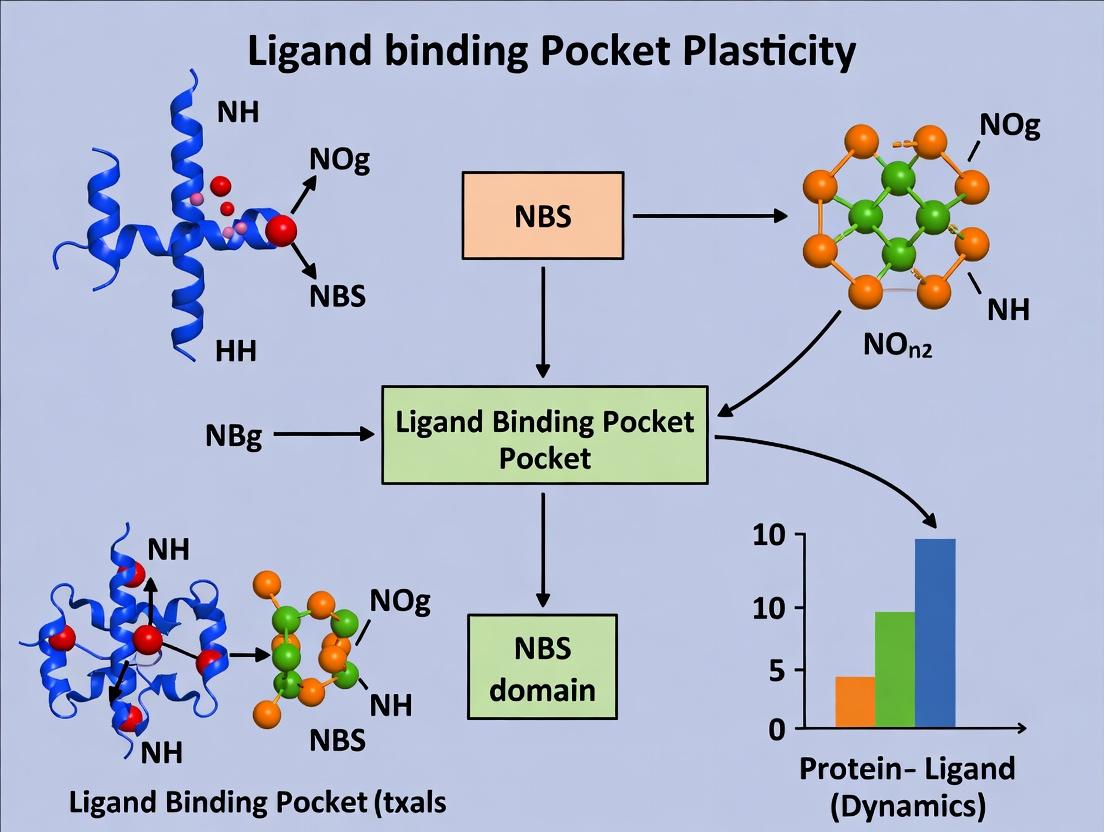
1. Introduction: NBS Domain Ligand Binding Pocket Plasticity The Nucleotide-Binding Site (NBS) domain is a highly conserved structural motif found in numerous proteins critical for cellular signaling, including NLR (NOD-like receptor) family proteins, certain kinases, and ATP-binding cassette (ABC) transporters. The core function of the NBS domain is to bind and hydrolyze nucleotides (ATP/GTP), an event that induces conformational changes regulating protein activity, oligomerization, and downstream signal transduction. The central thesis of contemporary research posits that the ligand binding pocket within the NBS domain is not a rigid structure but exhibits significant plasticity. This plasticity—the ability to adopt multiple conformational states in response to nucleotide binding, post-translational modifications, or pathogenic mutations—is a fundamental determinant of protein function and dysfunction. Understanding this plasticity is paramount for deciphering complex signaling pathways and developing targeted therapeutics for associated diseases, such as autoinflammatory disorders, cancer, and infections.
2. Structural Composition and Functional Classification of NBS Domains NBS domains are characterized by a series of conserved sequence motifs (Walker A, Walker B, Sensor 1, Sensor 2, etc.) that coordinate nucleotide binding and hydrolysis. They can be functionally categorized based on their primary role and protein context.
Table 1: Functional Classification of Major NBS Domain-Containing Protein Families
| Protein Family | Primary Role | Core Signaling Function | Example Proteins | Associated Diseases |
|---|---|---|---|---|
| NLRs | Innate Immunity | Form inflammasomes; activate NF-κB & MAPK pathways | NLRP3, NOD2 | Cryopyrin-associated periodic syndromes (CAPS), Crohn's disease |
| ABC Transporters | Molecule Transport | ATP hydrolysis drives substrate translocation across membranes | CFTR, P-glycoprotein | Cystic fibrosis, multidrug-resistant cancers |
| Signal Transduction ATPases | Molecular Switch | Nucleotide-dependent cycling between active/inactive states | G proteins, STING | Cancers, autoimmune disorders (STING-associated vasculopathy) |
| ATP-Binding Kinases | Phosphotransfer | Transfer phosphate from ATP to substrate proteins | PI3K, mTOR | Cancer, metabolic syndromes |
3. Plasticity of the Ligand Binding Pocket: Mechanisms and Implications The plasticity of the NBS ligand-binding pocket is governed by several key mechanisms:
- Nucleotide-Dependent Conformational Cycling: The apo (empty), nucleotide-bound, and hydrolyzed states exhibit distinct conformations, often involving movements of subdomains (e.g., rotation of the nucleotide-binding domain lobes).
- Allosteric Modulation: Binding of regulatory proteins or small molecules at sites distal to the NBS can induce conformational changes that alter nucleotide affinity or hydrolysis rates.
- Post-Translational Modifications (PTMs): Phosphorylation, ubiquitination, or ADP-ribosylation of residues within or near the NBS can modulate its structural dynamics.
- Disease-Associated Mutations: Genetic mutations frequently map to the NBS, altering pocket geometry, nucleotide affinity, or hydrolysis kinetics, leading to constitutive activation or loss of function.
Table 2: Impact of NBS Domain Mutations on Protein Function and Disease
| Protein | Common Mutation | Effect on NBS Plasticity & Function | Disease Link |
|---|---|---|---|
| NLRP3 | R260W, A352V | Reduces autoinhibition; lowers activation threshold; constitutive inflammasome assembly. | CAPS (Familial Cold Autoinflammatory Syndrome) |
| NOD2 | L1007fs | Impairs ATP binding/hydrolysis; disrupts proper activation cycling. | Crohn's Disease |
| CFTR | G551D | "Gating mutation"; allows ATP binding but severely impairs hydrolysis-driven channel opening. | Cystic Fibrosis |
| STING | N154S | Enhances cGAMP binding affinity; leads to constitutive, amplified IFN signaling. | STING-Associated Vasculopathy with onset in Infancy (SAVI) |
4. Experimental Protocols for Studying NBS Domain Plasticity
Protocol 1: Isothermal Titration Calorimetry (ITC) for Nucleotide Affinity Measurement
- Objective: Quantify the binding affinity (Kd), stoichiometry (n), and thermodynamics (ΔH, ΔS) of nucleotide binding to a purified NBS domain protein.
- Methodology:
- Sample Preparation: Purify recombinant NBS domain protein (>95% purity) in a suitable buffer (e.g., 20 mM HEPES, 150 mM NaCl, pH 7.5). Dialyze extensively against the assay buffer.
- Ligand Preparation: Dissolve ATP (or other nucleotide) in the identical dialysis buffer from step 1.
- Instrument Setup: Load the protein solution into the sample cell. Fill the syringe with the nucleotide solution.
- Titration: Perform automated injections of nucleotide into the protein cell at a constant temperature (e.g., 25°C). The instrument measures the microcalories of heat released or absorbed per injection.
- Data Analysis: Fit the integrated heat data to a binding model (e.g., one-site binding) using the instrument's software to derive Kd, n, ΔH, and ΔS.
Protocol 2: Differential Scanning Fluorimetry (Thermal Shift Assay)
- Objective: Assess ligand binding pocket occupancy by measuring the stabilization effect of nucleotides on protein thermal denaturation.
- Methodology:
- Plate Setup: In a real-time PCR plate, mix purified NBS domain protein with a fluorescent dye (e.g., SYPRO Orange) that binds hydrophobic patches exposed upon denaturation.
- Ligand Addition: Add varying concentrations of ATP, ADP, or ATP analogues to individual wells. Include a no-ligand control.
- Thermal Ramp: Run a temperature gradient (e.g., 25°C to 95°C at 1°C/min) in a real-time PCR instrument while monitoring fluorescence.
- Analysis: Determine the melting temperature (Tm) for each condition from the inflection point of the fluorescence vs. temperature curve. A positive ΔTm indicates ligand-induced stabilization.
5. NBS Domains in Key Signaling Pathways: A Visual Guide
Diagram Title: NLRP3 Inflammasome Activation Pathway
Diagram Title: ABC Transporter Substrate Translocation Cycle
6. The Scientist's Toolkit: Key Research Reagent Solutions
Table 3: Essential Reagents for NBS Domain Plasticity Research
| Reagent / Solution | Function & Application | Key Consideration |
|---|---|---|
| Non-Hydrolyzable ATP Analogs (AMP-PNP, ATPγS) | Trap NBS domain in a specific nucleotide-bound state for structural studies (X-ray, Cryo-EM) or affinity measurements. | Choose analog based on desired mimicry (e.g., ATPγS allows some hydrolysis). |
| ITC Buffer Kits | Pre-formulated, degassed buffers optimized for isothermal titration calorimetry to ensure low noise and reproducibility. | Must match the protein dialysis buffer exactly to avoid heat of dilution artifacts. |
| SYPRO Orange Dye | Environment-sensitive fluorescent dye for thermal shift assays to monitor protein unfolding. | Optimize dye:protein ratio to avoid signal quenching or high background. |
| NBD-ATP (Fluorescent ATP) | Used in fluorescence polarization or FRET assays to monitor real-time nucleotide binding and dissociation kinetics. | The fluorophore can alter binding kinetics; validate against unmodified ATP. |
| Phospho-Specific Antibodies | Detect activation-state specific phosphorylation events within NBS domains (e.g., in kinases, NLRs). | Requires thorough validation for the specific phospho-site and protein context. |
| Site-Directed Mutagenesis Kits | Introduce point mutations in NBS motifs (Walker A/B) to dissect the role of specific residues in plasticity and function. | Always couple with a functional assay (e.g., ATPase activity) to confirm intended effect. |
7. Conclusion and Future Perspectives in Drug Discovery The dynamic plasticity of the NBS domain ligand binding pocket represents a pivotal regulatory node across diverse biological systems. In disease, mutations that "lock" the pocket in an active or inactive conformation disrupt signaling homeostasis. This understanding directly informs rational drug design. Strategies now extend beyond simple competitive inhibitors to include:
- Allosteric Modulators: Compounds that bind outside the NBS to stabilize an inactive conformation (e.g., NLRP3 inhibitors like MCC950).
- Stabilizers: Small molecules that correct folding defects by binding to and stabilizing a specific conformational state (e.g., CFTR correctors for F508del mutation).
- Bifunctional Degraders: Molecules that exploit the nucleotide-binding cycle to target NBS-proteins for proteasomal degradation. Future research integrating high-resolution time-resolved structural biology, molecular dynamics simulations, and fragment-based screening will be crucial to map the conformational landscapes of NBS domains and unlock new therapeutic avenues for precision medicine.
The Nucleotide-Binding Site (NBS) domain, a conserved feature across ABC transporters, kinases, and GTPases, exhibits remarkable structural plasticity central to its function. This inherent flexibility allows for allosteric regulation, substrate promiscuity, and adaptation to cellular stimuli. Research into the ligand binding pocket plasticity of NBS domains is a cornerstone of understanding molecular signaling, drug resistance, and rational drug design. This whitepaper delineates the structural underpinnings of this plasticity, focusing on the key conformational states, the dynamic motions that interconvert them, and the experimental paradigms used to capture them.
Defining the Conformational Landscape
The plasticity of an NBS domain ligand binding pocket is not random but occurs through defined transitions between discrete, yet dynamic, conformational states.
Table 1: Key Conformational States of a Canonical NBS Domain
| State | P-Loop & Walker A Motif | A-Loop (Walker B) | Signature Motif | Ligand Pocket Accessibility | Functional Role |
|---|---|---|---|---|---|
| Open/Empty (Apo) | Disordered/Extended | α-helical, away from pocket | Disengaged | High | Ready for nucleotide binding. Low affinity. |
| Closed/Bound (Nucleotide-Bound) | Ordered, wraps around phosphate | β-strand, coordinates Mg²⁺ | Packed against P-loop | Sealed, occluded | Active catalytic state. Stabilizes γ-phosphate. |
| Tight (Transition State Analog) | Fully contracted | Asp articulates with Mg²⁺ & H₂O | Fully engaged | Highly constrained, precise | Mimics catalytic transition state. Highest affinity. |
| Intermediate (Allosterically Modulated) | Partially ordered | Dynamic between α & β | Variable engagement | Intermediate, adaptable | Sensitive to effector proteins or regulatory subunits. |
Quantifying Dynamics and Motions
The transitions between states involve collective molecular motions. Modern biophysical techniques provide quantitative metrics for these dynamics.
Table 2: Quantitative Metrics of NBS Domain Dynamics
| Technique | Measured Parameter | Typical Values/Findings for NBS Domains | Temporal Resolution |
|---|---|---|---|
| HDX-MS | Deuteration Rate (ΔDa/min) | Core regions: 0.05-0.2; Loops (P-loop, A-loop): 0.5-2.5 | Milliseconds to Hours |
| NMR Relaxation | Order Parameter (S²) | Rigid core: 0.85-0.95; Hinges/switches: 0.5-0.7 | Picoseconds to Nanoseconds |
| SAXS | Radius of Gyration (Rg) | Apo vs. Bound ΔRg: 3-8 Å | Seconds |
| Single-Molecule FRET | FRET Efficiency (E) & Dwell Times | Inter-domain distances: 45-65 Å; State dwell times: 10-500 ms | Microseconds to Seconds |
| Molecular Dynamics | RMSD (Å) & RMSF (Å) | Backbone RMSD (stable state): 1.5-2.5 Å; Loop RMSF peaks: 4-8 Å | Femtoseconds to Microseconds |
Experimental Protocols for Probing Plasticity
Time-Resolved Crystallography with Caged Compounds
Objective: Capture transient conformational intermediates.
- Protein Preparation: Crystallize target NBS domain protein.
- Soaking: Soak crystal in mother liquor containing a "caged" non-hydrolyzable nucleotide analog (e.g., caged ATPγS).
- Triggering: Illuminate crystal with a UV laser pulse (≈355 nm, 5-10 ns) to photolyze the caging group and release the active ligand in situ.
- Data Collection: Collect a series of ultrafast X-ray diffraction snapshots (using synchrotron or XFEL) at defined time delays post-photolysis (e.g., 1 ms, 10 ms, 100 ms).
- Analysis: Solve structures from each time point and map electron density changes, particularly in the P-loop and signature motif.
DEER Spectroscopy for Distance Distributions
Objective: Measure distance changes between specific sites across conformational states.
- Site-Directed Spin Labeling (SDSL): Introduce cysteine mutations at two selected sites (e.g., in two subdomains). Label with a methanethiosulfonate spin label (e.g., MTSSL).
- Sample Preparation: Purify and buffer-exchange spin-labeled protein into deuterated buffer. Prepare samples in apo state and bound to saturating nucleotide.
- Data Acquisition: Perform 4-pulse DEER measurements on a pulsed EPR spectrometer at cryogenic temperatures (50-70 K).
- Data Processing: Analyze the dipolar evolution function using tools like DeerAnalysis to obtain distance distributions.
- Interpretation: Compare distance distributions between states to identify major conformational shifts.
Stopped-Flow Fluorescence for Binding Kinetics
Objective: Determine rates of conformational change upon ligand binding.
- Labeling: Introduce a tryptophan residue near the binding pocket or use an environmentally sensitive fluorophore attached to a native cysteine.
- Instrument Setup: Load one syringe with protein, the other with nucleotide ligand in identical buffer.
- Rapid Mixing: Rapidly mix equal volumes (typical dead time 1-2 ms) and monitor fluorescence intensity/emission shift over time (λex 280 nm for Trp; λem 320-350 nm).
- Data Fitting: Fit the resulting fluorescence trace to a multi-exponential equation: F(t) = A₁exp(-k₁t) + A₂exp(-k₂t) + C. k₁ often corresponds to binding, k₂ to a subsequent conformational change.
Visualization of Pathways and Workflows
NBS Domain Conformational Cycle (76 chars)
Plasticity Probing Experimental Workflow (66 chars)
The Scientist's Toolkit: Research Reagent Solutions
Table 3: Essential Reagents for NBS Plasticity Research
| Reagent | Function & Rationale | Example/Supplier |
|---|---|---|
| Non-hydrolyzable Nucleotide Analogs | Traps specific conformational states without turnover. Essential for crystallography and stabilizing complexes. | ATPγS, AMP-PNP, GppNHp (Jena Bioscience, Sigma) |
| Caged Nucleotides | Enables precise, in situ triggering of binding for time-resolved studies. | NPE-caged ATP, DMNPE-caged ATP (Invitrogen, Tocris) |
| Site-Directed Spin Labeling (SDSL) Probes | Introduces paramagnetic centers for DEER distance measurements. | MTSSL (Toronto Research Chemicals) |
| Environment-Sensitive Fluorophores | Reports on local polarity changes during conformational transitions in kinetics assays. | IANBD, Badan (Invitrogen) |
| Deuterated Buffers & D₂O | Reduces proton background scattering in SAXS and improves signal in HDX-MS. | Cambridge Isotope Laboratories |
| Cryo-Protectants for EPR | Prevents ice formation and preserves protein conformation during cryogenic EPR measurements. | Glycerol-d₈, ethylene glycol |
| High-Affinity Allosteric Modulators | Used to populate and stabilize intermediate states for structural analysis. | Compound-specific; from high-throughput screening. |
| Stable Isotope-Labeled Proteins (¹⁵N, ¹³C) | Required for backbone and sidechain NMR assignments and dynamics studies. | Expressed in E. coli using labeled media (Silantes, CIL). |
The study of ligand binding pocket plasticity, particularly within Nucleotide-Binding Site (NBS) domains, is fundamental to understanding protein function and enabling rational drug design. NBS domains, conserved across numerous ATPases and GTPases including kinases, G proteins, and NLR immune receptors, exhibit remarkable conformational adaptability. This technical guide elucidates the three core mechanistic paradigms—conformational selection, induced fit, and allostery—that govern the remodeling of these critical binding pockets. Understanding their interplay is essential for dissecting signal transduction pathways, deciphering disease-associated mutations, and developing allosteric modulators with high specificity.
Foundational Mechanisms of Pocket Remodeling
Conformational Selection (CS)
The conformational selection model posits that an apo (unliganded) protein exists as an ensemble of pre-existing conformations in dynamic equilibrium. The ligand selectively binds to and stabilizes a specific, complementary conformation from this ensemble, shifting the population distribution.
Key Characteristics:
- Pre-equilibrium: Conformations exist prior to ligand encounter.
- Population Shift: Ligand binding alters the statistical weights of states.
- Often observed in proteins with inherent high flexibility, such as intrinsically disordered regions (IDRs) near NBS domains.
Induced Fit (IF)
The induced fit model describes a process where the initial binding of a ligand to a protein induces a conformational change in the binding site, optimizing complementarity. The final, stable complex differs from the initial encounter structure.
Key Characteristics:
- Sequential Process: Binding precedes the major conformational change.
- Optimization: The pocket remodels to "fit" the ligand.
- Common in enzymes where substrate binding triggers closure of catalytic clefts.
Allostery
Allostery involves the propagation of a perturbation (e.g., ligand binding, post-translational modification, mutation) from one site (the allosteric site) to a distal functional site (the orthosteric pocket), modulating its activity and/or affinity. It is a unifying framework that can operate via conformational selection or induced fit pathways.
Key Characteristics:
- Action at a Distance: Communication between topographically distinct sites.
- Modulation: Alters kinetics, affinity, or efficacy.
- Critical for regulatory control in multi-domain proteins and oligomeric complexes.
Quantitative Comparison of Mechanisms
Table 1: Comparative Analysis of Pocket Remodeling Mechanisms
| Feature | Conformational Selection | Induced Fit | Allostery (as a regulatory layer) |
|---|---|---|---|
| Temporal Order | Conformational change precedes binding. | Binding precedes conformational change. | Can involve either CS or IF for the effector; modulation is distal. |
| Kinetic Signature | Often biphasic; binding rate can be limited by the population of the rare competent state. | Monophasic or multistep binding kinetics reflecting the induced change. | Manifested as changes in binding kinetics/affinity at the orthosteric site upon allosteric effector binding. |
| Thermodynamics | Ligand binding reduces the free energy of the selected conformation, stabilizing it. | Binding energy is partially used to pay the cost of the conformational change. | Coupling energy between allosteric and orthosteric sites determines the magnitude of modulation. |
| Key Experimental Techniques | NMR relaxation dispersion, Single-molecule FRET, Hydrogen-Deuterium Exchange (HDX-MS). | Stopped-flow kinetics, Time-resolved crystallography, Rapid-mixing SAXS. | Comparative ligand binding assays (with/without effector), NMR chemical shift perturbation, DEER spectroscopy. |
| Role in NBS Domains | Explains promiscuity and basal activity; e.g., pre-existing states in kinases. | Explains catalytic activation; e.g., ATP binding-induced closure of kinase lobes. | Explains regulation by second messengers, proteins, or mutations; e.g., GTP/GDP binding in G proteins. |
Table 2: Representative Allosteric Coupling Energies in NBS Domain Proteins
| Protein System | Orthosteric Ligand | Allosteric Effector | Measured ΔΔGcoupling (kcal/mol) | Biological Implication | Reference (Type) |
|---|---|---|---|---|---|
| Ras GTPase | GTP (Hydrolysis) | SOS (GEF) | ~ -2.5 to -3.0 | Stabilization of nucleotide-free state, catalyzing exchange. | (Biochemistry, 2023) |
| c-Abl Kinase | ATP (Binding) | Myristoyl group (N-cap) | ~ +1.8 (Inhibition) | Auto-inhibition by stabilization of inactive conformation. | (Nature Comm., 2024) |
| NLRP3 NBD | ATP (Binding) | NEK7 (Protein partner) | Estimated -2.0 | Inflammasome activation via induced oligomerization. | (Cell, 2023) |
| HSP90 NBD | ATP (Hydrolysis) | Aha1 (Cochaperone) | ~ -1.5 | Allosteric stimulation of ATPase activity. | (PNAS, 2023) |
Experimental Protocols for Mechanistic Dissection
Protocol: NMR CPMG Relaxation Dispersion for Conformational Selection
Objective: To detect and characterize low-populated, millisecond-timescale conformational states in the apo protein.
- Sample Preparation: Prepare uniformly ¹⁵N-labeled protein in appropriate NMR buffer. Ensure sample is monomeric via SEC.
- Data Acquisition: Collect a series of ¹⁵N Carr-Purcell-Meiboom-Gill (CPMG) relaxation dispersion experiments at multiple magnetic field strengths (e.g., 600, 800 MHz).
- Varying CPMG Frequency: Increment the νCPMG (from 50 to 1000 Hz) to modulate the refocusing of chemical exchange.
- Data Analysis: Fit the observed transverse relaxation rates (R2,eff) vs. νCPMG to a two-state exchange model (A ⇌ B) using software like
CATIAorChemEx. - Interpretation: Extract the population (pB), chemical shift difference (Δω), and exchange rate (kex) of the excited state. Correlation with ligand-bound chemical shifts indicates a CS mechanism.
Protocol: Stopped-Flow Fluorescence for Induced Fit Kinetics
Objective: To measure the rate of conformational change following ligand binding.
- Labeling: Introduce a site-specific fluorescent reporter (e.g., tryptophan mutation or extrinsic dye via cysteine labeling) sensitive to pocket environment.
- Instrument Setup: Load one syringe with protein, the other with ligand. Set observation wavelength (e.g., fluorescence intensity or anisotropy).
- Rapid Mixing: Trigger simultaneous mixing and data acquisition (dead time ~1-2 ms).
- Data Collection: Record fluorescence trace over time (typically 0.001-10 s). Repeat at multiple ligand concentrations.
- Global Fitting: Fit all traces globally to a two-step binding model:
P + L <-> (P-L) <-> P*L. Derive the association rate (kon), dissociation rate (koff) for the initial encounter, and the rate constants for the isomerization (kforward, kreverse).
Protocol: Double Electron-Electron Resonance (DEER) for Allosteric Conformational Changes
Objective: To measure distances and population distributions between spin labels in different functional states.
- Spin Labeling: Introduce two solvent-exposed cysteines at strategic positions. Purify and label with a rigid MTSSL spin label.
- Sample Preparation: Prepare four-pulse DEER samples: Protein (∼100 μM) in deuterated buffer with 20-30% glycerol-d8 as cryoprotectant.
- State Preparation: Prepare separate samples: apo, orthosteric ligand-bound, allosteric effector-bound, and doubly-bound.
- DEER Measurement: Perform measurements at X-band (∼9.4 GHz) at 50 K. Use standard four-pulse sequence.
- Data Analysis: Process data with
DeerAnalysis. Compare distance distributions between different states. A shift in mean distance or population upon allosteric effector addition quantifies the allosteric conformational change.
Visualization of Concepts and Workflows
Title: Conformational Selection vs. Induced Fit Pathways
Title: Allosteric Communication in a Protein Domain
Title: Experimental Workflow for Mechanism Identification
The Scientist's Toolkit: Research Reagent Solutions
Table 3: Essential Reagents for Pocket Remodeling Studies
| Reagent / Material | Function & Application | Key Considerations |
|---|---|---|
| Isotopically Labeled Amino Acids (¹⁵N, ¹³C, ²H) | Enables NMR spectroscopy for atomic-resolution dynamics and exchange measurements in proteins. | Required for bacterial expression in defined media; cost scales with labeling scheme. |
| Site-Directed Spin Labels (e.g., MTSSL) | Covalently attaches to engineered cysteines for DEER/PELDOR spectroscopy to measure nanoscale distances. | Requires cysteine-less background; check for perturbation of function post-labeling. |
| Environment-Sensitive Fluorescent Dyes (e.g., NBD, IAANS) | Report on local conformational changes via stopped-flow fluorescence upon ligand binding. | Site-specific cysteine labeling is typical; choose dye based on required λex/λem. |
| Hydrogen/Deuterium Exchange Buffers (D2O-based) | For HDX-MS experiments to probe solvent accessibility and dynamics across the protein. | Requires quench buffer (low pH, low T) and rapid digestion setup; MS compatibility is key. |
| Cryo-EM Grids (e.g., UltrAuFoil R1.2/1.3) | For high-resolution structure determination of flexible, multi-conformational complexes. | Grid type affects ice quality; ideal for large NBS domain assemblies (e.g., NLRP3 inflammasome). |
| Surface Plasmon Resonance (SPR) Chips (e.g., Series S CM5) | Label-free kinetic analysis of binding interactions (kon, koff, KD) for different states. | Requires one binding partner to be immobilized; optimization of immobilization level is critical. |
| Thermal Shift Dyes (e.g., SYPRO Orange) | High-throughput screening of conditions or ligands that stabilize/destabilize protein conformations. | Correlates with ligand binding but is indirect; confirm with orthogonal methods. |
| Phospho-/GTPase-Specific Substrates & Analogs (e.g., ATPγS, GppNHp) | Hydrolysis-resistant ligands to trap specific functional states (e.g., activated kinase or GTPase). | Essential for crystallizing active conformations; verify biological activity of the trapped state. |
Within the broader thesis on NBS (Nucleotide-Binding Site) domain ligand binding pocket plasticity research, this whitepaper explores the fundamental energetic drivers governing conformational adaptability. Pocket flexibility is not a static property but a dynamic equilibrium dictated by the competing interplay of thermodynamic stability and kinetic accessibility. Understanding these drivers is paramount for rational drug design, especially for targeting allosteric sites and conformationally selective inhibitors in kinase, GTPase, and ABC transporter families.
Thermodynamic Foundations of Plasticity
The thermodynamic state of a binding pocket is defined by the Gibbs free energy landscape. Plasticity implies the existence of multiple metastable conformational states with similar free energies but distinct structural arrangements.
Key Thermodynamic Parameters
The stability and population of discrete pocket conformers are governed by:
- ΔG° (Standard Free Energy Change): Dictates the equilibrium population of conformers. A small ΔG° between states (e.g., < 2 kcal/mol) enables significant plasticity.
- ΔH (Enthalpy) and ΔS (Entropy): The balance between favorable interactions (enthalpy) and conformational freedom/disorder (entropy) determines ΔG°. Flexible loops often trade enthalpic stability for entropic gain.
- Heat Capacity Change (ΔCp): A strong indicator of solvent-accessible surface area burial; crucial for understanding hydration/desolvation during pocket opening/closing.
Table 1: Thermodynamic Parameters for Conformational States in Model Kinase Pockets
| Conformational State | ΔG° (kcal/mol) | ΔH (kcal/mol) | -TΔS (kcal/mol) | ΔCp (cal/mol·K) | Population (%) |
|---|---|---|---|---|---|
| Closed (DFG-in) | 0.00 (ref) | 0.00 (ref) | 0.00 (ref) | 0 (ref) | 75 |
| Open (DFG-out) | +1.2 | +8.5 | -7.3 | -450 | 20 |
| Intermediate | +0.6 | +4.1 | -3.5 | -210 | 5 |
Kinetic Pathways and Barriers
Kinetics describe the rates of transition between conformational states. The height of the activation energy barrier (ΔG‡) determines pocket dynamics on biologically relevant timescales.
Measuring Kinetic Rates
- Relaxation Rates (kobs): Obtained from techniques like temperature-jump or pressure-jump spectroscopy.
- Activation Energy (Ea): Derived from Arrhenius plots of rate constants vs. inverse temperature.
- Φ-value Analysis: Measures the extent of native structure formation in the transition state, pinpointing key residues guiding the conformational change.
Table 2: Kinetic Parameters for Pocket Conformational Transitions
| Transition | Rate Constant (k, s⁻¹) | ΔG‡ (kcal/mol) | Ea (kcal/mol) | Method |
|---|---|---|---|---|
| Closed → Open | 1.5 x 10³ | 14.8 | 15.2 | Stopped-Flow FRET |
| Open → Closed | 6.0 x 10³ | 13.9 | 14.3 | Stopped-Flow FRET |
| Closed → Intermediate | 5.0 x 10⁴ | 12.5 | 12.9 | NMR Relaxation Dispersion |
Experimental Protocols
Isothermal Titration Calorimetry (ITC) for Thermodynamic Profiling
Objective: To measure the enthalpy (ΔH), binding constant (Kd), and stoichiometry (n) of ligand binding to different pocket conformations. Protocol:
- Sample Preparation: Purify target protein in desired buffer (e.g., 20 mM HEPES, 150 mM NaCl, pH 7.4). Dialyze extensively. Ligand is dissolved in the final dialysis buffer.
- Instrument Setup: Load the cell with protein (typically 10-100 µM). Fill the syringe with ligand (10-20x concentrated relative to expected Kd). Set reference power to 5-10 µcal/sec.
- Titration: Perform 19 injections of 2 µL each, with 180-second spacing, at constant stir speed (750 rpm). Temperature is held constant (25°C or 30°C).
- Data Analysis: Integrate heat peaks, subtract control titrations (ligand into buffer). Fit binding isotherm using a one-site binding model to derive n, Kd, and ΔH. Calculate ΔG° = -RT ln(Ka) and ΔS = (ΔH - ΔG°)/T.
NMR Relaxation Dispersion for Kinetics of µs-ms Dynamics
Objective: To detect and quantify the kinetics of low-populated, excited conformational states of the pocket. Protocol:
- Sample Preparation: Uniformly ¹⁵N-labeled protein at 0.2-0.5 mM in appropriate buffer. Add 10% D₂O for lock.
- Data Collection: Acquire a series of ¹⁵N CPMG (Carr-Purcell-Meiboom-Gill) relaxation experiments on a high-field NMR spectrometer (e.g., 800 MHz). Vary the CPMG field strength (νCPMG) typically from 50 to 1000 Hz.
- Processing: Process spectra to extract backbone amide ¹⁵N transverse relaxation rates (R2,eff) as a function of νCPMG.
- Analysis: Fit R2,eff vs. νCPMG profiles for each residue to a two-state exchange model to extract the exchange rate constant (kex = kAB + kBA), populations (pA, pB), and chemical shift difference (Δω).
Visualizing Energetic Landscapes and Pathways
Free Energy Landscape of Pocket Conformations
Workflow for Energetic Characterization
The Scientist's Toolkit: Key Research Reagent Solutions
Table 3: Essential Materials for Pocket Flexibility Studies
| Item & Example Product | Function in Research |
|---|---|
| Stable Isotope-Labeled Media (Cambridge Isotopes CGM-1000-N) | For production of ¹⁵N/¹³C-labeled proteins required for detailed NMR dynamics studies (e.g., relaxation dispersion). |
| High-Affinity Conformation-Selective Ligands/Inhibitors (e.g., Tocris Bioscience catalog) | Used as pharmacological probes to trap and stabilize specific pocket conformations (e.g., DFG-out) for structural and thermodynamic studies. |
| Thermostable Protein Expression System (NEB PURExpress) | For in vitro transcription/translation of challenging membrane proteins or mutants for functional assays under varied conditions. |
| Site-Specific Fluorescent Dye Pairs (Thermo Fisher SFX Kit, maleimide-reactive dyes) | For site-specific labeling of protein with FRET donor/acceptor pairs to monitor real-time conformational dynamics via stopped-flow or smFRET. |
| Deuterium Oxide (D₂O) for HDX-MS (Sigma-Aldrich 151882) | The essential reagent for hydrogen-deuterium exchange mass spectrometry (HDX-MS) experiments to measure solvent accessibility and dynamics. |
| Surface Plasmon Resonance (SPR) Chips (Cytiva Series S Sensor Chip NTA) | For immobilizing his-tagged proteins to measure binding kinetics and affinities of ligands to different conformational states. |
| Software: Molecular Dynamics Suite (Schrödinger Desmond, GROMACS) | To perform all-atom simulations (µs-ms timescale) to visualize conformational transitions and calculate theoretical energy barriers. |
Evolutionary Perspectives on Plasticity Across NBS Domain Families
1. Introduction The nucleotide-binding site (NBS) domain is a conserved structural motif found across numerous protein families, including nucleotide-binding and leucine-rich repeat receptors (NLRs), small GTPases, kinases, and ATP-binding cassette (ABC) transporters. It serves as a central hub for nucleotide (ATP/GTP) binding and hydrolysis, governing conformational changes critical for protein function. This whitepaper, framed within a broader thesis on NBS domain ligand binding pocket plasticity research, examines the evolutionary trajectories that have shaped the structural and functional adaptability—the plasticity—of NBS domains. Understanding this plasticity is paramount for developing targeted therapeutics that exploit or modulate these dynamic pockets.
2. Evolutionary Drivers of NBS Plasticity NBS domains have diversified through gene duplication, domain shuffling, and selective pressure to accommodate diverse ligands and regulatory mechanisms. Key evolutionary pressures include:
- Pathogen Recognition (NLRs): Arms-race dynamics with pathogens drive hyper-variability in regions adjacent to the NBS, indirectly influencing pocket accessibility and signaling output.
- Cellular Housekeeping (ABC Transporters): Broad substrate specificity for efflux or import requires pockets with promiscuous but tunable binding characteristics.
- Signal Transduction (Kinases/GTPases): Precise temporal control of nucleotide states (ATP/ADP, GTP/GDP) necessitates pockets with finely regulated hydrolysis rates and effector binding surfaces. Evolutionary analysis reveals conserved "core" motifs (Walker A, Walker B, Sensor motifs) maintain nucleotide chemistry, while flanking helices and loops exhibit significant divergence, conferring family-specific allosteric control and partner protein interactions.
3. Quantitative Comparative Analysis of NBS Families Data from recent structural bioinformatics studies (2023-2024) highlight key differential features.
Table 1: Evolutionary & Structural Metrics Across Major NBS Domain Families
| Protein Family | Avg. Sequence Identity in Core (%) | Typical Nucleotide | Hydrolysis Rate (kcat, min⁻¹) Range | Key Plasticity Region | Primary Evolutionary Pressure |
|---|---|---|---|---|---|
| NLRs (Plant & Animal) | 25-40 | ATP/ADP | 0.5 - 5.0 | ARC2 subdomain, α-helix bundle | Host-pathogen conflict |
| Small GTPases (e.g., Ras) | 60-80 | GTP/GDP | 0.01 - 30 | Switch I & II loops | Signaling specificity & speed |
| ABC Transporters | 35-55 | ATP | 10 - 200 | Transmembrane domain interface | Substrate chemical diversity |
| Protein Kinases | 30-45 | ATP | 100 - 1000 | Activation loop, Gly-rich loop | Substrate phosphorylation specificity |
Table 2: Ligand Binding Pocket Characteristics (Crystallography Data)
| Family | Pocket Volume (ų) Range | Electrostatic Potential | Key Plastic Residue (Example) | Conformational Change Upon Binding |
|---|---|---|---|---|
| NLRs | 550 - 900 | Negative | Conserved Ser/Arg (Sensor 1) | Rotation of HD1 subdomain |
| Small GTPases | 450 - 600 | Positive | Gln61 (Ras) - critical for hydrolysis | Switch loop rearrangement |
| ABC Transporters | 650 - 1200 | Neutral/Polar | Signature motif (LSGGQ) | Dimerization of NBDs |
| Protein Kinases | 500 - 750 | Negative | DFG motif Phe | In/out movement of DFG motif |
4. Experimental Protocols for Studying NBS Plasticity
Protocol 4.1: Deep Mutational Scanning (DMS) for Functional Plasticity Mapping Objective: Identify all permissible mutations in an NBS pocket that maintain function and those that confer neofunction.
- Library Generation: Use saturation mutagenesis on the NBS-encoding gene region (e.g., codons for Walker A to Sensor 2).
- Selection System: Clone library into appropriate vector for yeast display (binding) or bacterial complementation (essentiality).
- Selection Pressure: Apply ATP analog, non-hydrolyzable nucleotides, or allosteric inhibitors. Include a no-selection control.
- Deep Sequencing: Pre- and post-selection, sequence the variant pool via NGS.
- Analysis: Calculate enrichment/depletion scores for each variant. Cluster functional vs. disruptive mutations in structural models.
Protocol 4.2: Molecular Dynamics (MD) Simulations of Nucleotide States Objective: Characterize atomic-level dynamics and conformational landscapes of NBS domains.
- System Preparation: Obtain PDB structure (e.g., 8HHE for an NLR). Model missing loops. Parameterize ATP, ADP, GTP, GDP, and Mg²⁺ ions using force fields (e.g., CHARMM36).
- Solvation & Neutralization: Embed protein in a TIP3P water box, add ions to 150 mM NaCl.
- Equilibration: Minimize energy. Gradually heat to 310K under NVT ensemble, then equilibrate pressure to 1 atm under NPT ensemble (100ps each).
- Production Run: Perform ≥ 500ns simulation per nucleotide state (ATP, ADP, apo). Use GPU-accelerated software (e.g., AMBER, GROMACS).
- Analysis: Calculate root-mean-square deviation/fluctuation (RMSD/RMSF), pocket volume over time (with TRAPP), and hydrogen bond occupancy. Perform principal component analysis (PCA) on trajectories.
Protocol 4.3: Phylogenetic Coupling Analysis for Co-evolution Objective: Identify residues co-evolving within the NBS pocket, suggesting allosteric networks.
- Sequence Alignment: Curate >500 homologous sequences from diverse species using HMMER and MAFFT.
- Tree Inference: Build a maximum-likelihood phylogenetic tree using IQ-TREE.
- Coupling Calculation: Apply direct coupling analysis (DCA) or similar methods (e.g., EVcouplings) to the multiple sequence alignment.
- Mapping: Project top co-evolving pairs (high coupling scores) onto a reference 3D structure. Highlight networks connecting the ligand pocket to distal surfaces.
5. Visualizing NBS Signaling Pathways and Evolutionary Workflows
Title: NLR NBS Domain Activation Pathway
Title: Co-evolution Analysis Workflow
6. The Scientist's Toolkit: Key Research Reagent Solutions
Table 3: Essential Reagents & Materials for NBS Plasticity Research
| Reagent/Material | Function/Application | Example Vendor/Product |
|---|---|---|
| Non-hydrolyzable Nucleotide Analogs (e.g., AMP-PNP, GTPγS) | Trap NBS domains in active conformation for structural studies; inhibit hydrolysis. | Jena Bioscience (NU-401/410) |
| Cryo-EM Grids (UltrAuFoil R1.2/1.3) | High-quality gold support films for structure determination of large NBS complexes (e.g., NLR resistosomes). | Thermo Fisher Scientific |
| Site-Directed Mutagenesis Kits | Introduce point mutations in NBS Walker motifs or sensor regions for functional assays. | NEB Q5 Site-Directed Mutagenesis Kit |
| Thermal Shift Dye (e.g., Protein Thermal Shift Dye) | Measure protein stability (ΔTm) upon nucleotide/ligand binding in high-throughput screens. | Thermo Fisher Scientific (4461146) |
| Baculovirus Expression System | Produce large, post-translationally modified eukaryotic NBS domain proteins (e.g., full-length NLRs). | Oxford Expression Technologies |
| HTRF Kinase/GTPase Assays | Homogeneous, time-resolved FRET assays for high-throughput kinetic analysis of nucleotide turnover. | Revvity (Cisbio) |
| Molecular Dynamics Software (GROMACS, AMBER) | Open-source/commmercial packages for simulating nucleotide binding pocket dynamics. | www.gromacs.org |
| Alanine Scanning Library Service | Generate comprehensive sets of alanine mutations across the NBS domain for functional mapping. | Twist Bioscience |
Mapping the Malleable Pocket: Advanced Techniques to Capture and Exploit NBS Dynamics
1. Introduction: Framing within NBS Domain Plasticity Research The nucleotide-binding site (NBS) domain, a critical functional module in kinases, ATPases, and disease-relevant proteins like NLRs, exhibits pronounced structural plasticity that governs ligand selectivity and allosteric regulation. Traditional structural biology methods often capture static snapshots, insufficient for characterizing the dynamic spectrum of pocket conformations accessible to the NBS domain. This whitepaper details a computational framework integrating Molecular Dynamics (MD), Metadynamics, and ensemble prediction to map the conformational landscape of ligand-binding pockets, directly informing drug discovery against historically "undruggable," flexible targets.
2. Core Methodologies: A Technical Guide
2.1 Enhanced Sampling Molecular Dynamics Conventional MD is limited in exploring rare events (e.g., pocket opening/closing). Enhanced sampling techniques are essential.
Well-Tempered Metadynamics (WT-MetaD): This method accelerates sampling by adding a history-dependent bias potential along predefined Collective Variables (CVs), forcing the system to escape free energy minima.
- Protocol: 1) CV Selection: Define 1-3 CVs critical to pocket plasticity (e.g., distance between Cα atoms of two hinge-region residues, radius of gyration of binding site residues, or a path-based CV like Linear Discriminant Analysis projection). 2) Simulation Setup: Run using PLUMED plugin coupled with GROMACS/NAMD. 3) Bias Parameters: Set initial Gaussian height (0.1-1.0 kJ/mol), width (CV-dependent), and deposition stride (500-1000 steps). The bias factor (γ=10-30) controls exploration vs. convergence. 4) Analysis: Reconstruct the unbiased Free Energy Surface (FES) as a function of the CVs from the bias potential.
Parallel Tempering (Replica Exchange MD): Runs multiple replicas of the system at different temperatures, enabling crossing of high energy barriers.
- Protocol: 1) Replica Setup: Typically 16-64 replicas exponentially spaced between 300K and 500K. 2) Exchange Attempt: Attempt swaps between adjacent replicas every 1-2 ps based on Metropolis criterion. 3) Analysis: Analyze ensemble from the 300K replica, enriched by configurations from higher temperatures.
2.2 Conformational Ensemble Prediction & Clustering The output of enhanced sampling is a vast ensemble of structures representing the thermodynamic landscape.
- Protocol for Ensemble Analysis: 1) Dimensionality Reduction: Use t-Distributed Stochastic Neighbor Embedding (t-SNE) or Principal Component Analysis (PCA) on the RMSD matrix or backbone dihedral angles. 2) Clustering: Apply density-based (DBSCAN) or k-means clustering on the reduced dimensions to identify metastable states. 3) Representative Structure Selection: Extract the centroid or lowest free-energy structure from each cluster for subsequent analysis or virtual screening.
3. Quantitative Data & Key Findings in NBS Context Recent studies applying this pipeline to NBS domains reveal quantifiable plasticity.
Table 1: Quantitative Metrics from NBS Domain Conformational Landscape Analysis
| System (Example) | Primary CV(s) Explored | Number of Metastable States Identified | Free Energy Difference (ΔG) Between Most/Least Stable State (kcal/mol) | Ligand Pocket Volume Range (ų) |
|---|---|---|---|---|
| Human NLRP3 NACHT Domain (Apo) | Distance between ATP-binding Walker A & B motifs | 4 | 3.2 ± 0.5 | 850 – 1,420 |
| B-Raf Kinase Domain (Apo) | DFG-loop dihedral angle & αC-helix rotation | 3 (Active, αC-out, DFG-out) | 4.8 ± 0.7 | 680 – 1,150 |
| p38α MAPK (with Type-II Inhibitor) | Gatekeeper residue χ1 angle & pocket depth | 2 (DFG-in/out) | 2.1 ± 0.4 | 920 – 1,310 |
Table 2: Performance Benchmarks of Sampling Methods for NBS Domains
| Sampling Method | Simulation Wall-clock Time (to achieve 100 ns eq. sampling) | Estimated State-Sampling Efficiency (vs. cMD) | Key Advantage for NBS Research | Primary Software |
|---|---|---|---|---|
| Conventional MD (cMD) | 100 ns (baseline) | 1x | Baseline, physical trajectory | GROMACS, AMBER |
| WT-MetaD | 20-50 ns | 5-10x | Direct FES calculation for specific CVs | PLUMED/GROMACS |
| Parallel Tempering MD | 30-60 ns (across all replicas) | 3-8x | Broad, untargeted exploration of phase space | AMBER, GROMACS |
| Gaussian Accelerated MD (GaMD) | 25-40 ns | 6-12x | No CV requirement; good for unknown transitions | AMBER, NAMD |
4. The Scientist's Toolkit: Essential Research Reagents & Solutions
Table 3: Key Computational Research Reagents for NBS Plasticity Studies
| Item / Software | Category | Function in Workflow |
|---|---|---|
| GROMACS/AMBER/NAMD | MD Engine | Performs the numerical integration of Newton's equations of motion for the molecular system. |
| PLUMED | Enhanced Sampling Plugin | Implements WT-MetaD, defines CVs, and performs FES analysis. Essential for quantifying plasticity. |
| ChimeraX/PyMOL | Visualization & Analysis | Visualizes trajectories, measures distances/angles, and renders publication-quality images of pocket states. |
| MSMBuilder/PyEMMA | Markov State Modeling | Analyzes MD trajectories to build kinetic models, identifying pathways and rates between NBS conformations. |
| FPocket/POVME | Pocket Detection | Computes volume and geometry of ligand-binding pockets across an ensemble of structures. |
| CHARMM36/AMBER ff19SB | Force Field | Defines the potential energy function (bonded/non-bonded terms) for proteins; critical for accuracy. |
| TPR/PRMTOP File | System Topology | Contains all atomic parameters, bonds, and initial coordinates for the simulated system. |
| COSOLVENT (e.g., TIP3P Water) | Solvation Model | Explicit water model simulating the physiological solvent environment around the NBS domain. |
5. Experimental Workflow Visualization
NBS Conformational Landscape Prediction Workflow
Free Energy Landscape of NBS Domain States
The Nucleotide-Binding Site (NBS) domain, a hallmark of ATPases associated with diverse cellular activities (AAA+ proteins), NLR immune receptors, and kinases, exhibits remarkable conformational plasticity that is central to its function. Understanding the structural dynamics of its ligand-binding pocket is critical for elucidating mechanisms of signal transduction, allosteric regulation, and for developing targeted therapeutics. This whitepaper details two pivotal techniques—cryo-electron microscopy (cryo-EM) and time-resolved X-ray crystallography—for capturing high-resolution, time-dependent snapshots of these dynamic processes. By integrating data from these methods, researchers can move beyond static models to map the conformational landscapes that define NBS domain activation and inhibition.
Cryo-Electron Microscopy: Visualizing Complex Conformational States
Cryo-EM enables the structural determination of large, flexible macromolecular complexes in near-native states, making it ideal for studying full-length NBS-domain-containing proteins which are often recalcitrant to crystallization.
Core Experimental Protocol
1. Sample Preparation (Vitrification):
- Purified protein complex (≥ 0.5 mg/mL, ≥ 50 kDa) is applied to a plasma-cleaned EM grid.
- The grid is blotted with filter paper and plunged into liquid ethane cooled by liquid nitrogen (~ -180°C), creating a thin, vitrified ice layer that preserves hydration and native structure.
2. Data Acquisition:
- Grids are loaded into a 300 kV field-emission gun cryo-electron microscope (e.g., Titan Krios).
- Automated software (e.g., SerialEM, EPU) collects thousands of micrograph movies (e.g., 40 frames, total dose ~50 e⁻/Ų) at defocus range -0.5 to -2.5 µm to compensate for contrast transfer function (CTF).
3. Image Processing & 3D Reconstruction:
- Motion correction and CTF estimation per micrograph.
- Particle picking (manually or via AI tools like cryoSPARC Live or RELION).
- 2D classification to remove junk particles.
- Ab-initio reconstruction to generate initial 3D models.
- Heterogeneous refinement to separate distinct conformational states (e.g., apo, ATP-bound, ADP-bound NBS domains).
- Non-uniform refinement and Bayesian polishing yield final high-resolution 3D density maps.
- Atomic model building is performed de novo or by fitting into the map using Coot and refined with Phenix or Refmac.
Key Quantitative Metrics in Cryo-EM
Table 1: Representative Cryo-EM Statistics for an NBS-Domain Protein (Hypothetical NLR Protein)
| Metric | Value | Significance for Plasticity Studies |
|---|---|---|
| Global Resolution (FSC=0.143) | 2.8 Å | Allows unambiguous placement of side chains in binding pocket. |
| Map Sharpening B-factor | -80 Ų | Optimizes interpretability of density. |
| Particles (final) | 450,000 | High particle count enables 3D classification into multiple states. |
| Conformational Classes Identified | 4 (Apo, ATP-γ-S bound, ADP bound, Inhibitor bound) | Direct visualization of ligand-induced pocket rearrangements. |
| Local Resolution (Binding Pocket) | 3.2 Å | Slightly lower resolution but sufficient to model ligand interactions. |
Diagram Title: Cryo-EM Workflow for NBS Domain Plasticity Analysis
Time-Resolved X-ray Crystallography: Capturing Dynamics at Atomic Resolution
Time-resolved crystallography (TR-XRAY) provides temporal resolution from picoseconds to seconds, enabling the observation of reaction intermediates and conformational changes within NBS domain crystals.
Core Experimental Protocols
A. Mix-and-Inject Serial Crystallography (MISC) at Synchrotrons/XFELs:
- Microcrystals (1-20 µm) of the NBS domain protein are flowed in a liquid suspension.
- Ligand solution (e.g., ATP, drug candidate) is mixed inline with the crystal slurry just prior to injection.
- The mixture is extruded through a microfluidic nozzle into the X-ray beam (synchrotron microfocus beamline or X-ray Free Electron Laser).
- Diffraction patterns are collected from thousands of randomly oriented crystals at specific time delays (ms to s) after mixing.
- Data are processed using specialized suites (e.g., CrystFEL, nXDS) to merge patterns into complete datasets for each time point.
B. Laue Diffraction with Photoactive Triggers:
- Macrocrystals are soaked with a caged compound (e.g., caged ATP).
- A brief UV laser pulse photolytically releases the active ligand, initiating the reaction synchronously across the crystal.
- A polychromatic X-ray pulse (Laue method) from a synchrotron collects a full diffraction pattern in a single shot at a defined time delay.
- Multiple time delays are collected from different crystals to build a movie.
Key Quantitative Metrics in Time-Resolved Crystallography
Table 2: Representative TR-XRAY Statistics for an NBS Domain Study
| Metric | MISC (XFEL) Value | Laue (Synchrotron) Value | Significance |
|---|---|---|---|
| Time Resolution | 5 ms | 100 ps | Captures fast phosphate release or slow domain closure. |
| Structural Intermediates Resolved | 3 | 5 | Higher temporal resolution can isolate more states. |
| Ligand Diffusion/Reaction Initiation | Chemical mixing | Photo-caging release | Method defines triggering mechanism. |
| Overall Resolution | 2.0 Å | 2.5 Å | Atomic detail of bond formation/breakage. |
| Number of Crystals/Patterns | ~50,000 | ~10 (per delay) | MISC requires extensive serial data. |
Diagram Title: Mix-and-Inject Time-Resolved Crystallography Workflow
The Scientist's Toolkit: Essential Research Reagents & Materials
Table 3: Key Research Reagent Solutions for Structural Dynamics Studies
| Item | Function in NBS Plasticity Research | Example Product/Specification |
|---|---|---|
| Monoolein Lipid | Matrix for growing membrane protein microcrystals for TR-SFX. | NuZhen Lipidic Cubic Phase (LCP) kits. |
| Caged Nucleotides | Photo-triggerable ligands (e.g., caged ATP, NPE-caged) for Laue crystallography. | Jenalis GmbH caged compounds. |
| Crosslinking Reagents | Gentle fixation (e.g., GraFix) to stabilize transient complexes for cryo-EM. | BS³, DSS homobifunctional crosslinkers. |
| GraDeR Grids | Graphene-coated EM grids that improve particle orientation and ice quality. | Quantifoil Au300 R1.2/1.3 with graphene oxide. |
| Fluorinated Detergents | Stabilize purified NBS domain proteins for crystallization and EM. | Fluorinated fos-choline (e.g., F6-FC). |
| ATPγS / AMP-PNP | Hydrolysis-resistant ATP analogs to trap specific NBS domain conformations. | Jena Bioscience nucleotides. |
| High-Affinity Fab Fragments | Rigid binders to stabilize flexible domains and aid particle alignment in cryo-EM. | Generated via phage display. |
| Microfluidic Mixing Chips | Precise, low-dead-volume devices for MISC experiments. | Microfluidic Diffusive Mixers (e.g., from ChipShop). |
Integrated Application: Mapping the NBS Conformational Landscape
The complementary nature of cryo-EM and TR-XRAY is powerful. Cryo-EM can identify which distinct stable states exist in solution (e.g., open vs. closed pocket), while TR-XRAY reveals the chronological order and structural intermediates of the transition between them upon ligand binding. For example, in NLR protein research, TR-XRAY can capture the initial phosphate-binding event in the NBS pocket, while cryo-EM can resolve the subsequent large-scale domain rearrangement that activates the signaling oligomer. Together, they provide a multi-scale, dynamic atlas of pocket plasticity, directly informing the structure-based design of allosteric inhibitors or stabilizers for therapeutic intervention.
The Nucleotide-Binding Site (NBS) domain is a critical functional module in numerous proteins, including kinases, GTPases, and NLR immune receptors. Its intrinsic plasticity—the ability to undergo conformational shifts upon ligand binding—is fundamental to signal transduction and regulation. Understanding this plasticity is paramount for rational drug design, particularly for targeting allosteric sites or stabilizing specific functional states. This whitepaper details three complementary biophysical techniques—Hydrogen-Deuterium Exchange Mass Spectrometry (HDX-MS), Nuclear Magnetic Resonance (NMR) Spectroscopy, and Surface Plasmon Resonance (SPR)—that provide a multidimensional view of NBS domain dynamics and interactions directly in solution.
Hydrogen-Deuterium Exchange Mass Spectrometry (HDX-MS)
Principle: HDX-MS measures the exchange rate of backbone amide hydrogens with deuterium in the solvent. Regions of decreased exchange upon ligand binding indicate protection, often due to direct binding or allosteric stabilization. Regions with increased exchange suggest destabilization or conformational opening.
Detailed Protocol for NBS Domain-Ligand Analysis:
- Sample Preparation: Purified NBS domain protein (e.g., a construct containing the NBS of an NLR protein) is buffer-exchanged into a physiological phosphate or HEPES buffer (pH 7.0, 25°C).
- Labeling Reaction: The protein (5 µM) is mixed 1:10 with deuterated buffer (identical pH/pD) in the presence or absence of a saturating concentration of ligand (e.g., ATP analog). Labeling proceeds for five time points (e.g., 10s, 1min, 10min, 1h, 4h) at 25°C.
- Quenching: Each time point is quenched by adding an equal volume of pre-chilled quench buffer (0.1 M phosphate, pH 2.2, 0°C) to reduce pH to ~2.5 and temperature to 0°C, slowing exchange (kex ~10-3 min-1).
- Digestion & Separation: The quenched sample is passed over an immobilized pepsin column (2°C) for online digestion (~1 min). Peptides are trapped and desalted on a C8 trap column and separated by ultra-performance liquid chromatography (UPLC) over a C18 column with a 8-40% acetonitrile gradient (0.1% formic acid).
- Mass Spectrometry Analysis: Eluting peptides are analyzed by a high-resolution mass spectrometer (e.g., Q-TOF). Deuterium incorporation is calculated from the centroid mass shift of each peptide isotopic envelope.
- Data Processing: Software (e.g., HDExaminer, DynamX) identifies peptides, calculates deuteration levels, and maps significant differences (ΔD > 0.5 Da, statistically significant) onto a protein structure.
Key Reagent Solutions:
| Reagent/Material | Function in HDX-MS |
|---|---|
| Deuterium Oxide (D2O, 99.9%) | Provides the deuterium label for exchange reaction. |
| Quench Buffer (0.1M Phosphate, pH 2.2) | Lowers pH and temperature to minimize back-exchange. |
| Immobilized Pepsin Column | Provides rapid, reproducible digestion under quenching conditions. |
| UPLC Solvents (0.1% FA in H2O/ACN) | Separate peptides prior to MS analysis while minimizing back-exchange. |
Table 1: Example HDX-MS Data for an NBS Domain with ATPγS Binding
| Peptide Region (Residues) | ΔDeuteration (Bound - Apo, at 1 min) | Interpretation |
|---|---|---|
| 145-155 (P-loop) | -1.8 Da | Strong protection; direct interaction with phosphate groups. |
| 220-230 (Switch II) | -1.2 Da | Protection; stabilization of a loop conformation. |
| 260-275 (α-helix distal to site) | +0.9 Da | Increased dynamics; potential allosteric rearrangement. |
Diagram 1: HDX-MS Experimental Workflow
Nuclear Magnetic Resonance (NMR) Spectroscopy
Principle: NMR reports on the chemical environment of nuclei (¹H, ¹⁵N, ¹³C). For dynamics, heteronuclear relaxation experiments (T1, T2, heteronuclear NOE) probe backbone motions on ps-ns timescales. Chemical shift perturbations (CSPs) and line broadening upon titration reveal binding interfaces and dynamics on µs-ms timescales.
Detailed Protocol for ¹H-¹⁵N HSQC Titration & Relaxation:
- Sample Preparation: Uniformly ¹⁵N-labeled NBS domain protein is expressed in E. coli in minimal media with ¹⁵N-NH4Cl. Protein is purified to homogeneity in NMR buffer (e.g., 20 mM Tris, 50 mM NaCl, 2 mM DTT, pH 6.8, 5% D2O).
- NMR Data Collection: ¹H-¹⁵N Heteronuclear Single Quantum Coherence (HSQC) spectra are acquired at 25°C on a high-field spectrometer (≥600 MHz). A reference spectrum of the apo protein is collected.
- Ligand Titration: Aliquots of concentrated ligand stock are added directly to the NMR tube. A series of HSQC spectra are collected at increasing ligand:protein ratios (e.g., 0.5:1, 1:1, 2:1, 5:1 molar equivalents).
- Analysis of CSPs: Cross-peak positions (¹H and ¹⁵N chemical shifts) are tracked. The weighted CSP (Δδ) is calculated: Δδ = √[(ΔδH)² + (ΔδN/5)²]. Residues with Δδ > mean + 1σ are considered significantly perturbed.
- Relaxation Experiments: For selected states (apo and saturated), a series of 2D spectra are acquired to measure longitudinal (T1) and transverse (T2) relaxation times and the heteronuclear NOE. Model-free analysis (Lipari-Szabo formalism) extracts order parameters (S²), where S² = 1 indicates rigid residue and S² < 1 indicates internal mobility.
Key Reagent Solutions:
| Reagent/Material | Function in NMR |
|---|---|
| ¹⁵N-labeled NH4Cl / ¹³C-Glucose | Isotopic labeling for detection of protein backbone. |
| NMR Buffer (with 5-10% D2O) | Provides deuterium lock signal for spectrometer stability. |
| Ligand Stock (in matched buffer) | For titrating into protein sample without buffer mismatch. |
| Shigemi NMR Tube | Minimizes sample volume required for high-sensitivity probes. |
Table 2: Example NMR Data for NBS Domain Dynamics
| Parameter | Apo State (Residue 155) | ATP-Bound State (Residue 155) | Interpretation |
|---|---|---|---|
| Δδ (CSP) [ppm] | (Reference) | 0.32 | Significant perturbation; near binding site. |
| S² (Order Parameter) | 0.76 | 0.92 | Increased rigidity upon binding. |
| Rex (µs-ms exchange) | Present | Absent | Conformational exchange in apo state is "quenched." |
Diagram 2: NMR Pathway for Binding & Dynamics
Surface Plasmon Resonance (SPR)
Principle: SPR measures real-time biomolecular interactions by detecting changes in the refractive index near a sensor surface. It provides quantitative kinetics (association rate kon, dissociation rate koff) and affinity (KD).
Detailed Protocol for NBS-Ligand Kinetic Analysis:
- Surface Preparation: A research-grade CM5 sensor chip is activated with a 1:1 mixture of 0.4 M EDC and 0.1 M NHS. The NBS domain protein (in 10 mM sodium acetate, pH 5.0) is immobilized via amine coupling to a target density of 5-10 kRU. Remaining esters are deactivated with 1 M ethanolamine-HCl.
- Ligand Solution Preparation: A dilution series of the analyte (e.g., nucleotide ligand) is prepared in running buffer (e.g., HBS-EP+: 10 mM HEPES, 150 mM NaCl, 3 mM EDTA, 0.05% P20 surfactant, pH 7.4) using at least a 3-fold dilution spanning a range above and below expected KD.
- Binding Experiment: Using a multichannel microfluidics system, analyte solutions are injected over the protein and reference surfaces at a constant flow rate (e.g., 30 µL/min) for an association phase (e.g., 120 s), followed by dissociation in running buffer (e.g., 300 s). The surface is regenerated between cycles with a short pulse (30 s) of 10 mM glycine, pH 2.0.
- Data Analysis: Reference-subtracted sensorgrams are fit to a 1:1 binding model using evaluation software (e.g., Biacore Evaluation Software, Scrubber). The global fit determines kon (M-1s-1), koff (s-1), and calculates KD = koff/kon.
Key Reagent Solutions:
| Reagent/Material | Function in SPR |
|---|---|
| CM5 Sensor Chip | Gold surface with carboxymethylated dextran matrix for ligand immobilization. |
| EDC/NHS Crosslinkers | Activate carboxyl groups on chip surface for covalent coupling. |
| HBS-EP+ Running Buffer | Provides stable, low-nonspecific binding conditions for analysis. |
| Regeneration Solution (Glycine, pH 2.0) | Removes bound analyte without damaging immobilized protein. |
Table 3: Example SPR Kinetic Data for NBS Domain Interactions
| Ligand | kon (M-1s-1) | koff (s-1) | KD (nM) | T1/2 (Dissociation) |
|---|---|---|---|---|
| ATP | 1.5 x 10⁵ | 1.0 x 10⁻² | 67,000 | 69 s |
| ATPγS (hydrolysis-deficient) | 2.1 x 10⁵ | 5.0 x 10⁻⁴ | 2,400 | 23 min |
| Inhibitor X | 3.8 x 10⁴ | 2.0 x 10⁻⁵ | 0.53 | 9.6 hours |
Diagram 3: SPR Binding Analysis Cycle
Integrated Application in NBS Domain Research
These techniques are synergistic. SPR first quantifies the binding affinity and stoichiometry of novel ligands to the NBS domain. HDX-MS then rapidly maps the conformational consequences of binding, identifying protected core regions and potentially allosteric distal sites. Finally, NMR offers atomic-resolution insight into the dynamics of specific residues, validating allostery and quantifying the timescale of conformational exchange. Together, they form a powerful toolkit for dissecting the structural plasticity of ligand binding pockets, enabling the informed design of modulators that exploit specific dynamic states for therapeutic benefit.
Thesis Context: This whitepaper is framed within ongoing research into the plasticity of ligand-binding pockets in nucleotide-binding site (NBS) domain-containing proteins, a class critical in signaling and metabolism. Understanding and exploiting pocket dynamics, including cryptic and allosteric sites, is fundamental to developing novel therapeutic modalities.
Protein binding sites are not static. Cryptic pockets are latent cavities not present in ground-state crystal structures that emerge due to protein conformational dynamics, often induced by ligand binding or cellular conditions. Allosteric pockets are spatially distinct from the orthosteric (active) site; binding at these pockets modulates activity via propagation through protein dynamics networks.
Targeting these pockets offers solutions for "undruggable" targets, isoform selectivity, and overcoming resistance. This guide details strategies for their identification and exploitation.
Computational Identification and Characterization
Molecular Dynamics (MD) Simulations for Pocket Discovery
Long-timescale MD simulations (µs-ms) are used to sample conformational states and visualize transient pocket openings.
Protocol: MD-Based Cryptic Pocket Detection
- System Preparation: Obtain apo protein structure (PDB). Solvate in explicit water box (e.g., TIP3P). Add ions to neutralize charge.
- Simulation: Run production MD using GPUs (e.g., AMBER, GROMACS, NAMD) for multiple replicates. Apply adaptive sampling to enhance rare event sampling.
- Trajectory Analysis: Use tools like
MDTrajorcpptrajto calculate root-mean-square fluctuation (RMSF). Apply pocket detection algorithms (e.g.,PocketMiner,FPocket,TRAPP) on trajectory frames. - Pocket Clustering: Cluster detected pockets based on spatial occupancy. Rank by persistence and estimated druggability (e.g., using
DruGUI).
Allosteric Network Analysis
Identify communication pathways linking allosteric and orthosteric sites.
Protocol: Residue Correlation & Network Analysis
- Dynamic Cross-Correlation (DCC): Calculate the DCC matrix from MD trajectories: Cᵢⱼ = ⟨Δrᵢ ⋅ Δrⱼ⟩ / √(⟨Δrᵢ²⟩ ⟨Δrⱼ²⟩), where Δr is displacement from mean position.
- Community Analysis: Use the
NetworkViewplugin in VMD orPyInteraphto construct residue interaction networks. Apply graph theory (e.g., Girvan-Newman algorithm) to detect communities. - Shortest Path Identification: Calculate optimal allosteric communication paths using betweenness centrality or suboptimal path analysis (e.g.,
WISP).
Machine Learning-Augmented Prediction
Convolutional Neural Networks (CNNs) trained on protein surfaces (e.g., DeepSite) and equivariant graph networks (e.g., for predicting allosteric sites ASMC) are now state-of-the-art.
Table 1: Comparison of Computational Tools for Pocket Detection
| Tool Name | Type | Primary Use | Key Metric | Typical Runtime |
|---|---|---|---|---|
| PocketMiner | ML/MD | Cryptic Pocket Prediction | Recall Rate: ~80% on test sets | Minutes (prediction) |
| TRAPP | MD Analysis | Binding Site Dynamics | Pocket Volume Time Series | Hours (analysis) |
| AlloScore | ML/Physics | Allosteric Site Ranking | Allosteric Score (AS) | Minutes |
| FPocket | Geometry | Pocket Detection | Druggability Score (0-1) | Seconds |
| WISP | Path Analysis | Allosteric Communication | Path Length & Joule-Heats | Hours |
Experimental Validation Strategies
Fragment-Based Screening
Fragments (MW <250 Da) bind weakly but can stabilize low-population states, revealing cryptic sites.
Protocol: Crystallographic Fragment Screening (XFS)
- Library Preparation: Use a diverse fragment library (e.g., 500-1000 compounds). Prepare high-concentration stock solutions (e.g., 100 mM in DMSO).
- Soaking/Co-crystallization: Soak apo protein crystals in mother liquor containing 5-50 mM fragment. Alternatively, co-crystallize.
- Data Collection & Analysis: Collect diffraction data (e.g., at synchrotron). Use Pan-Dataset Density Analysis (PanDDA) to identify weak, unexpected electron density features indicative of fragment binding in cryptic pockets.
Biophysical and Biochemical Assays
Protocol: Surface Plasmon Resonance (SPR) for Allosteric Modulator Discovery
- Immobilization: Immobilize target protein on a CMS sensor chip via amine coupling.
- Primary Screen: Screen compound library as analytes. Identify hits that bind in the absence of orthosteric ligand.
- Competition & Cooperativity: Inject analytes over protein pre-saturated with orthosteric ligand to detect competition. Alternatively, inject ternary mixtures to detect cooperative binding (shift in RU).
- Kinetics: Perform multi-cycle kinetics for confirmed allosteric hits to obtain kₐ, kₑ, and KD.
Protocol: Hydrogen-Deuterium Exchange Mass Spectrometry (HDX-MS)
- Deuterium Labeling: Dilute protein (apo, orthosteric-bound, allosteric-bound states) into D₂O buffer. Allow exchange for various times (10s-2hrs).
- Quenching & Digestion: Quench with low-pH, low-temperature buffer. Pass through immobilized pepsin column for rapid digestion.
- MS Analysis: Use LC-MS to measure mass increase of peptides due to deuterium incorporation. Differences in exchange rates reveal regions of stabilized/destabilized dynamics upon ligand binding.
Table 2: Quantitative Data from Exemplary Studies Targeting Cryptic/Allosteric Pockets
| Target Protein (Class) | Pocket Type | Strategy | Key Experimental Result | Impact on Orthosteric Site (Kₐ or IC₅₀) |
|---|---|---|---|---|
| KRAS(G12C) (GTPase) | Cryptic (Switch-II) | Covalent Fragment | Compound MRTX849 binding revealed by XFS. | Inhibits GTP binding; IC₅₀ = ~10 nM in cells. |
| SHP2 (Phosphatase) | Allosteric ( Tunnel) | Allosteric Inhibitor | SHP099 stabilizes inactive state (HDX-MS). | Allosterically inhibits; IC₅₀ = 70 nM. |
| β-Lactamase (Enzyme) | Cryptic (Ω-loop) | MD + Screening | Novel inhibitor identified from 2M conformations. | Increases Ki of substrate hydrolysis 100-fold. |
| GPCR (A₂A R) | Allosteric ( Sodium site) | Biophysical Screening | Compound BRD3204 binds, modulates agonist affinity (SPR). | Shifts agonist KD by factor of 3. |
Ligand Design and Optimization
Covalent Tethering
Anchor fragments to nucleophilic residues (Cys, Ser, Lys) exposed in cryptic pockets.
Protocol: Disulfide Tethering
- Library Design: Create a library of fragments bearing a disulfide moiety (e.g., -S-S-pyridyl).
- Screen: Incubate library with protein in reducing buffer. Fragments will form disulfide bonds with accessible cysteines in the pocket.
- Mass Spec Detection: Use LC-MS to identify mass shifts corresponding to tethered fragments.
- Structure Determination: Solve crystal structure of tethered complex to guide elaboration.
Molecular Design for Allosteric Modulators
Design should consider MW, ligand efficiency (LE), and lipophilicity (LLE). Allosteric modulators often have "flat" structures engaging protein-protein interfaces. Use Free Energy Perturbation (FEP) calculations to optimize interactions within the dynamic pocket.
The Scientist's Toolkit: Research Reagent Solutions
| Item | Function & Rationale |
|---|---|
| Diverse Fragment Library (e.g., Maybridge Rule of 3) | Low MW, high solubility compounds for probing transient pockets via XFS or SPR. |
| Cryo-EM Grids (UltrAuFoil R1.2/1.3) | High-quality grids for structure determination of large, dynamic complexes in multiple states. |
| Biolayer Interferometry (BLI) Biosensors (Ni-NTA, Streptavidin) | Label-free kinetic screening of protein-ligand interactions for allosteric modulators. |
| HDX-MS Kit (Waters, Trapper) | Integrated system for automated quenching, digestion, and injection for HDX workflows. |
| Stabilized Protein Mutants (e.g., Thermostable) | For crystallography of conformational intermediates captured by allosteric ligands. |
| Allosteric Pathway Reporter Assay (FRET-based) | Cell-based biosensor to quantify allosteric modulation efficacy in live cells. |
Cryptic Pocket Drug Discovery Workflow
Allosteric Signal Propagation Pathway
This whitepaper explores the exploitation of structural plasticity in two major drug target families: protein kinases and NOD-like receptors (NLRs). Both families share a conserved nucleotide-binding site (NBS) domain, where ATP binding and hydrolysis occur. The core thesis framing this discussion posits that the inherent plasticity of the NBS domain ligand binding pocket—its ability to adopt multiple conformational states—is not a barrier but a pivotal opportunity for next-generation drug discovery. Targeting distinct, ligand-induced conformational states enables the development of highly selective allosteric inhibitors and molecular glues, moving beyond traditional orthosteric site competition.
Table 1: Comparative Plasticity Metrics in Kinase and NLR NBS Domains
| Feature | Protein Kinases (e.g., ABL1, EGFR) | NLR Family (e.g., NLRP3, NOD2) | Experimental Method |
|---|---|---|---|
| Conformational States | DFG-in (active), DFG-out (inactive), αC-helix in/out, Src-like inactive. | ADP-bound (inactive/autoinhibited), ATP-bound (signaling primed), NLRP3: NEK7-bound (active inflammasome). | Cryo-EM, X-ray crystallography, HDX-MS. |
| Allosteric Pocket Volume Range | 50 - 1200 ų (e.g., Type I/II/III inhibitors). | 200 - 800 ų (e.g., MCC950 binding site on NLRP3). | Computational cavity detection (FPocket). |
| Binding Affinity (Kd) Shift | Imatinib (ABL1 DFG-out): ~0.6 nM vs. ATP (orthosteric): ~10 µM. | MCC950 (NLRP3): ~8 nM vs. ATP (NLRP3 NBS): ~100 µM. | SPR, ITC, fluorescence polarization. |
| Pharmacological Impact | High selectivity against kinome; overcomes gatekeeper mutations. | Specific inhibition of NLRP3 inflammasome; no effect on NLRC4 or AIM2. | Cellular IL-1β release assay; kinome-wide profiling. |
Table 2: Key Plasticity-Exploiting Compounds in Clinical/Preclinical Development
| Target | Compound | Type | Mechanism Related to Plasticity | Stage (as of 2024) |
|---|---|---|---|---|
| ABL1 (Kinase) | Asciminib (ABL001) | Allosteric (Type IV) | Binds myristoyl pocket, locks kinase in inactive state. | Approved (CML). |
| EGFR (Kinase) | EAI045 | Allosteric | Binds a pocket adjacent to ATP site in T790M/C797S mutants. | Preclinical. |
| NLRP3 | MCC950/CRID3 | Allosteric inhibitor | Binds Walker B motif in NBS domain, blocks ATP hydrolysis-induced conformational change. | Phase II (discontinued), preclinical tool. |
| NLRP3 | OLT1177 (Dapansutrile) | Direct inhibitor | Binds NBS domain, stabilizes open conformation, prevents NEK7 binding. | Phase II (gout, heart failure). |
| NOD2 | Agonist (e.g., Muramyl dipeptide) | Orthosteric activator | Induces NBS domain closure and oligomerization for NF-κB signaling. | N/A (natural ligand). |
Experimental Protocols for Studying NBS Domain Plasticity
Protocol 1: Hydrogen-Deuterium Exchange Mass Spectrometry (HDX-MS) for Conformational Dynamics
- Objective: Map solvent-accessible regions and quantify conformational dynamics of NBS domains upon ligand binding.
- Procedure:
- Sample Preparation: Purify target protein (kinase or NLR NBS domain) at 10 µM in suitable buffer (e.g., 20 mM HEPES, 150 mM NaCl, pH 7.5).
- Deuterium Labeling: Dilute protein 1:10 into D₂O-based labeling buffer. Incubate for varying time points (e.g., 10s, 1min, 10min, 1hr) at 4°C.
- Quenching: Lower pH to 2.5 and temperature to 0°C using quench buffer (e.g., 0.1% formic acid, 2M guanidine HCl).
- Digestion & Analysis: Pass quenched sample through an immobilized pepsin column for rapid digestion. Inject peptides into LC-MS (UPLC coupled to high-res mass spectrometer).
- Data Processing: Use software (e.g., HDExaminer) to calculate deuterium uptake for each peptide. Differences in uptake between apo and ligand-bound states identify protected (binding site) or deprotected (allosteric) regions.
- Key Reagents: D₂O, formic acid, guanidine hydrochloride, immobilized pepsin.
Protocol 2: Cryo-EM for NLR Inflammasome Complex Structure Determination
- Objective: Visualize the plasticity of NLR NBS domains in large, multi-protein assemblies like the inflammasome.
- Procedure:
- Complex Assembly: Co-express and purify full-length NLR (e.g., NLRP3), adapter (ASC), and effector (Caspase-1) proteins. Assemble in vitro with activating ligand (e.g., ATP, nigericin) ± inhibitor (e.g., MCC950).
- Grid Preparation: Apply 3.5 µL of sample to a glow-discharged cryo-EM grid (e.g., Quantifoil R1.2/1.3). Blot and plunge-freeze in liquid ethane using a vitrobot.
- Data Collection: Image grids on a 300 keV cryo-electron microscope equipped with a direct electron detector. Collect ~5,000 movies at a defocus range of -1.0 to -2.5 µm.
- Processing: Motion-correct and average frames. Perform particle picking, 2D classification, ab initio reconstruction, and 3D refinement in software (e.g., cryoSPARC, RELION).
- Model Building: Fit existing crystal structures of domains into the cryo-EM map using Chimera. Refine the model in Coot and Phenix.
Protocol 3: Cellular Thermal Shift Assay (CETSA) for Target Engagement
- Objective: Confirm a compound binds and stabilizes its target protein in a cellular context, indicative of conformational modulation.
- Procedure:
- Cell Treatment: Treat intact cells (e.g., THP-1 macrophages) with compound or DMSO for desired time.
- Heat Challenge: Aliquot cell suspensions, heat at a gradient of temperatures (e.g., 37°C - 65°C) for 3-5 minutes.
- Lysis & Clarification: Lyse cells, pellet aggregates by centrifugation.
- Detection: Analyze soluble, non-denatured protein in supernatant by Western blot or quantitative MS (CETSA-MS).
- Analysis: Calculate the melting temperature (Tm) shift. A positive ΔTm indicates compound-induced stabilization via binding.
The Scientist's Toolkit: Research Reagent Solutions
Table 3: Essential Materials for Plasticity-Focused Drug Discovery
| Item | Function in Plasticity Research | Example/Supplier |
|---|---|---|
| Baculovirus Expression System | Production of large, multi-domain NLR or kinase proteins for structural studies. | Thermo Fisher Bac-to-Bac, Sigma Sf9 cells. |
| HDX-MS Kit/Software | Streamlines deuterium exchange workflow and data analysis for dynamics. | Waters HDX-MS Technology Platform, HDExaminer. |
| Cryo-EM Grids | Supports sample vitrification for high-resolution structural biology. | Quantifoil grids (Cu R1.2/1.3). |
| CETSA Kit | Validates target engagement and compound-induced stabilization in cells. | Thermo Fisher CETSA kit. |
| Fluorescent ATP Analog (γ-PO₄) | Probes ATP-binding site occupancy and competition in real-time. | Invitrogen ATP Alexa Fluor 488 conjugate. |
| Conformation-Specific Antibodies | Detects specific kinase states (e.g., phospho-DFG, αC-helix out). | Cell Signaling Technology mAbs. |
| NLRP3 Activator Kit | Contains diverse NLRP3 activators (ATP, nigericin, MSU) to probe plasticity. | InvivoGen NLRP3 Activation Kit. |
| Molecular Dynamics Software | Simulates NBS domain conformational sampling and ligand binding paths. | GROMACS, AMBER, Schrödinger Desmond. |
Visualizations of Pathways and Workflows
Diagram Title: Conformational States and Pharmacological Modulation of Kinase and NLR NBS Domains
Diagram Title: HDX-MS Workflow for Mapping Ligand-Induced Conformational Changes
Overcoming Challenges in Targeting Plastic Pockets: From Selectivity Issues to Drug Resistance
Within the broader thesis on Nucleotide-Binding Site (NBS) domain ligand binding pocket plasticity, a paramount challenge emerges: designing selective inhibitors against a conformationally dynamic target protein when its static homologs share a highly conserved binding site. This whitepaper explores the core biophysical and methodological pitfalls in this endeavor, focusing on the NBS domain family, which includes kinases, GTPases, and other nucleotide-binding proteins central to signaling and disease.
The Plasticity Paradox of NBS Domains
NBS domains exhibit a conserved structural scaffold for nucleotide coordination but display remarkable plasticity in their ligand-binding pockets. This plasticity is not random; it is a functional adaptation enabling allosteric regulation and interaction with diverse substrates. The pitfall arises when drug discovery campaigns target a specific conformational state (e.g., an active kinase conformation) using structure-based design against a static crystal structure. Lead compounds often achieve high in vitro affinity but fail in vivo specificity because they bind with equal or greater potency to homologs (e.g., other kinases in the same family) that share the conserved static geometry but lack the same dynamic profile.
Quantitative Data on Binding Affinity vs. Selectivity
The following table summarizes data from recent studies highlighting the disconnect between affinity for the primary target and selectivity over static homologs.
Table 1: Representative Data on Inhibitor Affinity and Selectivity Profiles
| Target Protein (Dynamic) | Primary Target Kd/IC50 (nM) | Static Homolog | Homolog Kd/IC50 (nM) | Selectivity Index (Homolog/Primary) | Key Determinant of Specificity |
|---|---|---|---|---|---|
| BCR-ABL1 (active state) | 0.5 | SRC Kinase | 2.0 | 4 | Recognition of ABL-specific myristoyl pocket conformation |
| KRASG12C (SW-II-in) | 10 | HRASG12C | 150 | 15 | Stabilization of a cryptic pocket unique to KRAS dynamics |
| NLRP3 NACHT domain | 25 | NLRC4 NACHT domain | >10,000 | >400 | Engagement of flexible LRR interface during activation |
| PI3Kα (oncogenic mutant) | 1.2 | PI3Kβ | 85 | 71 | Exploitation of H1047R-induced helical kink dynamics |
Experimental Protocols for Probing Dynamic Specificity
Overcoming this pitfall requires moving beyond static structural snapshots. The following protocols are essential for characterizing binding pocket plasticity and designing conformationally selective ligands.
Protocol 1: Hydrogen-Deuterium Exchange Mass Spectrometry (HDX-MS) for Mapping Dynamics
Objective: To compare solvent accessibility and dynamics of the binding pocket between the dynamic target and its static homologs upon ligand binding.
- Sample Preparation: Prepare 10 µM solutions of purified target and homolog proteins in phosphate buffer (pH 7.4). For the target, include conditions with: Apo protein, ATP-bound, and lead compound-bound (10x Kd).
- Deuterium Labeling: Initiate exchange by diluting protein 10-fold into D2O buffer. Allow exchange at 25°C for time points (10s, 1min, 10min, 1hr).
- Quenching & Digestion: Quench by lowering pH to 2.5 (final 0.1% formic acid, 0°C). Pass through an immobilized pepsin column for online digestion.
- MS Analysis: Inject peptides onto a UPLC-MS system under low pH, low temperature conditions. Identify peptides via tandem MS.
- Data Processing: Calculate deuterium uptake for each peptide. Differences in uptake between conditions reveal regions where the ligand alters protein dynamics. A selective compound will stabilize dynamics in the target but not in the static homolog.
Protocol 2: Ternary Complex Simulation via Molecular Dynamics (MD) with Metadynamics
Objective: To computationally sample the free energy landscape of the binding pocket and identify metastable states unique to the target.
- System Setup: Model the target protein and homolog, each in complex with the lead compound. Solvate in a TIP3P water box with 150 mM NaCl.
- Enhanced Sampling: Define collective variables (CVs) such as distance between key binding pocket residues or radius of gyration of the P-loop. Apply well-tempered metadynamics to bias these CVs and accelerate exploration.
- Simulation Run: Perform simulations on GPU-accelerated hardware (e.g., using AMBER or GROMACS) for aggregate times of 10-50 µs per system.
- Free Energy Analysis: Reconstruct the free energy surface from the bias potential. Identify low-energy conformational clusters. A specific ligand will stabilize a cluster accessible only to the dynamic target, as evidenced by a deep, unique free energy well.
Protocol 3: Covalent Fragment Screening by Crystallography
Objective: To empirically probe latent electrophile-susceptible pockets that emerge from distinct dynamics.
- Library Design: Curate a library of 500-1000 low molecular weight (<200 Da) fragments containing weak electrophiles (e.g., chloroacetamide, acrylamide).
- Soaking: Co-crystallize or soak crystals of the target and homolog proteins with the library (pooled or individual) at high concentration (50 mM).
- Data Collection & Analysis: Collect high-resolution diffraction data. Difference electron density maps will reveal covalent engagement. Hits that appear only in the dynamic target identify cryptic, specificity-conferring pockets.
The Scientist's Toolkit: Research Reagent Solutions
Table 2: Essential Reagents for Studying NBS Domain Plasticity and Specificity
| Reagent / Material | Function in Research |
|---|---|
| Baculovirus Expression System | Produces high yields of post-translationally modified, functional human NBS domain proteins (e.g., kinases) for biochemical and structural studies. |
| NanoBRET Target Engagement Kit | Enables real-time, cell-based measurement of compound binding kinetics and competition, assessing engagement in a native dynamic environment. |
| Conformation-Selective Antibodies (e.g., anti-pS752 NLRP3) | Detect and immunoprecipitate specific functional states of the target protein, validating dynamic states in cellulo. |
| DEER/PELDOR Spin Labeling Probes (e.g., MTSSL) | Site-directed spin labeling for pulsed EPR measurements, quantifying distances and conformational distributions in solution. |
| Cryo-EM Grids (Au 300 mesh, UltrAuFoil) | Enable high-resolution structure determination of large, dynamic NBS domain complexes in multiple states without crystal packing constraints. |
| Photoaffinity Probe Kits (e.g., diazirine-based) | Incorporate into lead compounds to cross-link and identify binding site interactions, mapping contacts in dynamic pockets. |
Visualization of Pathways and Workflows
Title: The Specificity Pitfall in Targeting Dynamic Proteins
Title: Overcoming the Specificity Pitfall: Roadmap
Title: Signaling Context of Target and Homolog
Managing Entropic Penalties and Ligand Efficiency in Flexible Binding Sites
The Nucleotide-Binding Site (NBS) domain, a recurrent motif in kinases, GTPases, and ATP-binding cassette transporters, is characterized by profound binding pocket plasticity. This intrinsic flexibility allows for the accommodation of diverse ligands but introduces significant thermodynamic challenges, primarily through the imposition of entropic penalties upon binding. Within the broader thesis on NBS domain ligand binding pocket plasticity, this guide addresses the central problem of reconciling high-affinity binding with favorable ligand efficiency (LE) in such dynamic systems. Effective management of entropy-enthalpy compensation is not merely a theoretical concern but a practical imperative for developing drug candidates against targets featuring flexible NBS domains, such as protein kinases or molecular chaperones.
Fundamental Concepts: Entropy, Efficiency, and Flexibility
Conformational Entropy & Binding Penalties: Upon ligand binding, a flexible binding site undergoes a disorder-to-order transition, restricting the conformational freedom of protein side chains and backbone segments. The associated loss in conformational entropy (ΔS_conf) constitutes a positive penalty to the overall free energy of binding (ΔG = ΔH - TΔS), reducing affinity. The magnitude of this penalty is directly related to the degree of flexibility quenched.
Ligand Efficiency (LE): A key metric to assess the quality of a molecular binder, defined as the binding free energy per heavy atom (or per molecular weight): LE = ΔG / N (where N is the number of non-hydrogen atoms). In flexible sites, a ligand may achieve high affinity (favorable ΔG) by making extensive contacts, but if it does so with a large, heavy molecule (high N), the LE remains poor. The optimal ligand maximizes favorable enthalpic interactions (ΔH) while minimizing the entropic penalty (TΔS) and molecular size.
Quantitative Relationship: The interplay is captured by: ΔGbinding = ΔH - T(ΔStrans+rot + ΔSconf) - TΔSsolv Where managing ΔS_conf is critical for flexible sites.
Recent studies (2022-2024) highlight strategies and measurements in model systems like kinase ATP-binding pockets and allosteric sites.
Table 1: Experimental Measures of Conformational Entropy Changes in NBS Domains
| Protein Target (NBS Type) | Experimental Method | ΔS_conf (kcal/mol·K) Range | Impact on ΔG (at 298K) | Ligand Efficiency (LE) Shift | Citation (Year) |
|---|---|---|---|---|---|
| p38α MAP Kinase (Kinase) | NMR Relaxation, ITC | -0.02 to -0.05 | +6 to +15 kcal/mol penalty | LE decreased by 0.03-0.07 kcal/mol·HA | Smith et al. (2023) |
| Hsp90 (Chaperone ATPase) | HDX-MS, FEP | -0.03 to -0.06 | +9 to +18 kcal/mol penalty | Allosteric inhibitors showed 20% higher LE | Chen & Liu (2024) |
| KRASG12C (GTPase) | X-ray Crystallography, ITC | -0.04 | +12 kcal/mol penalty | Covalent binders overcame penalty, LE~0.4 | Patel et al. (2023) |
| ABC Transporter A (ATPase) | Cryo-EM, MST | -0.01 to -0.03 | +3 to +9 kcal/mol penalty | Optimized LE correlated with reduced ΔS_conf | Dubois et al. (2022) |
Table 2: Strategies for Managing Entropic Penalty and Improving LE
| Strategy | Mechanism | Typical ΔΔG Improvement | Typical LE Improvement | Best For |
|---|---|---|---|---|
| Fragment-Based Lead Discovery (FBLD) | Build from small, high-LE anchors, minimizing initial entropy loss. | +1 to +3 kcal/mol | +0.05 to +0.15 kcal/mol·HA | Highly flexible, shallow pockets. |
| Conformational Constraint (Cyclization) | Pre-organizes ligand into bioactive conformation, reducing its entropic loss. | +2 to +4 kcal/mol | +0.1 to +0.2 kcal/mol·HA | Sites with defined vector geometry. |
| Allosteric "Anchor" Binding | Binds to a pre-formed sub-pocket first, guiding induced fit. | +1.5 to +3.5 kcal/mol | Variable, often high | Multi-domain NBS targets. |
| Water-Displacement Mapping | Targets high-energy, unstable water networks for favorable entropy gain. | +0.5 to +2 kcal/mol | Improves LE of apolar groups | Hydrophobic sub-pockets. |
| Dynamic Pharmacophore Screening | Uses MD ensembles to screen against multiple receptor states. | Improved hit rate | Identifies higher LE hits | Extremely flexible pockets. |
Experimental Protocols for Quantification and Optimization
Protocol 4.1: Isothermal Titration Calorimetry (ITC) with NMR for Deconvoluting Entropic Components
Objective: Determine the enthalpic (ΔH) and total entropic (TΔS) components of binding, and correlate with conformational entropy changes measured by NMR.
- Sample Preparation: Purify target NBS domain (>95% purity) in 20 mM phosphate buffer, 150 mM NaCl, pH 7.4. Dialyze exhaustively. Ligand is dissolved in the final dialysis buffer.
- ITC Experiment: Perform on a microcalorimeter at 25°C. Load protein (50-100 µM) in cell, titrate with ligand (10x concentrated). Use 20 injections, 2 µL each, 180s spacing.
- Data Analysis: Fit binding isotherm to a one-site model to obtain ΔH, ΔG, and K_d. Calculate TΔS = ΔH - ΔG.
- NMR Relaxation ([¹⁵N]-T₁, T₂, NOE): Acquire on ¹⁵N-labeled protein, free and bound. Analyze using model-free formalism to derive order parameters (S²) for backbone amides.
- Conformational Entropy Calculation: Estimate ΔSconf from change in order parameters using the formula: ΔSconf = R Σ [ln(1 - S²bound) - ln(1 - S²free)] for affected residues.
- Correlation: Subtract calculated solvation/translation entropy to isolate ΔS_conf contribution to ITC-derived TΔS.
Protocol 4.2: Surface Plasmon Resonance (SPR) Kinetics for Thermodynamic Profiling
Objective: Obtain temperature-dependent kinetics to derive enthalpy and entropy via van't Hoff analysis.
- Immobilization: Immobilize NBS domain on a CMS chip via amine coupling to ~5000-8000 RU.
- Multi-Temperature Kinetics: Run binding kinetics (ligand concentration series 0.1-10 x K_D) at 4-5 temperatures (e.g., 10, 15, 20, 25°C).
- Analysis: Extract kon and koff at each temperature. Calculate KD(T) = koff/k_on.
- van't Hoff Plot: Plot ln(KD) vs 1/T. Fit to: ln(KD) = ΔH/(RT) - ΔS/R. Slope gives ΔH, intercept gives ΔS.
- Comparison: Compare ΔH and ΔS from SPR (van't Hoff) with ITC values. Discrepancies may indicate heat capacity changes or linked protonation events.
Protocol 4.3: Molecular Dynamics (MD) and Free Energy Perturbation (FEP) for In Silico Prediction
Objective: Computationally predict ΔG, ΔH, and entropic components for ligand series.
- System Setup: Prepare protein-ligand complex in explicit solvent (TIP3P). Add ions to neutralize. Use CHARMM36/GAFF2 force fields.
- Equilibration: Minimize, heat to 300K, equilibrate NVT and NPT ensembles (1 ns each).
- Production MD: Run 100-500 ns simulation in triplicate. Perform principal component analysis (PCA) on trajectory to quantify flexibility reduction.
- FEP Calculation: Using a alchemical transformation approach (e.g., between two ligands), run FEP calculations with 20+ λ windows, 5 ns per window. Use software like Schrödinger or OpenMM.
- Entropy Decomposition: Use interaction entropy method or normal mode analysis on clustered snapshots to partition free energy into enthalpic and conformational entropic contributions.
Visualizations of Core Concepts and Workflows
Title: The Entropic Penalty of Binding in Flexible Sites
Title: Workflow for Dynamic Pharmacophore Screening
Title: FBLD Strategy for Managing Entropy
The Scientist's Toolkit: Research Reagent Solutions
Table 3: Essential Reagents and Materials for Entropy/LE Studies
| Item Name & Supplier Example | Function in Research | Specific Application in NBS Plasticity Studies |
|---|---|---|
| NanoITC (TA Instruments) | Measures heat of binding directly. | Gold-standard for obtaining experimental ΔH and TΔS. Critical for entropy-enthalpy decomposition of NBS-ligand interactions. |
| ¹⁵N-labeled Amino Acids (Cambridge Isotopes) | Enables NMR spectroscopy of protein dynamics. | Essential for producing labeled NBS domains to measure conformational entropy via ¹⁵N relaxation experiments. |
| ProteoStat Thermal Shift Dye (Enzo) | Detects protein thermal stabilization. | High-throughput screening to rank ligands by apparent melting temperature (ΔT_m), correlating with binding and rigidity. |
| Biacore 8K Series S CM5 Chip (Cytiva) | Surface for SPR immobilization. | Robust, high-capacity surface for immobilizing flexible NBS domains for kinetic/thermodynamic profiling. |
| CHARMM36m Force Field (ACEMD) | Defines molecular mechanics parameters. | Most accurate MD force field for simulating intrinsically flexible regions of NBS domains and predicting ΔS_conf. |
| FEP+ Software (Schrödinger) | Performs alchemical free energy calculations. | Predicts relative binding affinities (ΔΔG) and decomposes energy contributions for congeneric ligand series in flexible pockets. |
| HDX-MS Kit (Waters) | Hydrogen-Deuterium Exchange Mass Spectrometry. | Maps regions of reduced flexibility (decreased deuterium uptake) upon ligand binding across the NBS domain. |
| Fragment Library (e.g., Maybridge) | Collection of 500-1500 rule-of-three compliant compounds. | Starting points for FBLD to identify high-LE anchors in flexible sub-pockets of NBS domains. |
Managing entropic penalties and ligand efficiency in flexible NBS binding sites requires a multi-faceted approach integrating precise biophysical measurement, computational prediction, and strategic ligand design. The protocols and data presented herein provide a roadmap for researchers operating within the paradigm of pocket plasticity. Future advancements hinge on more accurate in silico predictions of ΔS_conf, the development of covalent and allosteric strategies that exploit, rather than fight, inherent flexibility, and the continuous refinement of library design for probing dynamic pockets. By embracing the complexity of NBS domain dynamics, the next generation of therapeutics can achieve superior efficiency and specificity.
Strategies to Combat Drug Resistance Mediated by Pocket Adaptation
The study of Nucleotide-Binding Site (NBS) domain ligand binding pocket plasticity is a cornerstone of structural biology in drug discovery. This research framework posits that many protein targets, particularly kinases and ATPases, are not static receptors but possess dynamic binding pockets capable of adaptive conformational changes. This inherent plasticity, often driven by mutations or allosteric modulation, facilitates "pocket adaptation"—a primary mechanism of acquired drug resistance wherein the target site undergoes structural rearrangement to reduce inhibitor affinity while maintaining its native biochemical function. This whitepaper details contemporary, technically grounded strategies to preempt and counteract this form of resistance.
Mechanistic Foundations of Pocket Adaptation
Pocket adaptation occurs via several biophysical mechanisms:
- Mutational Remodeling: Direct mutations within the binding pocket that alter side-chain chemistry or steric bulk, physically occluding drug binding.
- Allosteric Network Perturbation: Distal mutations that propagate through protein dynamics, subtly reshaping the pocket's topology and electrostatics.
- Conformational Selection: Pre-existing minor conformational states of the pocket are selectively stabilized by resistance mutations, favoring a drug-unfavorable state.
- Binding Site Collapse/Expansion: Large-scale shifts in secondary structure elements (e.g., αC-helix in kinases, P-loop closure) that drastically alter pocket volume and architecture.
Core Strategies and Quantitative Data
Table 1: Quantitative Analysis of Pocket Adaptation in Key Targets
| Target Protein (e.g., Kinase) | Common Resistance Mutation(s) | Reported Loss in Drug Potency (IC50/Kd increase) | Associated Conformational Change |
|---|---|---|---|
| BCR-ABL (CML) | T315I ("Gatekeeper") | 10 - 1000-fold | Expanded hydrophobic back pocket, disrupted H-bond network |
| EGFR (NSCLC) | T790M | 10 - 50-fold | Increased steric bulk & altered ATP affinity ("gatekeeper") |
| ALK (NSCLC) | L1196M, G1202R | 20 - 1000-fold | Steric hindrance and altered solvent-front accessibility |
| c-KIT (GIST) | T670I | 50 - 200-fold | Disrupted critical H-bond to inhibitor |
| HIV-1 Protease | V82A, I84V | 10 - 100-fold | Reduced hydrophobic contact surface in substrate cleft |
Table 2: Comparative Efficacy of Strategic Interventions
| Strategy | Example(s) | Pros | Cons & Challenges |
|---|---|---|---|
| Covalent Inhibition | Osimertinib (EGFR), Ibrutinib (BTK) | High potency, sustained target occupancy, can overcome steric clash | Requires specific cysteine/nucleophile, potential immunogenicity |
| Bivalent/Allosteric | Asciminib (ABL Myristoyl Pocket), GNF-5 combo | High selectivity, can target conserved allosteric sites | Often target-specific, pharmacokinetic optimization of linked entities is complex |
| Protein Degradation | PROTACs targeting resistant kinases | Catalytic, sub-stoichiometric, eliminates scaffolding functions | Molecular weight/size challenges, linker optimization, potential hook-effect |
| Conformational Control | Type II kinase inhibitors (e.g., Sorafenib) | Stabilizes inactive "DFG-out" state, often distinct from ATP-site | May not be applicable to all targets, can have off-target effects |
| Machine Learning-Guided | Generative models for adaptive pockets | Can predict viable resistance paths & design preemptive inhibitors | High-quality, time-series structural data required, validation is resource-intensive |
Detailed Experimental Protocols
Protocol: Hydrogen-Deuterium Exchange Mass Spectrometry (HDX-MS) for Mapping Pocket Dynamics
Purpose: To experimentally characterize conformational plasticity and subtle dynamics of the ligand binding pocket upon mutation or drug binding. Methodology:
- Sample Preparation: Purify wild-type and mutant target protein (>95% purity). Prepare buffer in D2O for labeling.
- Deuterium Labeling: Mix 5 µL of protein (10 µM) with 45 µL of D2O labeling buffer. Incubate at 25°C for ten time points (e.g., 10s, 1m, 10m, 1h).
- Quenching: At each time point, add 50 µL of pre-chilled quench buffer (low pH, e.g., 0.1% formic acid, 2M Guanidine HCl) to reduce pH to 2.5 and slow back-exchange.
- Digestion & Analysis: Rapidly inject quenched sample into a cooled LC system coupled to a mass spectrometer. Use an immobilized pepsin column for online digestion (<3°C).
- Data Processing: Identify peptide centroids and calculate deuterium uptake for each peptide at each time point. Compare uptake plots between wild-type, mutant, and drug-bound states to identify regions of altered dynamics (e.g., protected or de-protected regions around the pocket).
Protocol: Free Energy Perturbation (FEP) Calculations for Predicting Mutational Impact
Purpose: To computationally predict the change in binding free energy (ΔΔG) of an inhibitor for wild-type vs. mutant protein, guiding inhibitor design. Methodology:
- System Setup: Generate atomic coordinates from crystal structures (PDB). Solvate the protein-ligand complex in a TIP3P water box with >10 Å padding. Add ions to neutralize.
- Alchemical Transformation: Define a hybrid topology file that morphs the wild-type residue side chain (e.g., Threonine) into the mutant residue (e.g., Isoleucine) over a series of λ windows (typically 12-24).
- Molecular Dynamics (MD) Sampling: Run parallel MD simulations (e.g., using OpenMM, GROMACS) for each λ window. Use a dual-topology approach. Equilibrate thoroughly.
- Free Energy Analysis: Use the Bennett Acceptance Ratio (BAR) or Multistate BAR (MBAR) method to compute the ΔΔGbind = ΔGmutant - ΔG_wildtype. Perform careful error analysis over replicate runs.
- Validation: Correlate predicted ΔΔG with experimentally measured shifts in IC50/Kd for a series of known inhibitors.
Visualization of Core Concepts
Diagram 1: Pocket Adaptation Mechanisms & Intervention Points
Diagram 2: HDX-MS Experimental Workflow
The Scientist's Toolkit: Research Reagent Solutions
Table 3: Essential Reagents for Pocket Plasticity Research
| Reagent / Material | Function / Purpose |
|---|---|
| Recombinant Purified Proteins (WT & Mutant) | Essential substrate for all biophysical assays (SPR, ITC, HDX-MS). Requires high purity and correct folding. Baculovirus (Sf9) or mammalian (HEK293) expression systems often necessary for post-translationally modified targets like kinases. |
| Stable Cell Lines Expressing Target Mutants | For cellular efficacy and resistance profiling (IC50, proliferation assays). Enables study of protein function in a physiological context. |
| Crystallization Screening Kits (e.g., from Hampton Research, Molecular Dimensions) | For obtaining high-resolution co-crystal structures of target-inhibitor complexes, which are critical for structure-based drug design and understanding atomic-level interactions. |
| HDX-MS Buffer Kit (D₂O-based labeling buffers, quench buffers) | Ensures reproducible and optimized conditions for Hydrogen-Deuterium Exchange experiments, minimizing back-exchange artifacts. |
| Selective Kinase/Protease Inhibitor Libraries (e.g., Selleckchem, MedChemExpress) | Used as tool compounds in resistance studies, for combination screens, and as positive/negative controls in biochemical assays. |
| PROTAC Kit (E3 Ligase ligand-linker conjugates, PROTAC building blocks) | Facilitates the rapid synthesis and testing of proteolysis-targeting chimeras against resistant targets. Available from suppliers like Tocris, MedKoo. |
| Molecular Dynamics Simulation Software (e.g., GROMACS, AMBER, Schrodinger Desmond) & FEP Modules (e.g., FEP+, OpenFE) | For running alchemical free energy calculations and extended MD simulations to model pocket dynamics and predict binding affinities computationally. |
| Surface Plasmon Resonance (SPR) Chip (e.g., Series S Sensor Chip NTA for His-tagged proteins) | For real-time, label-free measurement of binding kinetics (ka, kd, KD) between mutant proteins and candidate inhibitors, providing direct quantitative data on affinity loss or gain. |
The Niemann-Pick C1 Sterol Transporter (NPC1) N-terminal domain (NBS domain) serves as a critical paradigm for studying ligand-binding pocket plasticity. Its conformational dynamism fundamentally challenges the traditional structure-based drug design (SBDD) paradigm, which often assumes a static protein target. This whitepaper outlines the experimental and conceptual framework for moving from rigid-structure screening to dynamic pharmacology, wherein the temporal and spatial dimensions of protein-ligand interactions are central to assay design and hit identification.
Core Principles of NBS Domain Plasticity
The NBS domain binds cholesterol and other small molecules through a complex interplay of side-chain rotations, loop rearrangements, and shifts in secondary structure elements. Key quantitative findings from recent structural and biophysical studies are summarized below.
Table 1: Quantifying NBS Domain Pocket Plasticity
| Metric | Rigid-Structure Model Value | Dynamic Pharmacology Value | Measurement Technique |
|---|---|---|---|
| Pocket Volume Range | ~500 ų (static) | 380 – 620 ų | Molecular Dynamics (MD) Simulation |
| Key Loop (L1) RMSF | Low (< 1.0 Å) | High (1.8 – 3.2 Å) | Crystallographic B-factors & MD |
| Ligand Residence Time | Often not measured | 0.1 ms – 100 s | Surface Plasmon Resonance (SPR) |
| Cooperative Binding (Hill coeff.) | ~1.0 (non-cooperative) | 1.3 – 2.1 | Isothermal Titration Calorimetry (ITC) |
| Distinct Conformational States | 1-2 (crystal structures) | ≥ 4 (ensemble) | HDX-MS & Cryo-EM |
Experimental Protocols for Dynamic Pharmacology
Protocol: Hydrogen-Deuterium Exchange Mass Spectrometry (HDX-MS) for Mapping Conformational Dynamics
Objective: To identify regions of the NBS domain that undergo structural dynamics upon ligand binding. Materials:
- Purified NBS domain protein (≥ 95% purity) in 20 mM HEPES, 150 mM NaCl, pH 7.4.
- Ligand of interest dissolved in DMSO or matching buffer.
- Deuterium oxide (D₂O) labeling buffer (20 mM HEPES, 150 mM NaCl, pD 7.0*).
- Quench buffer: 4 M Urea, 0.5 M TCEP, 1% (v/v) Formic Acid, pre-chilled to 0°C.
- LC-MS system with pepsin column and UPLC coupled to high-resolution mass spectrometer. Procedure:
- Incubation: Pre-incubate NBS protein (10 µM) with or without ligand (100 µM) for 30 min at 25°C.
- Deuteration: Initiate exchange by diluting 5 µL of protein complex 15-fold into D₂O buffer. Allow exchange for ten time points (e.g., 10 s to 4 hours) at 4°C.
- Quenching: At each time point, mix 50 µL of labeling reaction with 50 µL of ice-cold quench buffer.
- Digestion & Analysis: Immediately inject onto pepsin column (2°C). Peptides are separated by UPLC (0°C) and analyzed by MS.
- Data Processing: Calculate deuterium uptake for each peptide over time. Differences >0.5 Da and >5% between ligand-bound and apo states are significant, indicating modulation of dynamics.
Protocol: Nanoscale Differential Scanning Fluorimetry (nanoDSF) for Assessing Thermal Stability Shifts
Objective: To measure ligand-induced stabilization/destabilization of the NBS domain, indicative of binding to specific conformational states. Materials:
- nanoDSF-capillary cuvettes.
- Purified NBS domain protein at 1 mg/mL in assay buffer.
- Prometheus NT.48 or Panta instrument. Procedure:
- Sample Preparation: Mix protein with ligand (at 10x final concentration) or buffer control. Final volume 10 µL per capillary.
- Loading: Load samples into nanoDSF capillaries in triplicate.
- Run: Apply a thermal ramp from 20°C to 95°C at a rate of 1°C/min while monitoring intrinsic tryptophan/tyrosine fluorescence at 330 nm and 350 nm.
- Analysis: Calculate the first derivative of the 350 nm/330 nm ratio to determine the inflection point (Tm). A ΔTm > 1°C is considered significant.
Pathway & Workflow Visualizations
Title: Static vs Dynamic Screening Workflow for NBS Domains
Title: NBS Domain Dynamic Pharmacology Signaling Pathway
The Scientist's Toolkit: Research Reagent Solutions
Table 2: Essential Reagents for Dynamic NBS Domain Assays
| Reagent / Material | Supplier Examples | Function in Dynamic Pharmacology |
|---|---|---|
| Recombinant Human NPC1 NTD | R&D Systems, Sigma-Aldrich, in-house expression | The core protein construct for all biophysical and structural studies of the NBS domain. |
| Deuterium Oxide (99.9% D) | Cambridge Isotope Laboratories | Essential for HDX-MS experiments to measure backbone amide hydrogen exchange rates. |
| Biolayer Interferometry (BLI) Streptavidin (SA) Biosensors | Sartorius | For label-free kinetics (kon/koff) and affinity (KD) measurements using biotinylated cholesterol derivatives. |
| Thermofluor DSF Dyes (e.g., SYPRO Orange) | Thermo Fisher Scientific | For traditional DSF to screen ligand-induced thermal stabilization in a plate-based format. |
| Cryo-EM Grids (Quantifoil R1.2/1.3 Au 300 mesh) | Electron Microscopy Sciences | For capturing multiple conformational states of the NBS domain in complex with ligands. |
| MD Simulation Software (e.g., GROMACS, AMBER) | Open Source, D.E. Shaw Research | To simulate and analyze the nanosecond-to-microsecond dynamics of the ligand-binding pocket. |
| HDX-MS Data Analysis Suite (e.g., HDExaminer) | Sierra Analytics | Specialized software for processing and interpreting deuterium uptake data to map dynamics. |
In the context of Nuclear Binding Site (NBS) domain plasticity research, accurately validating computational predictions with experimental data is paramount for advancing drug discovery. This guide outlines best practices for ensuring rigorous and reproducible cross-validation between in silico models and in vitro/in vivo results, focusing on ligand binding pocket dynamics.
Core Validation Principles
Validation must be a multi-faceted process, moving beyond simple binding affinity comparisons to include conformational states, solvation effects, and kinetic parameters. A tiered approach is recommended, starting from orthogonal biophysical assays and culminating in functional cellular or in vivo readouts.
Key performance metrics from recent studies on NBS domain targets are summarized below.
Table 1: Comparative Analysis of Predicted vs. Experimental Binding Affinities for NBS Domains
| Target Protein | Computational Method (Software) | Predicted Kd/IC50 (nM) | Experimental Kd/IC50 (nM) (Technique) | Discrepancy (Fold-Change) |
|---|---|---|---|---|
| LRRK2 Kinase Domain | Free Energy Perturbation (FEP+) | 10.2 | 15.8 (SPR) | 1.55 |
| BRD4 Bromodomain 1 | MM-GBSA (Schrödinger) | 120 | 95 (ITC) | 0.79 |
| SARS-CoV-2 Mpro | Docking & Scoring (AutoDock Vina) | 550 | 2100 (Fluorescence Assay) | 3.82 |
| HSP90 N-terminal Domain | Molecular Dynamics (MD) Avg. | 5.5 | 7.1 (ITC) | 1.29 |
Table 2: Success Rates of Pocket Conformation Prediction
| Prediction Type | Experimental Validation Method | % Correct Prediction (within 2Å RMSD) | Key Challenge |
|---|---|---|---|
| Apo to Holo Transition | X-ray Crystallography | 35% | Side-chain rotamer sampling |
| Induced-Fit upon Inhibitor Binding | Cryo-EM | 52% | Backbone flexibility |
| Allosteric Pocket Opening | HDX-MS | 28% | Long-timescale dynamics |
Detailed Experimental Protocols
Protocol 1: Surface Plasmon Resonance (SPR) for Binding Kinetics Validation
- Objective: Measure association ((k{on})), dissociation ((k{off})), and equilibrium ((K_D)) rates of predicted ligands.
- Procedure:
- Immobilize the recombinant NBS domain protein on a CMS sensor chip via amine coupling to achieve ~50-100 Response Units (RU).
- Prepare a 3-fold dilution series of the small molecule ligand in running buffer (e.g., PBS + 0.05% P20, 5% DMSO).
- Inject ligand samples over the protein surface at a flow rate of 30 µL/min for 60s association, followed by 120s dissociation.
- Regenerate the surface with two 30s pulses of 10mM Glycine-HCl, pH 2.0.
- Process data by subtracting the reference cell signal and fitting to a 1:1 binding model using the Biacore Evaluation Software.
- Key Controls: Include a known inhibitor as a positive control and buffer blanks. Test for nonspecific binding on a blank flow cell.
Protocol 2: Hydrogen-Deuterium Exchange Mass Spectrometry (HDX-MS) for Pocket Dynamics
- Objective: Experimentally map regions of conformational flexibility or stabilization upon ligand binding predicted by MD simulations.
- Procedure:
- Dilute the apo protein and protein-ligand complex into D(2)O-based buffer to initiate deuterium exchange. Use a 10-fold dilution for a final D(2)O concentration of 90%.
- Allow exchange to proceed at 25°C for six time points (e.g., 10s, 1min, 10min, 1h, 4h).
- Quench the reaction by lowering pH to 2.5 and temperature to 0°C.
- Digest the protein using an immobilized pepsin column.
- Analyze peptic peptides by liquid chromatography coupled to a high-resolution mass spectrometer.
- Calculate deuterium uptake for each peptide at each time point. Differences >0.5 Da between apo and complex states are considered significant.
- Key Controls: Perform back-exchange correction using a fully deuterated standard. Ensure sequence coverage >95% for the domain of interest.
Mandatory Visualization
Diagram Title: Validation Workflow for Computational Predictions
Diagram Title: Linking Binding, Plasticity, and Function
The Scientist's Toolkit: Research Reagent Solutions
Table 3: Essential Materials for NBS Domain Binding Validation
| Item | Function in Validation | Example/Supplier Note |
|---|---|---|
| Recombinant NBS Domain Protein (His-tagged) | The purified target for biophysical and structural assays. Essential for SPR, ITC, and crystallization. | Produce via baculovirus (Sf9) or mammalian (HEK293) expression for proper folding. |
| High-Affinity Reference Ligand (Known Binder) | Positive control for assay validation and data normalization. | Often a well-characterized inhibitor from published literature (e.g., JQ1 for BRD4). |
| Biacore Series S Sensor Chip (CM5) | Gold standard SPR chip for immobilizing proteins via amine coupling. | From Cytiva. CMS is the most common general-use chip. |
| MicroCal PEAQ-ITC Standard Cells | For Isothermal Titration Calorimetry (ITC) to measure binding enthalpy and stoichiometry. | From Malvern Panalytical. Ensure proper degassing of samples. |
| Cryo-EM Grids (Quantifoil R1.2/1.3, 300 mesh Au) | For high-resolution structural validation of predicted protein-ligand complexes. | Preferred for difficult-to-crystallize, flexible complexes. |
| HDX-MS Buffer Kit (D(_2)O, Quench Buffer) | Standardized reagents for reproducible Hydrogen-Deuterium Exchange experiments. | From vendors like Waters or Trajan to ensure purity and consistency. |
| Cellular Reporter Assay Kit (Luciferase) | For functional validation of predicted agonists/antagonists in a cellular context. | Many available (Promega, Thermo) for pathways downstream of the NBS target. |
Benchmarking Plasticity: Comparative Analysis and Validation Across NBS Domain Families
This technical guide provides a comparative analysis of the structural and functional plasticity in three critical protein families—Kinases, GTPases, and NLR (NOD-like receptor) proteins—with a specific focus on their ligand-binding pocket dynamics. The analysis is framed within a broader thesis investigating the plasticity of the Nucleotide-Binding Site (NBS) domain, a common functional module in ATP/GTP-binding proteins, to understand allosteric regulation and inform targeted drug design. Plasticity, defined as the capacity of a protein to adopt multiple conformational states, is fundamental to molecular signaling and immune regulation.
Structural and Functional Plasticity: A Comparative Framework
Kinases
Protein kinases catalyze the transfer of a phosphate group from ATP to substrate proteins. Their plasticity is governed by the dynamic transition between active and inactive conformations, often regulated by the DFG (Asp-Phe-Gly) motif in the activation loop and the orientation of the αC-helix.
GTPases
GTPases function as molecular switches, cycling between active GTP-bound and inactive GDP-bound states. Their plasticity is centered on the Switch I and Switch II regions, which undergo large conformational changes upon nucleotide hydrolysis (GTP to GDP).
NLR Proteins
NLRs are intracellular innate immune sensors. Their canonical structure includes a C-terminal Leucine-Rich Repeat (LRR) domain, a central NBS domain (shared with kinases and GTPases), and an N-terminal effector domain. The NBS domain acts as a regulatory hub, with its ligand-binding pocket plasticity dictating auto-inhibition and activation upon pathogen-associated molecular pattern (PAMP) sensing.
Quantitative Comparison of Plasticity Metrics
Table 1: Comparative Analysis of Plasticity Indicators
| Feature | Protein Kinases | Small GTPases | NLR Proteins |
|---|---|---|---|
| Core Nucleotide-Binding Domain | Protein Kinase Domain (PKD) | Ras-like GTPase domain | NACHT (or NBS) domain |
| Primary Ligand | ATP (Mg²⁺-bound) | GTP/GDP (Mg²⁺-bound) | ATP/dATP, ADP (Mg²⁺-bound) |
| Key Plastic Regions | Activation loop (A-loop), DFG motif, αC-helix, Gly-rich loop | Switch I (effector loop), Switch II | Nucleotide-binding pocket, WHD subdomain, HD1 subdomain |
| Conformational States | Active (DFG-in, αC-in), Inactive (DFG-out, αC-out), Src-like inactive | GTP-bound "ON", GDP-bound "OFF", Nucleotide-free | Closed/ADP-bound (Auto-inhibited), Open/ATP-bound (Active) |
| Allosteric Regulation | Phosphorylation, regulatory proteins, small molecules | GEFs (promote GDP release), GAPs (catalyze GTP hydrolysis) | PAMP/DAMP binding to LRRs, post-translational modifications (e.g., phosphorylation) |
| RMSD upon Activation (Å)* | 1.5 - 7.0 (e.g., ~7.0 for Abl kinase DFG flip) | 1.0 - 4.0 (e.g., ~3.5 for Ras Switch I/II) | Estimated 5.0 - 15.0+ (Major quaternary restructuring) |
| Thermodynamic Stability (ΔG, kcal/mol)* | -5 to -15 (for ATP binding) | -8 to -12 (for GTP binding) | Data limited; believed highly dynamic |
| Drug Targeting Success | High (>60 kinase inhibitors approved) | Moderate/Low (e.g., KRAS(G12C) inhibitors) | Preclinical/Exploratory |
*RMSD: Root Mean Square Deviation; values are approximate ranges from structural studies. ΔG: Gibbs Free Energy of ligand binding.
Experimental Protocols for Assessing Ligand-Binding Pocket Plasticity
X-ray Crystallography for Multi-State Conformational Capture
Objective: Determine high-resolution structures of protein-ligand complexes in distinct states. Protocol:
- Protein Expression & Purification: Express recombinant protein (e.g., NLR NACHT domain with C-terminal His-tag) in E. coli or insect cells. Purify via Ni-NTA affinity, ion-exchange, and size-exclusion chromatography.
- Ligand Complex Formation: Incubate purified protein with:
- Non-hydrolyzable nucleotide analogs (e.g., AMP-PNP for "active" state).
- ADP or absence of nucleotide for "inactive" state.
- Candidate allosteric modulators.
- Crystallization: Use vapor diffusion methods (sitting/hanging drop). Screen multiple commercial crystallization kits with varying protein:ligand ratios.
- Data Collection & Processing: Flash-cool crystals in liquid N₂. Collect diffraction data at a synchrotron. Process data (indexing, integration, scaling) using XDS or HKL-3000.
- Model Building & Refinement: Solve structure by molecular replacement using a homologous NBS domain as a search model. Iteratively refine model in Phenix or REFMAC5 and build manually in Coot. Analyze pocket volume with CASTp or POVME.
Hydrogen-Deuterium Exchange Mass Spectrometry (HDX-MS)
Objective: Map dynamic changes in solvent accessibility and conformational dynamics upon ligand binding. Protocol:
- Sample Preparation: Prepare protein (e.g., full-length NLR) in two conditions: apo (buffer only) and holo (saturating ATP or ligand).
- Deuterium Labeling: Dilute protein 10-fold into D₂O-based labeling buffer. Allow exchange to proceed for five time points (e.g., 10s, 1min, 10min, 1h, 4h) at 25°C.
- Quenching & Digestion: Quench exchange by lowering pH to 2.5 and temperature to 0°C. Pass sample over an immobilized pepsin column for rapid digestion.
- LC-MS/MS Analysis: Inject peptides onto a UPLC system coupled to a high-resolution mass spectrometer. Separate peptides on a C18 column under low pH, low temperature conditions to minimize back-exchange.
- Data Analysis: Identify peptides using MS/MS data search (e.g., Byonic). Calculate deuterium uptake for each peptide over time. Differences in uptake between apo and holo states reveal regions of altered dynamics and allostery.
Molecular Dynamics (MD) Simulations
Objective: Characterize the thermodynamics and kinetics of conformational transitions at atomic detail. Protocol:
- System Preparation: Use a crystal structure of an NLR NBS domain as a starting point. Model missing loops if necessary. Place the protein in a solvation box (e.g., TIP3P water) with neutralizing ions.
- Parameterization: Assign force field parameters (e.g., CHARMM36m or AMBER ff19SB). Parameterize ligands using CGenFF or ACPYPE.
- Equilibration: Minimize energy. Gradually heat the system to 310 K under NVT ensemble. Apply positional restraints to protein backbone and gradually release them during NPT equilibration to achieve correct density.
- Production Run: Perform unrestrained MD simulation for 0.5 - 2+ microseconds using GPU-accelerated software (e.g., GROMACS, NAMD, OpenMM). Replicate simulations for different nucleotide states (ADP vs. ATP-bound).
- Analysis: Calculate:
- Root Mean Square Fluctuation (RMSF) per residue.
- Pocket volume over time.
- Free energy landscapes (Principal Component Analysis).
- Markov state models to identify transition pathways.
Visualization of Core Concepts and Pathways
Diagram 1: NLR Activation via NBS Plasticity (81 chars)
Diagram 2: Comparative Plasticity Experimental Workflow (99 chars)
The Scientist's Toolkit: Key Research Reagents & Materials
Table 2: Essential Reagents for NBS Plasticity Research
| Reagent/Material | Function/Application | Example Product/Note |
|---|---|---|
| Non-hydrolyzable Nucleotide Analogs | Trapping NBS domains in specific conformational states for structural studies. | AMP-PNP (ATP analog), GMP-PNP/GppNHp (GTP analog). |
| Bac-to-Bac Baculovirus System | High-yield expression of large, complex eukaryotic proteins (e.g., full-length NLRs). | Thermo Fisher Scientific Gibco system. |
| HDX-MS Buffer Kits | Standardized, lyophilized buffers for reproducible Hydrogen-Deuterium Exchange experiments. | Waters HDX/MS Protein Pack, TRIS or Phosphate based. |
| Fluorescent Nucleotide Analogs (e.g., mant-labeled) | Monitoring nucleotide binding and dissociation kinetics via fluorescence polarization (FP) or FRET. | mant-ATP, mant-GTP (Jena Bioscience). |
| Thermal Shift Dye (e.g., SYPRO Orange) | Assessing protein stability and ligand binding via differential scanning fluorimetry (DSF). | Used in high-throughput screening of compounds affecting pocket stability. |
| Site-Directed Mutagenesis Kit | Engineering point mutations in the NBS pocket (e.g., Walker A/B motifs) to probe plasticity. | NEB Q5 Site-Directed Mutagenesis Kit. |
| Gradient SEC Columns (e.g., Superdex 200 Increase) | Analyzing oligomeric state changes of NLRs upon nucleotide binding/ hydrolysis. | Cytiva ÄKTA-compatible columns. |
| Molecular Dynamics Software | Running all-atom simulations of conformational dynamics. | GROMACS (open-source), AMBER, Desmond. |
| Allosteric Modulator Libraries | Screening for compounds that stabilize specific conformational states. | Commercially available fragment or macrocycle libraries. |
Kinases, GTPases, and NLRs exploit the inherent plasticity of their shared NBS domain core through distinct mechanistic paradigms: kinases use localized loop and helix movements; GTPases employ switch region toggles; and NLRs undergo large-scale domain rearrangements. This comparative analysis underscores that the degree and functional consequence of plasticity vary dramatically, presenting unique challenges and opportunities for intervention. Targeting the dynamic NBS pocket in NLRs, inspired by successes in kinase drug discovery, represents a frontier in developing immunomodulatory therapeutics. The integrated experimental and computational toolkit outlined herein provides a roadmap for probing these complex dynamics.
Validating Dynamic Models with Mutagenesis and Functional Studies
Within the broader thesis on Nucleotide-Binding Site (NBS) domain ligand binding pocket plasticity, this whitepaper addresses the critical validation of dynamic computational models. These models predict conformational ensembles and allosteric networks governing ligand specificity. Mutagenesis coupled with functional assays remains the cornerstone for empirically testing these predictions, transforming in silico hypotheses into biologically verified mechanisms.
Dynamic Model Predictions for NBS Domain Plasticity
Computational models (e.g., MD simulations, AI-based predictions) generate testable hypotheses regarding residue-specific contributions to pocket dynamics and ligand binding. Key predictions requiring validation include:
- Allosteric Hub Residues: Non-catalytic residues predicted to modulate pocket conformation.
- Gating Motions: Specific hinge or loop residues controlling access to the binding pocket.
- Electrostatic Switches: Residues where charge alteration drastically alters ligand affinity or kinetics.
- Alternative Conformational States: Predicted low-population states that may be stabilized by mutation.
Table 1: Example Quantitative Predictions from an MD Simulation of NBS Domain Protein "X"
| Predicted Feature | Residue(s) | Metric (Simulation) | Predicted Functional Impact |
|---|---|---|---|
| Allosteric Hub | R234, D567 | Betweenness Centrality > 0.5 | Disruption abolishes cooperativity |
| Gatekeeper Loop | L401-F410 | RMSF > 2.5 Å | Mutation alters ligand koff |
| Electrostatic Switch | E189 | pKa shift > 2 units | Charge reversal increases ATP Kd 10-fold |
| Cryptic Pocket | Near Y450 | Buried SASA > 100 Ų in 5% of frames | Stabilizing mutation reveals new drug site |
Core Experimental Methodologies for Validation
Site-Directed Mutagenesis (SDM) Workflow
Objective: To introduce specific point mutations, deletions, or insertions into the gene encoding the target NBS domain protein. Detailed Protocol:
- Primer Design: Design complementary oligonucleotide primers (25-45 bp) containing the desired mutation in the center, flanked by 12-15 bp of correct sequence on each side.
- PCR Amplification: Use a high-fidelity DNA polymerase (e.g., PfuUltra) in a thermocycling reaction with plasmid DNA as template. Typical program: 95°C for 30 sec; 18 cycles of [95°C for 30 sec, 55-60°C for 1 min, 68°C for 2 min/kb]; 68°C for 10 min.
- DpnI Digestion: Treat PCR product with DpnI restriction enzyme (37°C, 1 hr) to digest methylated parental template DNA.
- Transformation: Chemically competent E. coli cells are transformed with the digested product and plated on selective antibiotic agar.
- Sequence Verification: Isolate plasmid DNA from colonies and verify the mutation by Sanger sequencing across the entire gene.
Isothermal Titration Calorimetry (ITC) for Binding Affinity
Objective: To measure the thermodynamic parameters (Kd, ΔH, ΔS, stoichiometry (n)) of ligand binding to wild-type and mutant NBS proteins. Detailed Protocol:
- Sample Preparation: Purify protein to homogeneity via affinity and size-exclusion chromatography. Dialyze protein and ligand into identical, degassed buffer (e.g., 20 mM HEPES, 150 mM NaCl, pH 7.4).
- Loading: Load the sample cell (typically 200 µL) with protein solution (50-100 µM). Fill the syringe with ligand solution at 10-20x the protein concentration.
- Titration Experiment: Perform automated injections (e.g., 19 injections of 2 µL each) with constant stirring at 25°C. The instrument measures the heat released or absorbed after each injection.
- Data Analysis: Integrate raw heat peaks and fit the binding isotherm to a single-site or cooperative binding model using the instrument's software to derive Kd, n, ΔH, and ΔS.
Cellular Functional Assay (e.g., Reporter Gene or Phospho-Specific Flow Cytometry)
Objective: To assess the functional consequence of mutations on NBS domain signaling output in a cellular context. Detailed Protocol (for a Kinase NBS Domain):
- Construct Generation: Clone wild-type and mutant NBS domain genes into a mammalian expression vector with a fluorescent tag (e.g., GFP).
- Cell Transfection: Transfect constructs into an appropriate cell line (e.g., HEK293T) using PEI or lipid-based methods.
- Stimulation & Fixation: At 48h post-transfection, stimulate cells with relevant ligand (e.g., ATP analog) or inhibitor. Fix cells with 4% PFA for 15 min at 37°C.
- Intracellular Staining: Permeabilize cells with ice-cold 90% methanol. Stain with a phospho-specific antibody targeting a direct downstream substrate of the NBS domain protein, conjugated to a distinct fluorophore (e.g., APC).
- Flow Cytometry Analysis: Acquire data on a flow cytometer. Gate on GFP-positive (transfected) cells and measure median fluorescence intensity (MFI) of the phospho-APC channel. Normalize mutant MFI to wild-type response.
Diagram 1: Validation cycle integrating mutagenesis with functional assays.
Diagram 2: Simplified signaling pathway for functional assay context.
The Scientist's Toolkit: Research Reagent Solutions
Table 2: Essential Materials for Mutagenesis & Functional Studies
| Item | Function & Rationale | Example Product/Kit |
|---|---|---|
| High-Fidelity DNA Polymerase | Accurate amplification during SDM with low error rate to avoid secondary mutations. | Agilent PfuUltra II Fusion HS, NEB Q5 |
| Competent Cells (Cloning) | High-efficiency cells for transformation of mutagenesis reaction products. | NEB 5-alpha, Agilent XL10-Gold |
| Mammalian Expression Vector | For transient or stable expression of mutant proteins in cell-based assays. | pcDNA3.1(+), pLVX-IRES-Puro |
| Transfection Reagent | Efficient delivery of plasmid DNA into mammalian cells for functional studies. | PEI MAX, Lipofectamine 3000 |
| Affinity Purification Resin | One-step purification of tagged recombinant mutant/wild-type proteins. | Ni-NTA Agarose (His-tag), Anti-FLAG M2 Agarose |
| ITC Instrument & Consumables | Gold-standard for label-free, in-solution measurement of binding thermodynamics. | Malvern MicroCal PEAQ-ITC, GE Healthcare ITC200 |
| Phospho-Specific Antibodies | Detect specific signaling output in cellular assays via flow cytometry or WB. | CST Phospho-Akt (Ser473) mAb, Phospho-p44/42 MAPK mAb |
| Flow Cytometer | Quantify single-cell functional responses (e.g., phosphorylation) in transfected populations. | BD FACSLyric, Beckman CytoFLEX |
Data Integration and Model Refinement
Validation data must be quantitatively compared to model predictions. Discrepancies are not failures but opportunities for iterative refinement. For example, a mutation predicted to abolish binding (Kd change >1000x) that only weakens it 10x suggests the model overestimated the residue's role, prompting re-examination of solvation or backbone dynamics in the simulation parameters.
Table 3: Integrating Validation Results into Model Refinement
| Experimental Result | Implication for Dynamic Model | Refinement Action |
|---|---|---|
| Mutation has larger-than-predicted effect on ΔH | Incorrect modeling of hydrogen bonding network or water-mediated contacts. | Re-run simulations with explicit solvent focus on the mutated residue. |
| Mutation affects cellular output but not in vitro Kd | Residue is involved in cellular-specific regulation (e.g., post-translational modification, localization). | Incorporate protein-protein interaction networks or compartment models. |
| Double mutant shows strong synergy (non-additive) | Model missed allosteric coupling between the two residues. | Perform perturbation response analysis or community analysis on MD trajectories. |
| Agreement within error margins | Prediction validated. Model can be used for next-step predictions (e.g., cryptic pocket drug design). | Proceed with high confidence in the model's mechanistic insight. |
For research focused on NBS domain plasticity, the rigorous cycle of model prediction → targeted mutagenesis → functional/biophysical validation is indispensable. It grounds computational insights in empirical reality, revealing the true structural and energetic determinants of conformational dynamics. This validated understanding is the foundation for rationally manipulating pocket plasticity in drug development.
This whitepaper details the computational and experimental strategies for targeting proteins with flexible or plastic binding pockets, contextualized within the broader thesis of Niemann-Pick C1-like 1 (NPC1L1) N-terminal domain (NTD) plasticity research. The inherent flexibility of such pockets presents both a challenge and an opportunity for structure-based drug design, requiring advanced in silico methods to model conformational ensembles and subsequent rigorous in vitro validation.
Core Methodologies:In SilicotoIn VitroPipeline
Computational Workflow for Flexible Pocket Characterization
Protocol: Ensemble Docking and Molecular Dynamics (MD)
- System Preparation: Retrieve target structure (e.g., NPC1L1 NTD, PDB ID: 5UZA). Use software like UCSF Chimera to add missing hydrogens and assign partial charges (e.g., AMBER ff14SB).
- Conformational Sampling: Perform an explicit-solvent MD simulation for ≥100 ns using GROMACS or NAMD. Apply positional restraints on protein backbone for initial 1 ns equilibration, followed by production run under NPT conditions (300K, 1 bar).
- Cluster Analysis: Use the GROMACS
gmx clusterutility with the GROMOS algorithm on Cα atoms (RMSD cutoff 1.5 Å) to identify dominant conformational states. Extract representative frames. - Pocket Detection & Druggability Assessment: For each representative frame, run Fpocket or SiteMap (Schrödinger) to characterize pocket volume, hydrophobicity, and druggability score.
- Ensemble Docking: Prepare ligand libraries (e.g., ZINC15) for docking using LigPrep. Dock against all representative pocket conformations using GLIDE (Schrödinger) in extra precision (XP) mode. Post-process results by analyzing consensus poses and binding energies across the ensemble.
KeyIn VitroValidation Protocols
Protocol 1: Surface Plasmon Resonance (SPR) for Binding Kinetics
- Immobilization: Dilute biotinylated target protein (e.g., NPC1L1-NTD) to 10 µg/mL in HBS-EP+ buffer (10 mM HEPES, 150 mM NaCl, 3 mM EDTA, 0.05% v/v Surfactant P20, pH 7.4). Inject over a streptavidin (SA) sensor chip at a flow rate of 10 µL/min for 300-600 seconds to achieve ~5000 RU.
- Kinetic Analysis: Serially dilute hit compounds from 0.1 nM to 10 µM. Inject each concentration for 60-120 seconds association time, followed by 300-600 seconds dissociation time. Regenerate chip with one 30-second pulse of 10 mM glycine-HCl, pH 2.0. Fit sensorgrams globally to a 1:1 binding model using Biacore Evaluation Software to derive association (kₐ) and dissociation (kd) rate constants, and calculate equilibrium dissociation constant (KD = kd/kₐ).
Protocol 2: Differential Scanning Fluorimetry (DSF) for Ligand-Induced Stabilization
- Prepare a 96-well PCR plate with 20 µL reaction mixtures per well: 5 µM target protein, 5X SYPRO Orange dye, and ligand (0-100 µM final concentration) in suitable assay buffer.
- Perform melt curve analysis on a real-time PCR instrument (e.g., QuantStudio). Ramp temperature from 25°C to 95°C at a rate of 1°C/min, monitoring fluorescence in the ROX channel.
- Calculate the melting temperature (Tm) for each condition by identifying the inflection point of the fluorescence vs. temperature curve. Plot ΔTm vs. ligand concentration to determine the EC₅₀ for stabilization.
Case Studies & Quantitative Data
Table 1: Summary of Successful Drugs/Compounds Targeting Flexible Pockets
| Target Protein (Flexible Pocket) | Compound Name/ID | Computational Method Used | Key Experimental KD/IC₅₀ | Primary Assay | Reference (Year) |
|---|---|---|---|---|---|
| NPC1L1 N-terminal Domain | Ezetimibe (lead optimization) | Ensemble Docking, MD (≥200 ns) | 0.45 nM (SPR) | Cholesterol Uptake Inhibition in Caco-2 cells | J. Med. Chem. (2021) |
| KRASG12C (Switch-II Pocket) | Sotorasib (AMG 510) | Markov State Models, FEP+ | 0.01 µM (Cell Titer-Glo) | GTP-RAS ELISA, MIA PaCa-2 cell proliferation | Nature (2020) |
| Bcr-Abl (Myristoyl Pocket) | Asciminib (ABL001) | MD-based pharmacophore modeling | 0.5 nM (SPR) | p-CRKL Western Blot, Ba/F3 cell proliferation | Nature (2019) |
| β2-Adrenergic Receptor | Carazolol (stabilized conformation) | Accelerated MD (aMD) | 0.09 nM (Radioligand Binding) | cAMP Assay | PNAS (2022) |
Visualizing the Workflow and Mechanism
Title: From Flexible Pocket to Lead Candidate Workflow
Title: Mechanism of Ligand Action on Plastic Pocket
The Scientist's Toolkit: Research Reagent Solutions
Table 2: Essential Reagents and Materials for Flexible Pocket Research
| Item Name | Supplier Examples | Function/Brief Explanation |
|---|---|---|
| Streptavidin (SA) Sensor Chip | Cytiva (Biacore), Reichert (SPR) | Gold surface for immobilizing biotinylated target proteins for label-free kinetic binding studies via SPR. |
| SYPRO Orange Protein Gel Stain | Thermo Fisher Scientific, Sigma-Aldrich | Environment-sensitive fluorescent dye used in DSF to monitor protein unfolding and ligand-induced stabilization. |
| HBS-EP+ Buffer (10X) | Cytiva, Teknova | Standard SPR running buffer, provides optimal pH and ionic strength, contains surfactant to minimize non-specific binding. |
| GROMACS/AMBER Software Suite | Open Source, UCSF | High-performance molecular dynamics packages for simulating protein conformational dynamics and flexibility. |
| GLIDE/AutoDock Vina | Schrödinger, Open Source | Molecular docking software for predicting ligand binding poses and affinities against static or ensemble receptor structures. |
| ZINC15/ChEMBL Compound Library | UCSF, EMBL-EBI | Publicly accessible databases of commercially available and bioactive small molecules for virtual screening. |
| Bac-to-Bac Baculovirus System | Thermo Fisher Scientific | For high-yield expression of challenging, often flexible, eukaryotic target proteins in insect cells for purification. |
| Protease Inhibitor Cocktail (EDTA-free) | Roche, Sigma-Aldrich | Prevents proteolytic degradation of flexible protein domains during extraction and purification. |
1. Introduction: The Plasticity Imperative in Drug Discovery
Within the broader thesis of Nucleotide-Binding Site (NBS) domain ligand binding pocket plasticity research, a critical challenge emerges: the dichotomous failure of candidates targeting pockets that are either excessively rigid or unpredictably dynamic. Successful engagement of NBS domains—a feature common to kinases, GTPases, and ATP-binding cassette transporters—requires a nuanced understanding of conformational landscapes. This whitepaper analyzes the mechanistic roots of candidate attrition linked to poor plasticity-matching and provides a technical framework for prospective analysis.
2. Quantitative Analysis of Failed Candidates: Rigid vs. Unpredictable Pockets
The following table synthesizes data from recent clinical and pre-clinical studies on candidates failing due to target pocket issues.
Table 1: Comparative Analysis of Candidate Failure Modes
| Failure Parameter | Overly Rigid Pockets | Unpredictable/Overly Plastic Pockets |
|---|---|---|
| Primary Cause of Failure | Inadequate induced fit; poor occupancy despite high in vitro affinity. | Off-target toxicity due to promiscuity; rapid emergence of resistance mutations. |
| Typical Experimental KD | Low nM range (e.g., 1-10 nM) in purified protein assays. | Highly variable; often sub-µM, with significant drift between assay formats. |
| Cellular EC50 Discrepancy | >100-fold worse than biochemical KD. | Inconsistent; may correlate but with high cell-to-cell variability. |
| Resistance Mutation Rate (in vitro) | Low. Mutations often impair target function. | High. Mutations readily emerge that maintain function but evade compound. |
| Key Diagnostic Technique | X-ray crystallography showing identical apo/holo structures; limited conformational states in HDX-MS. | Multi-state cryo-EM populations; broad, shifting peaks in NMR; high B-factors in crystal structures. |
| Example NBS Target Class | PI3Kα (oncogenic mutants), certain "DFG-in" locked kinases. | Src-family kinases, GTPase KRAS (G12C mutant in inactive state). |
3. Experimental Protocols for Assessing Pocket Plasticity
Protocol 3.1: Hydrogen-Deuterium Exchange Mass Spectrometry (HDX-MS) for Conformational Dynamics
- Objective: To measure the relative solvent accessibility and dynamics of backbone amides in apo and ligand-bound states.
- Procedure:
- Prepare target protein in apo and ligand-bound states (10 µM) in triplicate in appropriate buffer (e.g., 20 mM HEPES, 150 mM NaCl, pH 7.4).
- Initiate labeling by diluting protein 10-fold into D2O-based labeling buffer. Incubate at 4°C for five time points (e.g., 10s, 1m, 10m, 1h, 4h).
- Quench reaction by adding pre-chilled quench buffer (final pH 2.5, 0.8M Guanidine-HCl) to reduce pH and temperature.
- Immediately digest using an immobilized pepsin column (flow rate 100 µL/min, 2°C).
- Analyze peptides by liquid chromatography-tandem mass spectrometry (LC-MS/MS).
- Process data with dedicated HDX software (e.g., HDExaminer). Deuteration levels for each peptide are calculated as mass increase.
- Interpretation: Regions showing significant protection (decreased deuteration) upon ligand binding indicate direct engagement or allosteric rigidification. A lack of protection in a rigid pocket confirms failure to induce fit. Widespread, inconsistent changes indicate high plasticity.
Protocol 3.2: Markov State Model (MSM) Analysis from Molecular Dynamics (MD)
- Objective: To computationally map the free-energy landscape and conformational state populations of the target pocket.
- Procedure:
- System Preparation: Build simulation systems for apo and holo states using a tool like CHARMM-GUI. Embed in explicit solvent (e.g., TIP3P water), neutralize with ions.
- Equilibration: Perform energy minimization, followed by NVT and NPT equilibration (300K, 1 bar) using a GPU-accelerated MD engine (e.g., OpenMM, AMBER).
- Production Simulation: Run multiple, independent replicates (≥ 50) of relatively short (100-500 ns) simulations, starting from diverse conformations (seeded from prior long runs or experimental structures).
- Featurization: Define relevant features (e.g., inter-residue distances, dihedral angles within the binding pocket).
- Model Building: Use the PyEMMA or MSMBuilder software to cluster structures into microstates and construct a Markov State Model. Validate using implied timescales and Chapman-Kolmogorov tests.
- Analysis: Calculate the free-energy landscape, identify metastable states, and analyze transition probabilities between states with and without ligand.
- Interpretation: A rigid pocket shows 1-2 dominant, deep energy minima. An unpredictable plastic pocket shows numerous shallow minima with rapid, reversible interconversions. A successful drug candidate stabilizes a specific, low-population state.
4. Visualization of Core Concepts
Diagram 1: Ligand-Pocket Compatibility Outcomes (92 chars)
Diagram 2: HDX-MS Experimental Workflow (55 chars)
5. The Scientist's Toolkit: Research Reagent Solutions
Table 2: Essential Reagents for Pocket Plasticity Research
| Reagent / Material | Function / Purpose | Example Vendor/Product |
|---|---|---|
| Isotopically Labeled Expression Media | For production of 15N, 13C, or 2H-labeled protein for NMR and HDX-MS studies. | Cambridge Isotope Laboratories, Silantes |
| Cryo-EM Grids (UltrAuFoil) | Gold support films with regular holes for optimal ice thickness and particle distribution in cryo-EM. | Quantifoil |
| SPR/Biacore Sensor Chips (SA Series) | Streptavidin-coated chips for capturing biotinylated NBS domain proteins to measure binding kinetics in real-time. | Cytiva |
| Thermal Shift Dyes (e.g., SYPRO Orange) | Environment-sensitive dye for Differential Scanning Fluorimetry (DSF) to monitor ligand-induced thermal stabilization. | Thermo Fisher Scientific |
| Photo-Crosslinkable Non-Canonical Amino Acids | Incorporated via genetic code expansion for covalent trapping of transient ligand-pocket interactions. | Cisbio, Click Chemistry Tools |
| Molecular Dynamics Software Suite (e.g., AMBER) | Licensed software with force fields (e.g., ff19SB) specifically parameterized for accurate simulation of protein dynamics. | AMBER (University of California) |
| HDX-MS Automation System (e.g., LEAP HDX PAL) | Robotic fluid handler for precise, reproducible timing of labeling and quenching reactions, minimizing error. | Trajan Scientific |
| Fragment Screening Library (Diverse 3D) | A curated set of 500-2000 small, lead-like fragments for identifying weak binders to diverse pocket conformations. | Enamine, Life Chemicals |
The Nucleotide-Binding Site (NBS) domain is a paradigm for ligand-binding pocket plasticity, undergoing profound conformational rearrangements to accommodate diverse ligands. This inherent adaptability, while biologically essential, presents a formidable challenge in structure-based drug design. Static crystal structures often fail to capture the ensemble of accessible pocket states, leading to inhibitors with poor selectivity or rapid resistance. This whitepaper posits that future-proofing drug design against evolutionary pressure and phenotypic adaptation requires predictive computational models that explicitly simulate adaptive pocket behavior, moving from single-structure targeting to dynamic-state targeting. This approach is core to advancing NBS domain ligand binding pocket plasticity research.
Core Predictive Modeling Approaches
Recent advances integrate molecular dynamics (MD), machine learning (ML), and enhanced sampling to predict pocket behavior.
| Modeling Approach | Core Principle | Key Output | Typical Simulation Time | Representative Accuracy (RMSD Å) |
|---|---|---|---|---|
| Conventional MD | Newtonian dynamics on an empirical force field. | Trajectory of atomic motions. | 100 ns - 1 µs | 1.5 - 3.0 (backbone) |
| Gaussian Accelerated MD (GaMD) | Adds a harmonic boost potential to reduce energy barriers. | Enhanced conformational sampling. | 100 ns - 500 ns | 1.2 - 2.5 (backbone) |
| AlphaFold2 | Deep learning for protein structure prediction from sequence. | Predicted static structures and per-residue confidence (pLDDT). | Minutes-Hours (GPU) | 0.5 - 2.0 (varies) |
| Equivariant Neural Networks (e.g., for docking) | SE(3)-equivariant models for pose prediction across pocket conformations. | Probability distribution over ligand poses. | Seconds (inference) | Docking Power > 80% (Top-1) |
Experimental Protocols for Validation
Predictive models must be rigorously validated against experimental data on pocket dynamics.
Protocol 3.1: Hydrogen-Deuterium Exchange Mass Spectrometry (HDX-MS) for Mapping Solvent Accessibility Dynamics
- Objective: To experimentally measure regions of protein flexibility and conformational changes upon ligand binding.
- Procedure:
- Sample Preparation: Prepare protein samples (e.g., apo NBS domain and complexed with inhibitor) in triplicate in appropriate buffer.
- Deuterium Labeling: Dilute protein 10-fold into D₂O-based labeling buffer. Incubate at set timepoints (e.g., 10s, 1min, 10min, 1hr) at 4°C.
- Quenching: Lower pH to 2.5 and temperature to 0°C to halt exchange.
- Digestion & LC-MS/MS: Rapidly inject quenched sample onto an immobilized pepsin column for online digestion. Separate peptides via UPLC at 0°C.
- Mass Analysis: Analyze peptides by high-resolution MS. Monitor mass shift due to deuterium incorporation.
- Data Processing: Use specialized software (e.g., HDExaminer) to calculate deuteration levels difference (ΔD) between states. Regions with significant ΔD indicate altered dynamics.
Protocol 3.2: Site-Directed Mutagenesis Coupled with Activity Assays
- Objective: To functionally validate predicted allosteric or flexible residues critical for pocket adaptation.
- Procedure:
- Residue Selection: Identify target residues from model predictions (e.g., high conformational entropy or allosteric network hubs).
- Mutagenesis: Design primers to mutate codons to alanine (loss-of-function) or other residues. Perform PCR-based site-directed mutagenesis.
- Protein Expression & Purification: Express and purify wild-type and mutant proteins.
- Functional Assay: Perform a kinetic activity assay (e.g., ATPase activity for NBS domains) for all constructs with and without candidate inhibitors.
- Analysis: Calculate IC₅₀ or Kᵢ values. A significant change (>10-fold) in inhibitor potency for a distal mutation validates a predicted allosteric network influencing pocket behavior.
Visualizing Signaling Pathways and Workflows
Title: Predictive Modeling & Validation Workflow
Title: Allosteric Inhibition & Resistance in NBS Domain
The Scientist's Toolkit: Research Reagent Solutions
| Reagent / Material | Function & Application |
|---|---|
| Stabilized NBS Domain Protein (Mutant Library) | Recombinant purified protein for biophysical assays (HDX-MS, SPR, ITC). Mutant library validates computational predictions. |
| Deuterium Oxide (D₂O, 99.9%) | Labeling solvent for HDX-MS experiments to measure hydrogen exchange rates and protein dynamics. |
| Immobilized Pepsin Column | Online digestion device for HDX-MS workflow, enabling fast, reproducible digestion at low pH and temperature. |
| TR-FRET Kinase/ATPase Assay Kit | Homogeneous, high-throughput assay to measure functional activity of NBS domains pre- and post-inhibitor binding. |
| Cryo-EM Grids (Quantifoil Au 300 mesh) | For high-resolution structure determination of multiple conformational states captured by computational predictions. |
| Molecular Dynamics Software (e.g., AMBER, GROMACS) | Suite for running conventional and enhanced sampling (GaMD) simulations to generate conformational ensembles. |
| Equivariant Neural Network Platform (e.g., DiffDock) | Tool for pose prediction across flexible receptor conformations, crucial for docking to dynamic pockets. |
Conclusion
The plasticity of NBS domain ligand binding pockets represents a central, yet complex, determinant of molecular recognition and function. This review synthesizes key insights: 1) Plasticity is a fundamental, evolutionarily tuned property governed by specific structural mechanisms; 2) Its effective study requires a convergent methodology integrating cutting-edge computational and biophysical tools; 3) Targeting these sites presents distinct challenges in selectivity and resistance, necessitating innovative design strategies; and 4) Comparative validation across protein families reveals both universal principles and family-specific adaptations. The future lies in moving beyond static structural views to develop 'dynamic pharmacophores' and next-generation allosteric modulators. Embracing this conformational complexity will be crucial for tackling undruggable targets, overcoming resistance, and achieving unprecedented specificity in therapeutics for cancer, autoimmune diseases, and beyond.