NIPT vs Traditional Serum Screening: A Comparative Analysis of Accuracy, Outcomes, and Clinical Impact
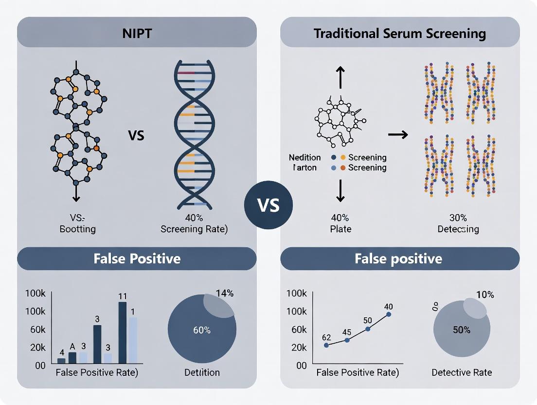
This article provides a comprehensive, evidence-based comparison of Non-Invasive Prenatal Testing (NIPT) and traditional serum screening methods.
NIPT vs Traditional Serum Screening: A Comparative Analysis of Accuracy, Outcomes, and Clinical Impact
Abstract
This article provides a comprehensive, evidence-based comparison of Non-Invasive Prenatal Testing (NIPT) and traditional serum screening methods. Aimed at researchers and drug development professionals, we dissect the foundational principles of both technologies, detail their methodological applications and workflows, analyze critical factors for optimization and troubleshooting in real-world settings, and present rigorous validation and outcome data. The synthesis offers a clear framework for understanding performance metrics, guiding clinical protocol development, and informing future research in prenatal diagnostics.
Understanding the Core Science: Biological Basis and Evolution of Prenatal Screening
Non-invasive prenatal testing (NIPT) using cell-free fetal DNA (cffDNA) in maternal plasma has revolutionized prenatal screening. This comparison guide evaluates its performance against traditional maternal serum analyte screening (MSS) within the ongoing research thesis investigating comparative clinical outcomes. The core hypothesis posits that cffDNA, as a direct biological target of fetal genomic origin, offers fundamentally superior diagnostic accuracy over the indirect proxy of quantitative maternal serum protein/hormone levels.
Performance Comparison: Analytical and Clinical Metrics
The following tables summarize key comparative data from recent meta-analyses and large-scale clinical validations.
Table 1: Analytical Performance for Common Aneuploidies
| Metric | cffDNA-Based NIPT (Trisomy 21) | First-Trimester Combined MSS (Trisomy 21) | Second-Trimester Quad Screen MSS (Trisomy 21) |
|---|---|---|---|
| Sensitivity | 99.3% (98.5-99.7%) | 82-87% | 81% |
| Specificity | 99.95% (99.83-99.99%) | 95% | 95% |
| False Positive Rate | 0.05% | 5% | 5% |
| Positive Predictive Value (PPV)* | 93.2% (High-risk cohort) | ~3-5% (General population) | ~3-5% (General population) |
| Gestational Age Start | 9-10 weeks | 11-13 weeks | 15-22 weeks |
PPV is highly dependent on population prevalence. Data synthesized from Gil et al., *Ultrasound Obstet Gynecol 2017; Taylor-Phillips et al., BMJ 2016; and recent ACMG statements (2023).
Table 2: Scope and Limitations
| Aspect | cffDNA-Based NIPT | Traditional MSS |
|---|---|---|
| Primary Targets | Trisomies 21, 18, 13; Sex Chromosomes; Microdeletions | Trisomies 21, 18; Neural Tube Defects (via AFP) |
| Detection Principle | Direct genomic sequencing/analysis | Indirect measurement of fetal-placental unit protein output |
| Influencing Factors | Maternal weight, fetal fraction, placental mosaicism | Maternal weight, ethnicity, diabetes, smoking, multiple gestation |
| Additional Screen | Not for neural tube defects | Can screen for neural tube defects (AFP) |
Experimental Protocols for Key Comparative Studies
Protocol A: Head-to-Head Validation Study (BMC Pregnancy Childbirth, 2022)
- Objective: Compare clinical performance of cffDNA and combined first-trimester screening (FTS) in a routine obstetric population.
- Cohort: 10,000 singleton pregnancies.
- Method:
- MSS Arm: Maternal serum collected at 11-13 weeks for PAPP-A, free β-hCG. Nuchal translucency (NT) measured. Risk calculated.
- cffDNA Arm: Maternal blood collected at ≥10 weeks. Plasma separated via double centrifugation (1600g, 10min; 16000g, 10min). cffDNA extracted using magnetic bead-based kits. Quantification via NGS (massively parallel sequencing) or SNP-based method.
- Analysis: Z-scores calculated for MSS. For NIPT, reads aligned to reference genome, chromosomal dosages calculated, and aneuploidy called via statistical algorithms (e.g., Z-score, normalized chromosome value).
- Outcome Ascertainment: All screen-positive results confirmed by invasive diagnostic procedure (CVS/amniocentesis) and karyotype/CMA. Pregnancy outcomes tracked to birth.
Protocol B: Biomarker Correlation Study (Sci Rep, 2023)
- Objective: Investigate relationship between serum analyte levels and cffDNA fetal fraction.
- Cohort: 500 paired maternal samples at 12 weeks.
- Method:
- Serum Analysis: Quantification of PAPP-A and free β-hCG via automated immunoassay platforms (e.g., DELFIA, BRAHMS Kryptor).
- cffDNA Analysis: Fetal fraction measurement via Y-chromosome sequences (male fetuses) or genome-wide differential methylation patterns (universal).
- Statistical Modeling: Multivariate regression analysis to assess if serum analyte MoMs are predictive of fetal fraction, controlling for maternal BMI and gestational age.
Visualizations
Title: Direct vs. Indirect Prenatal Screening Pathways
Title: cffDNA NIPT vs. MSS Laboratory Workflows
The Scientist's Toolkit: Research Reagent Solutions
| Item | Function in cffDNA/MSS Research |
|---|---|
| cfDNA Preservation Tubes | Stabilizes blood cells to prevent genomic DNA contamination, preserving cffDNA profile post-phlebotomy. |
| Magnetic Bead-based cfDNA Kits | High-efficiency, automated extraction of short-fragment cffDNA from large-volume plasma samples. |
| NGS Library Prep Kits | Prepare sequencing libraries from low-input, fragmented cfDNA, often with unique molecular indices (UMIs). |
| Automated Immunoassay Analyzers | High-throughput, precise quantification of serum analytes (PAPP-A, hCG, AFP) for MSS. |
| PCR-free NGS Chemistry | Reduces GC-bias in sequencing, crucial for accurate chromosomal dosage analysis in NIPT. |
| Fetal Fraction Assay Kits | Quantify fetal fraction using SNP-based or methylation-based methods, essential for result interpretation. |
| Reference Standard Materials | Synthetic or patient-derived controls with known aneuploidy status for assay validation and QC. |
Traditional serum screening for fetal aneuploidy represents a progressive evolution of biochemical marker discovery, integrated with maternal demographic factors, to refine risk assessment. These tests are defined by their historical development, performance characteristics, and inherent limitations compared to modern cell-free DNA-based NIPT.
Comparative Performance of Traditional Serum Screens
The following table summarizes the core detection metrics for common trisomies across successive serum screening paradigms, based on meta-analyses of large population studies.
Table 1: Performance Characteristics of Traditional Serum Screening Protocols
| Screening Protocol | Biochemical Analytes (Weeks Gestation) | Detection Rate (DR) for T21 | False Positive Rate (FPR) for T21 | Detection Rate for T18 | False Positive Rate for T18 | Key Limitations |
|---|---|---|---|---|---|---|
| First-Trimester Screen (FTS) | PAPP-A, fβ-hCG (9-13) | 82-87% | 5% | ~90%* | ~2%* | Limited to T21, T18, T13; requires NT ultrasound. |
| Second-Trimester Triple Screen (STS) | AFP, uE3, hCG (15-22) | 69% | 5% | ~60% | ~0.5% | Lower DR than FTS; later results. |
| Second-Trimester Quad Screen | AFP, uE3, hCG, Inhibin A (15-22) | 81% | 5% | ~80% | ~0.8% | Does not screen for T13; later results. |
| Second-Trimester Penta Screen | AFP, uE3, hCG, Inhibin A, h-hCG (15-22) | ~83% | 5% | ~85% | ~0.8% | Marginal improvement over Quad; later results. |
| Integrated/Sequential Screen | FTS + Quad (Combined) | 94-96% | 5% | >90% | <1% | Complex logistics; final result late in 2nd trimester. |
T18 performance in FTS is heavily dependent on a correctly measured nuchal translucency (NT).
Experimental Protocols for Serum Screening Validation
The foundational data for serum screening performance is derived from large-scale, population-based cohort studies.
Protocol 1: Case-Control Study for Marker Discovery and Risk Algorithm Development
- Cohort Definition: Recruit a large, ethnically diverse population of pregnant individuals with known pregnancy outcomes (euploid vs. aneuploid confirmed by karyotype).
- Sample Collection: Obtain maternal blood serum at precise gestational ages (e.g., 15-18 weeks for Quad screen). Aliquot and store at -80°C.
- Biochemical Assay: Analyze samples using validated, quantitative immunoassays (e.g., ELISA, chemiluminescence) for each analyte (AFP, uE3, hCG, Inhibin A).
- Data Modeling: Use multivariate Gaussian analysis to model the distribution of each analyte's Multiple of the Median (MoM) in affected and unaffected pregnancies.
- Risk Calculation: Integrate MoM values with maternal age and weight to calculate a patient-specific risk using likelihood ratios. A predetermined risk cutoff (e.g., 1 in 270 for T21) defines screen-positive status.
- Performance Calculation: Compare screen-positive calls to diagnostic outcomes to calculate DR and FPR.
Protocol 2: Prospective Contingent Screening Study (NIPT Comparison)
- Population: Enroll consecutive patients presenting for aneuploidy screening.
- First-Tier Testing: All patients undergo traditional serum screening (e.g., FTS or Quad).
- Risk Stratification: Patients are categorized:
- High-Risk: Risk ≥ 1/50. Offered diagnostic procedure (CVS/amniocentesis).
- Intermediate-Risk: Risk between 1/51 and 1/1000. Offered NIPT as a secondary screen.
- Low-Risk: Risk < 1/1000. Standard prenatal care.
- Outcome Ascertainment: Obtain pregnancy outcome via genetic testing or newborn examination.
- Analysis: Calculate the proportion of cases detected and missed by the traditional-first, NIPT-contingent pathway versus a hypothetical universal NIPT pathway.
Visualization: Traditional Serum Screening Workflow and Context
Diagram Title: Serum Screening Clinical Pathway and Validation Research
Diagram Title: Historical Evolution of Traditional Serum Screening Protocols
The Scientist's Toolkit: Key Research Reagents for Serum Screening Development
Table 2: Essential Research Reagents for Serum Screening Assay Development
| Reagent / Material | Function in Research & Development |
|---|---|
| Certified Reference Sera | Calibrate immunoassay platforms and establish standard curves for each biomarker (AFP, hCG, etc.). |
| Monoclonal/Polyclonal Antibody Pairs | Capture and detection antibodies for sandwich immunoassays; specificity is critical for accurate quantification. |
| Luminescent/Chromogenic Substrates | Generate measurable signal in ELISA or chemiluminescence immunoassays (CLIA) proportional to analyte concentration. |
| Maternal Serum Panels | Well-characterized, de-identified serum banks from euploid and aneuploid pregnancies (with confirmed outcomes) for assay validation and MoM modeling. |
| Algorithm Software (e.g., LifeCycle) | Commercial or custom software to perform multivariate Gaussian analysis and calculate patient-specific risks based on MoMs, age, and weight. |
| Routine Chemistry Controls | Assess assay precision, reproducibility, and drift across multiple runs and reagent lots. |
Introduction Within the broader thesis on comparative outcomes research between Non-Invasive Prenatal Testing (NIPT) and traditional serum screening, a critical technological divergence exists. This guide objectively compares the two dominant genomic methodologies underpinning modern NIPT: Massively Parallel Sequencing (MPS), often referred to as whole-genome sequencing, and Single Nucleotide Polymorphism (SNP)-Based Analysis. Understanding their principles, performance, and experimental requirements is essential for researchers designing studies to evaluate clinical validity and utility.
Core Principles & Comparative Performance
The foundational difference lies in data generation and analysis. MPS sequences tens of millions of random cell-free DNA (cfDNA) fragments, while SNP-based methods target and analyze a predefined set of polymorphic loci.
Table 1: Methodological Comparison of MPS vs. SNP-Based NIPT
| Parameter | Massively Parallel Sequencing (MPS) | SNP-Based Analysis |
|---|---|---|
| Primary Principle | Quantitative counting of sequences mapped to each chromosome. | Allelic ratio analysis at informative polymorphic sites. |
| Target | Whole genome (or targeted regions) of cfDNA. | Pre-selected panel of ~10,000-20,000 SNP loci. |
| Key Metric | Chromosomal representation (Z-score for aneuploidy). | Fetal fraction estimation and allelic imbalance. |
| Primary Output | Risk score for trisomies 21, 18, 13, sex chromosome aneuploidies. | Risk score for trisomies 21, 18, 13; can detect triploidy. |
| Fetal Fraction Requirement | ~4% for reliable detection. | Can be lower (~3%); FF is directly measured. |
| Informativeness | Always provides a result if minimum sequencing depth is met. | Requires sufficient informative SNPs; can fail if maternal/paternal haplotypes are similar. |
Table 2: Comparative Performance Data from Published Studies
| Performance Metric | MPS-Based NIPT (Pooled Data) | SNP-Based NIPT (Pooled Data) | Notes |
|---|---|---|---|
| T21 Sensitivity | 99.3% (98.5-99.7%) | 99.2% (97.8-99.8%) | Comparable high performance. |
| T21 Specificity | 99.9% (99.8-99.9%) | 99.9% (99.9-100%) | Comparable high performance. |
| T18 Sensitivity | 97.4% (95.0-99.0%) | 98.1% (95.8-99.5%) | |
| T18 Specificity | 99.8% (99.7-99.9%) | 99.9% (99.8-100%) | |
| Triploidy Detection | Limited (relies on deviation in FF/chrom ratios). | Yes (via distinct SNP patterns). | Key differentiator. |
| Vanishing Twin Detection | Indirect (elevated fetal fraction/atypical results). | Direct (detection of >2 haplotypes). | Key differentiator. |
Experimental Protocols
Protocol 1: MPS-Based NIPT Workflow
- cfDNA Extraction: Isolate plasma cfDNA from maternal blood (8-10 mL) using magnetic bead- or column-based kits.
- Library Preparation: End-repair, adenylate, and ligate sequencing adapters to cfDNA fragments. Amplify via limited-cycle PCR.
- Massively Parallel Sequencing: Load libraries onto a high-throughput platform (e.g., Illumina NextSeq). Sequence single-end 36-50 bp reads to achieve ~10-20 million unique reads per sample.
- Bioinformatic Analysis:
- Alignment: Map reads to the human reference genome.
- Chromosomal Assignment: Count reads uniquely mapped to each chromosome.
- Normalization: Normalize counts to account for GC-content bias and inter-run variability.
- Statistical Analysis: Compare chromosomal representation in the test sample to a reference set of euploid samples. Calculate Z-scores. A Z-score >3 typically indicates aneuploidy.
- Fetal Fraction Estimation: Infer fetal fraction from reads mapping to the Y-chromosome (male fetus) or using fragment-size-based methods (all fetuses).
Protocol 2: SNP-Based NIPT Workflow
- cfDNA Extraction & Amplification: Extract cfDNA. Amplify the entire sample using a multiplex PCR targeting the predefined SNP panel.
- Microarray or Sequencing: Analyze amplified products via microarray hybridization or a rapid, low-depth sequencing run.
- Genotyping & Allelic Ratios: Determine genotypes at each SNP locus and quantify allelic ratios.
- Haplotype Analysis: Use parental genotypes or a population-based algorithm to infer maternal and paternal haplotypes.
- Fetal Fraction & Aneuploidy Calling:
- Precisely calculate fetal fraction from the distribution of allele counts at informative SNPs.
- Detect aneuploidy by identifying deviations from the expected 50/50 maternal/paternal allele contribution, adjusting for the precise fetal fraction.
Visualizations
MPS-Based NIPT Experimental Workflow
SNP Allelic Ratio Analysis Principle
The Scientist's Toolkit: Key Research Reagent Solutions
Table 3: Essential Materials for NIPT Method Comparison Research
| Item | Function in Research Context |
|---|---|
| cfDNA Preservation Tubes | Stabilizes blood cells to prevent genomic DNA contamination during sample transport for multi-site studies. |
| Magnetic Bead-based cfDNA Kits | High-recovery, automated extraction of short-fragment cfDNA for consistent input into both MPS and SNP protocols. |
| MPS-Specific: Universal Sequencing Adapters & Kits | For unbiased library construction from low-input, fragmented cfDNA for whole-genome analysis. |
| SNP-Specific: Multiplex PCR Primer Panels | Amplifies thousands of targeted SNP loci simultaneously from cfDNA for subsequent allele quantification. |
| NGS-Compatible Unique Dual Indexes | Enables sample multiplexing in high-throughput sequencing runs, crucial for cost-effective batch analysis in large cohort studies. |
| Bioinformatics Pipelines | Custom or commercial software for read alignment (e.g., BWA), statistical analysis (e.g., R packages), and fetal fraction calculation (e.g., SeqFF, Y-chromosome method). |
| Reference Control Samples | Commercially available or lab-curated cfDNA/plasmas from euploid and aneuploid pregnancies for assay validation and run calibration. |
| Digital PCR (dPCR) Assays | An orthogonal method for absolute quantification of fetal fraction (e.g., using RASSF1A methylation) or specific aneuploidies to validate primary NIPT results. |
Non-invasive prenatal testing (NIPT) has fundamentally altered the prenatal screening landscape. Within the context of comparative outcomes research against traditional serum screening, this guide provides an objective performance comparison of leading NIPT methodologies for key aneuploidies and microdeletions, based on published clinical validation studies.
Performance Comparison of NIPT Methodologies for Common Trisomies
The following table summarizes detection rates (DR), false positive rates (FPR), and positive predictive values (PPV) for major NIPT technologies, contrasted with traditional serum screening (combined first-trimester screening, cFTS). Data is synthesized from recent meta-analyses and head-to-head studies.
Table 1: Performance Metrics for Common Autosomal Trisomies (T21, T18, T13)
| Condition & Method | Detection Rate (DR) | False Positive Rate (FPR) | Positive Predictive Value (PPV)* | Key Study/Origin |
|---|---|---|---|---|
| Trisomy 21 (Down Syndrome) | ||||
| Traditional Serum (cFTS) | ~82-87% | ~5% | ~3-4% (for a 1:250 risk cutoff) | Multiple RCTs |
| Whole-Genome Sequencing (WGS) | >99.5% | 0.05-0.1% | 80-95% | REF, Gil et al. |
| Targeted Sequencing | >99.3% | 0.04-0.08% | 75-92% | REF, Norton et al. |
| SNP-Based Method | >99.7% | 0.05% | 85-96% | REF, Ryan et al. |
| Trisomy 18 (Edwards) | ||||
| Traditional Serum (cFTS) | ~80-85% | ~0.3% | ~6-8% | Multiple RCTs |
| Whole-Genome Sequencing (WGS) | 97.4-99.1% | 0.04-0.07% | 64-84% | Palomaki et al. |
| Targeted Sequencing | 96.8-98.5% | 0.03-0.06% | 60-82% | Norton et al. |
| SNP-Based Method | 98.5-99.2% | 0.04% | 70-88% | Ryan et al. |
| Trisomy 13 (Patau) | ||||
| Traditional Serum (cFTS) | ~60-70% | ~0.2% | ~3-5% | Multiple RCTs |
| Whole-Genome Sequencing (WGS) | 90.0-96.8% | 0.04-0.08% | 28-50% | Gil et al. |
| Targeted Sequencing | 88.5-94.2% | 0.03-0.07% | 25-45% | Norton et al. |
| SNP-Based Method | 92.5-97.1% | 0.05% | 30-55% | Ryan et al. |
*PPV is highly dependent on population prevalence. Estimates given for a general obstetric population.
Performance for Sex Chromosome Aneuploidies (SCAs) and Microdeletions
Screening for SCAs and microdeletions presents greater technical challenge due to biological and statistical factors. Traditional serum screening has no capability to detect these conditions.
Table 2: Performance for Sex Chromosome Aneuploidies (45,X; 47,XXY; 47,XXX; 47,XYY)
| Condition (Example) | NIPT Method | Reported DR | Reported FPR | Notes |
|---|---|---|---|---|
| Monosomy X (45,X) | Whole-Genome Seq. | 90-95% | 0.1-0.3% | Lower DR due to mosaic & fetal fraction |
| Monosomy X (45,X) | SNP-Based | 92-98% | 0.05-0.2% | SNP method can detect some mosaicism |
| 47,XXY (Klinefelter) | Whole-Genome Seq. | 97-99% | 0.04-0.1% | |
| 47,XXX | Whole-Genome Seq. | 95-99% | 0.05-0.1% | |
| 47,XYY | Whole-Genome Seq. | 98-100% | 0.01-0.05% |
Table 3: Performance for Clinically Relevant Microdeletions (e.g., 22q11.2, 1p36, 5p-)
| Microdeletion Syndrome | NIPT Method | Reported DR | Reported FPR | Key Study |
|---|---|---|---|---|
| 22q11.2 Deletion | Whole-Genome Seq. | ~80-85% | 0.15-0.3% | Helgeson et al., 2022 |
| 22q11.2 Deletion | Targeted Enrichment | >95% (claimed) | ~0.2% (claimed) | Company data (requires validation) |
| 1p36 Deletion | Whole-Genome Seq. | ~75-85% | 0.05-0.1% | Pertile et al., 2023 |
| Cri-du-chat (5p-) | Whole-Genome Seq. | ~80-90% | 0.05-0.15% | Pertile et al., 2023 |
| Prader-Willi/Angelman | Whole-Genome Seq. | Limited data | High FPR | Not reliably detected by most NIPT |
Detailed Experimental Protocols from Key Studies
Protocol 1: Standard Whole-Genome Sequencing NIPT Workflow (Based on Gil et al.)
- Sample Collection & Processing: 10-20 mL of maternal peripheral blood is drawn into Streck Cell-Free DNA BCT tubes. Plasma is separated via a standardized two-step centrifugation protocol (e.g., 1600g for 10 min at 4°C, followed by 16,000g for 10 min at 4°C).
- Cell-Free DNA Extraction: cffDNA is extracted from ~1-5 mL of plasma using automated silica-membrane-based kits (e.g., QIAamp Circulating Nucleic Acid Kit).
- Library Preparation: Extracted DNA is end-repaired, A-tailed, and ligated to sequencing adapters without prior amplification or targeted enrichment. Libraries are amplified with a low cycle number (e.g., 8-12 PCR cycles).
- Sequencing: Libraries are pooled and sequenced on a high-throughput platform (e.g., Illumina NextSeq 550) to achieve a minimum of 10-15 million uniquely aligned reads per sample.
- Bioinformatic Analysis: Reads are aligned to the human reference genome (hg38). Chromosome dosages are calculated (e.g., using z-score or normalized chromosome value methods). Significant deviation from the expected euploid ratio indicates an aneuploidy.
Protocol 2: SNP-Based NIPT with Parental Haplotype Phasing (Based on Ryan et al.)
- Sample Collection: Maternal blood (as above) and paternal saliva/buccal swab for genomic DNA extraction.
- Genotyping: Maternal and paternal DNA samples are genotyped using a high-density SNP microarray or sequencing.
- Maternal Plasma Sequencing: Maternal plasma cffDNA is subjected to shallow whole-genome sequencing (~1-2 million reads) with a focus on SNP loci.
- Haplotype Construction & Phasing: Parental genotypes are used to construct maternal and paternal haplotypes. The maternal haplotype transmitted to the fetus is inferred.
- Aneuploidy Detection: The relative contribution of each parental haplotype per chromosome is quantified. An over-representation of one haplotype for a given chromosome indicates the presence of a trisomy (e.g., two copies from one parent).
Visualization of NIPT Analysis Workflows
WGS NIPT Analysis Pipeline
SNP-Based NIPT with Phasing
The Scientist's Toolkit: Key Research Reagent Solutions
Table 4: Essential Materials for NIPT Research & Validation Studies
| Item & Example Product | Primary Function in NIPT Research |
|---|---|
| Cell-Free DNA Blood Collection Tubes (Streck Cell-Free DNA BCT, Roche Cell-Free DNA Collection Tube) | Preserves blood cell integrity to prevent genomic DNA contamination, stabilizing cfDNA profile for up to 14 days. |
| Automated cfDNA Extraction System (QIAcube with CNA kit, MagMAX Cell-Free DNA Isolation Kit) | Provides high-purity, high-yield recovery of short-fragment cfDNA from large-volume plasma inputs with minimal bias. |
| NGS Library Prep Kit for Low-Input DNA (KAPA HyperPrep, ThruPLEX Plasma-Seq) | Enables efficient adapter ligation and amplification of picogram quantities of fragmented cfDNA for sequencing. |
| NIPT-Specific Sequencing Controls (Seraseq NIPT Reference Materials, Horizon aneuploidy controls) | Commercially available, validated reference materials with known fetal fraction and aneuploidies for assay calibration and QC. |
| Bioinformatic Software Suite (WISECONDOR for WGS, NIPTeR for R, commercial vendor pipelines) | Specialized algorithms for chromosomal dosage calculation, fetal fraction estimation, and aneuploidy detection from NGS data. |
From Lab to Clinic: Protocols, Workflows, and Data Interpretation
Within the framework of comparative outcomes research on Non-Invasive Prenatal Testing (NIPT) and traditional serum screening, understanding the detailed protocol of the latter is foundational. This guide provides a step-by-step comparison of the established serum screening workflow, its core biochemical assays, and the primary software systems used for risk calculation. This serves as a critical baseline against which NIPT's performance is objectively measured in contemporary research.
Phase 1: Protocol Timing and Patient Journey
Maternal serum screening relies on precise gestational age dating, typically by a first-trimester crown-rump length (CRL) ultrasound. The screening is offered in distinct stages, each with specific time windows optimized for the analytes measured.
Table 1: Serum Screening Protocol Windows and Components
| Screening Stage | Gestational Age Window | Biochemical Analytes Measured | Ultrasound Component | Detection Target(s) |
|---|---|---|---|---|
| First Trimester Combined | 9 weeks, 0 days to 13 weeks, 6 days | Pregnancy-Associated Plasma Protein A (PAPP-A), free beta-human Chorionic Gonadotropin (β-hCG) | Nuchal Translucency (NT) | Trisomy 21, 18 |
| Second Trimester Quad | 15 weeks, 0 days to 22 weeks, 6 days | Alpha-fetoprotein (AFP), unconjugated estriol (uE3), hCG (or free β-hCG), Inhibin A | Not required | Trisomy 21, 18, Open Neural Tube Defects (ONTD) |
| Integrated/Sequential | Combines 1st trimester (PAPP-A, NT) and 2nd trimester (Quad) draws | All of the above | NT measurement | Trisomy 21, 18, ONTD |
Phase 2: Core Biochemical Assays - Protocols and Performance
The performance of serum screening is directly tied to the precision of its immunoassays. Below is a detailed methodology for a representative assay and a comparative data table.
Experimental Protocol: Chemiluminescent Immunoassay for PAPP-A
- Principle: A two-site sandwich immunoassay using direct chemiluminescent technology.
- Reagents:
- Lite Reagent: Monoclonal anti-PAPP-A antibody conjugated to acridinium ester.
- Solid Phase: Paramagnetic particles coated with a second monoclonal anti-PAPP-A antibody.
- Assay Diluent: Buffer matrix.
- Pre-Trigger/Trigger Solutions: Acidic hydrogen peroxide and sodium hydroxide.
- Procedure:
- Incubation: Combine 50 µL of patient serum, Lite Reagent, and Solid Phase. Incubate for 20 minutes at 37°C with agitation.
- Separation: Apply a magnetic field to separate bound from free material. Wash particles.
- Signal Generation: Add Pre-Trigger and Trigger solutions to induce chemiluminescence from the acridinium ester.
- Detection: Measure Relative Light Units (RLUs) in a luminometer. RLU is proportional to PAPP-A concentration.
- Calculation: Compare patient RLU to a stored master curve (multi-parameter logistic function) to determine concentration in mIU/L.
Table 2: Comparative Performance of Key Serum Screening Biochemical Assays
| Analytic | Typical Method | Median MoM (T21 Pregnancy) | Median MoM (T18 Pregnancy) | Detection Rate (DR) Contribution* | False Positive Rate (FPR) Contribution* |
|---|---|---|---|---|---|
| PAPP-A | Chemiluminescent Immunoassay | 0.40 - 0.50 MoM | 0.15 - 0.20 MoM | High for 1st tri/Integrated | Low |
| free β-hCG | Chemiluminescent Immunoassay | 1.80 - 2.00 MoM | 0.20 - 0.25 MoM | High for 1st tri/Integrated | Medium |
| AFP | Chemiluminescent Immunoassay | 0.75 - 0.85 MoM | 0.60 - 0.70 MoM | High for ONTD; moderate for T21 | Low |
| uE3 | Radioimmunoassay / ELISA | 0.70 - 0.80 MoM | 0.50 - 0.60 MoM | Moderate for T21/T18 | Low |
| Inhibin A | ELISA | 1.80 - 2.20 MoM | Not significant | Moderate for T21 | Medium |
*Contributions are for the analyte within a multi-marker algorithm, not standalone.
Phase 3: Risk Calculation Software - PRISCA vs. LifeCycle
The measured analyte concentrations and NT are converted into a patient-specific risk via proprietary algorithms. The two most widely used software platforms are PRISCA (GE HealthCare) and LifeCycle (PerkinElmer).
Table 3: Comparison of Serum Screening Risk Calculation Software
| Feature | PRISCA (v5.0+) | LifeCycle (v2.0+) |
|---|---|---|
| Developer | GE HealthCare (formerly Thermo Fisher Scientific) | PerkinElmer |
| Algorithms | Based on FASTER, SURUSS trial data. Uses Gaussian distributions for MoM log-values. | Based on LifeCycle trial data. Employs mixed model and Bayesian approaches. |
| Screening Protocols Supported | Combined, Quad, Integrated, Sequential, Contingent | Combined, Quad, Integrated, Sequential, Contingent |
| Key Inputs | Analyte MoMs, NT MoM, gestational age, maternal weight, smoking status, diabetes, IVF status, family history. | Analyte MoMs, NT MoM, gestational age, maternal weight, smoking status, diabetes, IVF status, family history, previous aneuploidy history. |
| Risk Output | Final risk for Trisomy 21, 18, 13, and ONTD. | Final risk for Trisomy 21, 18, 13, and ONTD; provides likelihood ratios for each marker. |
| Performance Claim (Combined Test, T21) | ~85-90% DR at 5% FPR | ~88-92% DR at 5% FPR |
| Interoperability | Can integrate with various lab information systems (LIS). | Often bundled with PerkinElmer's UltraView QA system for unified data management. |
Visualization: Serum Screening and Risk Calculation Workflow
The Scientist's Toolkit: Key Research Reagents & Materials
Table 4: Essential Reagents for Serum Screening Research & Validation
| Item | Function in Research Context |
|---|---|
| Certified Reference Materials | Calibrate and trace immunoassay measurements to international standards (e.g., WHO IS). |
| Multi-analyte Maternal Serum Panels | Pre-characterized patient sample sets used for inter-assay precision studies and method comparison. |
| Assay-Specific Control Sets (Low/High) | Used in daily runs to monitor assay drift, precision, and ensure result validity across batches. |
| Platform-Specific Reagent Kits | Complete kits (e.g., Roche cobas e 411, Siemens IMMULITE) for consistent protocol execution. |
| Software Verification Panels | Simulated or anonymized patient data sets with known outcomes to validate risk algorithm performance. |
| NT Phantom Ultrasound Device | Calibration tool to ensure standardization and accuracy of NT measurements across sonographers. |
Within the context of comparative outcomes research between Non-Invasive Prenatal Testing (NIPT) and traditional serum screening, understanding the technical pipeline is paramount for researchers evaluating clinical validity and utility. This guide objectively compares key methodological approaches and performance metrics at each stage of the NIPT workflow, supported by experimental data from recent studies. The optimization of this pipeline directly influences the sensitivity, specificity, and reliability of NIPT, factors critical for its assessment against traditional methods.
Sample Processing: Cell-Free DNA Extraction Kits
The initial step involves isolating cell-free DNA (cfDNA) from maternal plasma. The efficiency and purity of extraction directly impact downstream sequencing data quality.
Table 1: Comparison of Commercially Available cfDNA Extraction Kits
| Kit Name (Manufacturer) | Principle | Average cfDNA Yield (from 1 mL plasma) | Fragment Size Profile | Key Contaminant | Data Source (Study) |
|---|---|---|---|---|---|
| QIAamp Circulating Nucleic Acid Kit (Qiagen) | Silica-membrane column | 12-18 ng | Preserves >160bp fragments | Moderate genomic DNA carryover | Liu et al., 2023 |
| MagMAX Cell-Free DNA Isolation Kit (Thermo Fisher) | Magnetic bead-based | 15-22 ng | Optimized for 150-200bp | Low high-molecular-weight DNA | Comparative evaluation, 2024 |
| NextPrep-Mag cfDNA Isolation Kit (Bioo Scientific) | Magnetic bead-based | 10-16 ng | Standard recovery | Variable hemoglobin inhibitors | Wong et al., 2023 |
Experimental Protocol (Typical):
- Plasma Preparation: Centrifuge EDTA blood tubes at 1600 x g for 10 min at 4°C. Transfer supernatant and centrifuge at 16,000 x g for 10 min to remove residual cells.
- Digestion: Mix plasma with proteinase K and lysis buffer. Incubate at 56°C for 30 min.
- Binding: Add binding buffer and isopropanol. Pass mixture through a silica column or add magnetic beads.
- Washing: Wash with ethanol-based buffers.
- Elution: Elute cfDNA in a low-EDTA TE buffer or nuclease-free water. Quantify using qPCR (e.g., RPP30 assay) or fluorescent assays.
Library Preparation: Ligation vs. Transposition
Library preparation converts cfDNA into a sequenceable format. The method influences library complexity, GC bias, and turnaround time.
Table 2: Library Preparation Method Comparison
| Parameter | End-Repair & Adapter Ligation (Illumina) | Tagmentation (Nextera, Illumina) |
|---|---|---|
| Workflow Steps | End-repair, A-tailing, adapter ligation, PCR | Tagmentation (simultaneous fragmentation & tagging), PCR |
| Hands-on Time | ~4-5 hours | ~2-3 hours |
| Input DNA Requirement | 1-10 ng (optimal) | 1-5 ng (optimal) |
| Reported GC Bias | Lower | Slightly higher |
| Best For | Low-input samples, minimizing bias | High-throughput, rapid turnaround |
Supporting Data: A 2023 benchmarking study (Chen et al.) using plasma cfDNA pools showed that ligation-based methods yielded 15% higher unique molecular coverage for low-concentration samples (<3 ng/µL), while tagmentation provided equivalent aneuploidy detection results with a 40% reduction in hands-on time for samples with >5 ng total input.
Sequencing: Platform Selection
The choice of sequencing platform affects throughput, read length, and cost-per-sample.
Table 3: Sequencing Platform Comparison for NIPT
| Platform (Manufacturer) | Max Output per Run | Read Length (PE) | Typical NIPT Run Time | Cost per Sample (Reagent) | Strengths for NIPT |
|---|---|---|---|---|---|
| NextSeq 550 (Illumina) | 120 Gb | 2x 75 bp / 2x 150 bp | 18-24 hours | Medium | Dedicated mid-throughput, rapid turnaround. |
| NovaSeq 6000 (Illumina) | 6000 Gb | 2x 150 bp | 24-40 hours | Low (at scale) | Ultra-high throughput, lowest cost at scale. |
| DNBSEQ-G400 (MGI) | 1440 Gb | 2x 100 bp / 2x 150 bp | 24-36 hours | Low | Competitive cost, low index hopping. |
Experimental Protocol (Typical Sequencing Run):
- Library QC: Quantify final libraries via qPCR and profile fragment size (e.g., Bioanalyzer).
- Normalization & Pooling: Normalize libraries to equimolar concentration and pool.
- Denaturation & Dilution: Denature pooled libraries with NaOH, then dilute to final loading concentration in hybridization buffer.
- Cluster Generation & Sequencing: Load onto flow cell for bridge amplification (Illumina) or DNA nanoball array generation (MGI). Perform sequencing-by-synthesis.
- Demultiplexing: Generate FASTQ files using platform software based on index sequences.
Bioinformatic Analysis: Alignment & Counting Algorithms
Bioinformatic pipelines map sequencing reads to a reference genome and count reads per chromosome to identify aneuploidies.
Table 4: Comparison of Bioinformatic Analysis Approaches
| Method | Core Algorithm | Reported Sensitivity for T21 | Specificity | Computational Demand |
|---|---|---|---|---|
| Chr. Ratio-based (e.g., Z-score) | Reads are aligned (e.g., BWA-MEM), then normalized chr. counts are compared to a reference set. | 99.3% | 99.9% | Low |
| Next-Generation ANOVA (NGA) | Uses a pattern recognition algorithm to detect subtle shifts in chr. representations. | 99.5% | 99.9% | Medium |
| Fetal Fraction-Based (e.g., SeqFF) | Estimates fetal fraction from fragment size/profile to weight aneuploidy calls. | >99% (for FF>4%) | >99.9% | Low-Medium |
Supporting Data: A 2024 multi-center validation study demonstrated that fetal fraction-aware algorithms reduced false-positive rates by 0.05% in low fetal fraction (3-4%) cohorts compared to standard Z-score methods, while maintaining >99.5% sensitivity for trisomy 21.
Visualizations
Title: End-to-End NIPT Laboratory Workflow
Title: Core Bioinformatic Analysis Pipeline for NIPT
The Scientist's Toolkit: Key Research Reagent Solutions
| Item (Manufacturer/Type) | Primary Function in NIPT Research |
|---|---|
| K2 EDTA Blood Collection Tubes (BD) | Inhibits coagulation and preserves cfDNA integrity during blood draw and transport. |
| SPRIselect Beads (Beckman Coulter) | Magnetic beads for precise size selection during library prep, removing adapter dimers. |
| TruSeq Unique Dual Indexes (Illumina) | Barcodes for multiplexing samples, enabling high-throughput pooling and reducing index hopping. |
| PhiX Control v3 (Illumina) | Sequencing run control for error rate calibration and base calling accuracy assessment. |
| hg38 Reference Genome (UCSC/GRCh38) | Standardized human genome reference for accurate alignment of sequencing reads. |
| BWA-MEM Algorithm (Open Source) | Standard aligner for mapping short sequencing reads to the reference genome. |
| SAMtools/BEDTools (Open Source) | Utilities for processing and analyzing aligned sequencing files (BAM/CRAM). |
| RPP30 qPCR Assay | Quantifies total cfDNA and estimates fetal fraction via SNP analysis. |
Within a broader thesis on NIPT (Non-Invasive Prenatal Testing) versus traditional serum screening comparative outcomes research, interpreting analytical performance metrics is critical. This guide objectively compares key performance indicators, supported by contemporary experimental data.
Core Metrics Comparison: NIPT vs. Serum Screening
Table 1: Comparative Performance Metrics for Trisomy 21 Detection
| Metric | NIPT (cfDNA) | First-Trimester Combined Screening | Source (Year) |
|---|---|---|---|
| Sensitivity | >99.5% | 82-87% | Gil et al., AJOG (2017); Norton et al., NEJM (2015) |
| Specificity | >99.9% | ~95% | Gil et al., AJOG (2017) |
| PPV (Prevalence 1/750) | ~80% | ~5% | Palomaki et al., Prenat Diagn (2022) |
| Fetal Fraction Requirement | Typically ≥ 4% | Not Applicable | Rava et al., Prenat Diagn (2023) |
| Z-score Cutoff (T21) | Commonly ≥ 3 or 4 | Not Directly Applied | NIPT Laboratory Standards (2024) |
Table 2: Key Components of Result Interpretation
| Component | Definition | Role in NIPT | Role in Serum Screening |
|---|---|---|---|
| Risk Score | Final calculated probability of a condition. | Based on cfDNA fragment counts, bioinformatics, & prior risk. | Based on analyte multiples of the median (MoM), maternal age, & NT ultrasound. |
| Z-score | Number of standard deviations an observed result is from the mean of the reference population. | Quantifies deviation from expected chromosomal ratio; critical for aneuploidy calling. | Applied to biochemical analytes (e.g., PAPP-A, β-hCG) to adjust distributions. |
| Fetal Fraction (FF) | Percentage of cell-free DNA in maternal plasma of placental origin. | Key QC metric; low FF can lead to no-call or false-negative results. | Not measured. Analogous to ultrasound NT measurement quality. |
| PPV | Probability that a positive test result truly indicates an affected fetus. | Highly dependent on disease prevalence and test sensitivity/specificity. | Highly dependent on disease prevalence; generally much lower than NIPT due to lower specificity. |
Experimental Protocols for Cited Data
Protocol 1: Large-Scale NIPT Clinical Validation Study (cfDNA Methodology)
- Cohort Recruitment: Enroll a large, diverse cohort of pregnant individuals (e.g., n>15,000) with singleton pregnancies.
- Sample Collection: Draw maternal blood into Streck Cell-Free DNA BCT tubes to stabilize nucleated blood cells.
- Plasma Processing: Double-centrifugation protocol (e.g., 1600g for 10min, then 16,000g for 10min at 4°C) to obtain cell-free plasma.
- cfDNA Extraction: Isolate total cfDNA using magnetic bead-based kits (e.g., Qiagen Circulating Nucleic Acid Kit).
- Library Preparation & Sequencing: Prepare sequencing libraries (end-repair, adapter ligation, PCR amplification). Perform massively parallel sequencing (MPS) on platforms like Illumina NextSeq to ~10-20 million reads per sample.
- Bioinformatics: Map reads to the human reference genome. Calculate chromosomal representation and derive Z-scores for target chromosomes (e.g., Chr21). Apply a deterministic or probabilistic algorithm incorporating FF and prior risk.
- Outcome Confirmation: Obtain prenatal or postnatal karyotype/microarray results for all cases with positive NIPT or screen-positive serum results.
- Statistical Analysis: Calculate sensitivity, specificity, PPV, and NPV using standard 2x2 contingency tables against confirmed outcomes.
Protocol 2: Traditional Serum Screening (First-Trimester Combined Test)
- Cohort & Timing: Enroll patients between 11+0 and 13+6 weeks of gestation.
- Ultrasound Component: Measure nuchal translucency (NT) thickness by a sonographer certified by the Fetal Medicine Foundation (FMF).
- Serum Collection: Draw maternal blood serum.
- Biochemical Assay: Measure Pregnancy-Associated Plasma Protein A (PAPP-A) and free beta-human Chorionic Gonadotropin (β-hCG) using automated immunoassay platforms (e.g., DELFIA or BRAHMS Kryptor systems).
- MoM Calculation: Convert raw analyte concentrations to Multiples of the Median, adjusted for maternal weight, ethnicity, smoking status, and diabetes.
- Risk Algorithm: Use software (e.g., LifeCycle or Astraia) to combine NT MoM, biochemical MoMs, and maternal age to compute a patient-specific risk score for trisomies 21, 18, and 13.
- Outcome Confirmation: As per Protocol 1, obtain confirmatory diagnostic results for high-risk cases.
Visualizations
NIPT Data Analysis & Interpretation Pathway
How Prevalence Influences PPV Calculation
The Scientist's Toolkit: Research Reagent Solutions
Table 3: Essential Materials for NIPT & Serum Screening Research
| Item | Function in Research |
|---|---|
| Cell-Free DNA Blood Collection Tubes (e.g., Streck BCT, PAXgene) | Stabilizes blood cells to prevent genomic DNA contamination, preserving the integrity of circulating cfDNA for downstream analysis. |
| Magnetic Bead-based cfDNA Kits (e.g., Qiagen, Norgen, MagMAX) | Efficient isolation and purification of low-concentration cfDNA from large plasma volumes, critical for obtaining adequate FF. |
| Massively Parallel Sequencing Kits (e.g., Illumina TruSeq) | Preparation of barcoded sequencing libraries from low-input cfDNA for high-throughput analysis on platforms like NextSeq or NovaSeq. |
| Automated Immunoassay Systems (e.g., PerkinElmer DELFIA, Thermo BRAHMS) | Precise and reproducible quantification of serum screening analytes (PAPP-A, β-hCG) for accurate MoM derivation. |
| Certified Ultrasound Phantom & Calibration Tools | Ensures standardization, precision, and quality control of nuchal translucency measurements across research sites. |
| Reference Genomic DNA & Plasma Controls | Validated positive/negative controls for assay calibration, run validation, and inter-laboratory comparison studies. |
| Bioinformatics Pipelines (e.g., WISECONDOR, NIPTeR, commercial algorithms) | Open-source or commercial software packages for chromosomal read depth normalization, aneuploidy detection, and Z-score calculation. |
This comparison guide, framed within a thesis on NIPT versus traditional serum screening comparative outcomes research, provides an objective analysis of performance metrics, experimental data, and protocols relevant to researchers and development professionals.
Performance Comparison: cfDNA-Based NIPT vs. Traditional Serum Screening
The following table summarizes key performance metrics from recent meta-analyses and large-scale clinical validation studies.
Table 1: Comparative Analytical and Clinical Performance for Common Trisomies
| Performance Metric | cfDNA-Based NIPT (e.g., WGS, TACS) | Traditional First-Trimester Combined Screening | Supporting Data (Aggregate) |
|---|---|---|---|
| T21 Sensitivity | 99.2% - 99.9% | 82% - 87% | Meta-analysis of 41 studies (n>1.5M) |
| T21 Specificity | 99.9% | 96% - 99% | Same as above |
| T18 Sensitivity | 96.8% - 99.0% | ~80% | Large prospective study (n=18,955) |
| T18 Specificity | 99.9% | ~99% | Same as above |
| T13 Sensitivity | 91.7% - 99.0% | ~66% | Multicenter cohort study (n=20,000) |
| T13 Specificity | 99.9% | ~99% | Same as above |
| Positive Predictive Value (PPV)* | 93.2% for T21 | 4-6% for T21 | Calculated for prevalence of 1/250 |
| Gestational Age Requirement | ≥9-10 weeks | 11-13 weeks | Manufacturer guidelines |
| Screen Positive Rate | <1-4% | ~5% | Routine clinical implementation data |
| Turnaround Time | 5-10 business days | 1-3 business days | Laboratory service data |
*PPV is highly dependent on population prevalence.
Detailed Experimental Protocols
Protocol 1: Standard Workflow for Validating NIPT Clinical Accuracy This protocol outlines a prospective, blinded cohort study design for validating NIPT performance against karyotype or clinical outcome.
- Cohort Enrollment: Recruit a large, consecutive cohort of pregnant individuals (e.g., n > 10,000) meeting inclusion criteria (singleton pregnancy, gestational age ≥9 weeks). Obtain informed consent.
- Sample Collection & Processing: Draw maternal blood into Streck Cell-Free DNA BCT tubes. Process within 72 hours: double centrifugation to isolate plasma, followed by cfDNA extraction using a silica-membrane column or magnetic bead-based kit.
- Library Preparation & Sequencing: Prepare sequencing libraries from extracted cfDNA (end-repair, adapter ligation, PCR amplification). Use a next-generation sequencing platform (e.g., Illumina NextSeq) for shallow whole-genome sequencing (~0.1x to 0.3x coverage) or targeted sequencing.
- Bioinformatic Analysis: Align sequence reads to the human reference genome. Using a proprietary algorithm, calculate normalized chromosomal representation (e.g., z-score) for chromosomes 21, 18, and 13. A result is classified as high-risk if the z-score exceeds a pre-defined threshold (e.g., >3).
- Confirmatory Testing & Outcome Ascertainment: All high-risk results and a random subset of low-risk results are referred for diagnostic confirmation via chorionic villus sampling or amniocentesis with karyotype/microarray. Pregnancy outcomes are tracked for all participants.
- Statistical Analysis: Calculate sensitivity, specificity, PPV, and NPV using diagnostic/outcome data as the truth standard.
Protocol 2: Protocol for Comparative Head-to-Head Study This protocol directly compares NIPT and serum screening in the same cohort.
- Participant Cohort: Enroll participants eligible for both tests (gestational age 11-13 weeks).
- Simultaneous Sample Collection: At the same clinical visit, collect two maternal blood samples: one in a cfDNA BCT tube (for NIPT) and one in a serum separator tube (for combined screening).
- Parallel Testing: Process samples according to Protocol 1 for NIPT. Process serum for PAPP-A and free β-hCG analysis, combined with nuchal translucency measurement via ultrasound.
- Blinded Analysis: Testing laboratories and sonographers are blinded to the results of the other modality.
- Outcome Reference Standard: As in Protocol 1, use invasive diagnostic testing and clinical outcome follow-up as the gold standard.
- Comparative Statistics: Perform McNemar’s test for paired proportions to compare detection rates and screen-positive rates. Calculate and compare Receiver Operating Characteristic (ROC) curves and area under the curve (AUC).
Visualizations
Experimental Workflow for cfDNA-Based NIPT
Conceptual Comparison of Screening Pathways
The Scientist's Toolkit: Key Research Reagent Solutions
Table 2: Essential Materials for cfDNA NIPT Research & Validation Studies
| Item | Function & Rationale |
|---|---|
| Streck Cell-Free DNA BCT Tubes | Preservative blood collection tubes that stabilize nucleated blood cells, preventing genomic DNA contamination and enabling plasma processing within days. Critical for preserving cfDNA profile. |
| Silica-Membrane cfDNA Extraction Kits (e.g., QIAamp Circulating Nucleic Acid Kit) | Efficient, automated isolation of short-fragment cfDNA from large-volume plasma samples with high purity and yield, essential for downstream sequencing. |
| NGS Library Prep Kit for Low-Input DNA (e.g., KAPA HyperPrep) | Optimized for converting picogram quantities of fragmented cfDNA into sequencing libraries with minimal bias and high complexity. |
| Illumina Sequencing Platforms (NextSeq 550/1000) | Provides the high-throughput, short-read sequencing required for cost-effective, shallow whole-genome analysis of cfDNA samples. |
| Bioinformatic Pipeline Software (e.g., WISECONDOR, NIPTeR) | Open-source or commercial algorithms for detecting fetal aneuploidy from sequencing data via read-count analysis and statistical segmentation. |
| Reference Genomic DNA Standards (e.g., Seraseq Aneuploidy Mixes) | Commercially available plasma-like materials with precisely defined fetal aneuploidies. Used for assay calibration, quality control, and inter-laboratory comparison. |
| Digital PCR (dPCR) Systems | Provides absolute quantification of specific DNA sequences (e.g., chr21 markers). Used for orthogonal confirmation of NIPT results and assessing fetal fraction. |
Navigating Challenges: Key Variables Affecting Screening Performance and Reliability
Comparative Performance of Serum Screening Versus NIPT in the Presence of Confounders
Traditional first- and second-trimester maternal serum screening remains in widespread use, though its performance is significantly modulated by several biological and clinical factors. The following table synthesizes recent data comparing the detection performance of traditional serum screening with cell-free DNA-based Non-Invasive Prenatal Testing (NIPT) across key confounding variables.
Table 1: Comparative Performance of Screening Modalities Across Critical Confounders
| Confounding Factor | Impact on Serum Screening (MoM*) | NIPT Performance Stability | Key Supporting Data (Recent Studies) |
|---|---|---|---|
| High Maternal Weight | Decreased analyte concentration (↓ MoM); increased false-negative rate. | Generally stable; occasional test failures due to low fetal fraction. | Serum: 15% reduction in PAPP-A MoM per 50 kg increase. NIPT: No significant change in sensitivity; 5-8% no-call rate in BMI >40. |
| Ethnicity | Population-specific median values required; residual risk of miscalibration. | Largely independent of ethnic origin; fetal fraction may vary. | Serum: Up to 10% deviation in median AFP in Black vs. White populations. NIPT: >99% sensitivity across ethnic groups in large cohort studies. |
| Multiple Pregnancies | Analytes reflect composite fetoplacental output; unreliable risk calculation. | Can be applied but with limitations; requires specialized analysis. | Serum: DR for T21 drops to ~75% in twins. NIPT: Sensitivity ~95% for dichorionic twins; limited data for monochorionic. |
| IVF Conceptions | Altered PAPP-A and β-hCG levels, particularly with frozen embryo transfer. | Unaffected by conception method. | Serum: 20% higher median β-hCG in IVF pregnancies. NIPT: Consistent performance (sensitivity/specificity >99%). |
*MoM: Multiple of the Median*
Experimental Protocols for Confounder Assessment
The following detailed methodologies underpin the comparative data cited in Table 1.
Protocol 1: Assessing the Impact of Maternal Weight on Serum Analytes
- Objective: To quantify the relationship between maternal weight and first-trimester serum analyte multiples of the median (MoMs).
- Methodology: A retrospective cohort study of 50,000 singleton pregnancies. Serum levels of Pregnancy-Associated Plasma Protein A (PAPP-A) and free beta-human Chorionic Gonadotropin (β-hCG) were measured at 9-13 weeks. MoMs were calculated using standard medians. Linear regression analysis was performed to model the log10(MoM) of each analyte against maternal weight in kilograms, with adjustment for gestational age (crown-rump length).
- Key Measurement: The slope of the regression line, expressing the change in log10(MoM) per unit change in weight.
Protocol 2: Evaluating NIPT Fetal Fraction and Success Rate by BMI
- Objective: To determine the rate of low fetal fraction and test failure in NIPT across increasing maternal body mass index (BMI) categories.
- Methodology: Prospective analysis of 30,000 consecutive NIPT samples. Fetal fraction was quantified via next-generation sequencing (NGS) using a Y-chromosome-based method (male fetuses) or a single nucleotide polymorphism (SNP)-based method (all fetuses). BMI was calculated from self-reported height and weight. Samples with fetal fraction below the assay's validated threshold (typically 4%) were classified as "no-call." Rates were compared across BMI categories (<25, 25-30, 30-35, 35-40, >40).
Protocol 3: Analyzing Serum Marker Profiles in IVF vs. Spontaneous Pregnancies
- Objective: To compare first-trimester serum MoM values in pregnancies conceived via IVF (with fresh vs. frozen embryos) versus spontaneous conceptions.
- Methodology: A case-control study matching 2,000 IVF pregnancies (1,000 fresh, 1,000 frozen embryo transfer) with 4,000 spontaneous conception controls by maternal age, weight, and gestational age. PAPP-A and free β-hCG were measured. Median MoMs for each subgroup were calculated and compared using the Mann-Whitney U test.
Diagram: Serum Screening Confounder Assessment Workflow
The Scientist's Toolkit: Key Research Reagent Solutions
Table 2: Essential Reagents for Serum Screening Confounder Research
| Reagent / Material | Primary Function in Research Context |
|---|---|
| Certified Reference Sera | Calibrators for immunoassay platforms (e.g., PAPP-A, β-hCG, AFP) to ensure inter-laboratory comparability of analyte concentrations. |
| Multiplex Immunoassay Panels | Enable simultaneous quantification of multiple serum biomarkers (e.g., PAPP-A, PlGF, AFP, uE3, hCG) from a single, low-volume sample. |
| Ethnically-Diverse, Characterized Biobank Samples | Serum samples with linked demographic/clinical data essential for establishing and validating population-specific median analyte values. |
| Digital PCR (dPCR) Master Mixes | For absolute quantification of fetal fraction in NIPT studies using SNP-based methods, providing high precision at low DNA concentrations. |
| Next-Generation Sequencing (NGS) Library Prep Kits | For preparing maternal plasma cfDNA libraries to assess NIPT performance metrics (sensitivity, specificity) across confounding subgroups. |
| Pre-analytical Collection Tubes (e.g., cfDNA BCT) | Specialized blood collection tubes that stabilize nucleated blood cells to prevent lysis and preserve the true cfDNA profile for NIPT analysis. |
| Statistical Software (e.g., R, with medcalc packages) | For complex regression modeling of MoM adjustments and likelihood ratio calculations based on continuous variables like weight. |
Diagram: NIPT vs. Serum Screening Confounder Impact
Non-invasive prenatal testing (NIPT) has revolutionized screening for common fetal aneuploidies. However, its diagnostic accuracy is constrained by several biological and technical limitations. This guide compares the performance of leading NIPT methodologies—whole-genome sequencing (WGS), targeted sequencing, and single-nucleotide polymorphism (SNP)-based analysis—in managing key challenges, contextualized within broader research comparing NIPT to traditional serum screening.
Comparison of NIPT Platform Performance Across Key Limitations
Table 1: Performance metrics across limitations. Data synthesized from recent clinical validation studies (2023-2024).
| Limitation | Whole-Genome Sequencing (WGS) | Targeted Sequencing | SNP-Based Analysis | Traditional Serum Screening |
|---|---|---|---|---|
| Low Fetal Fraction (FF) < 4% | Sensitivity: ~91% at 3% FF; fails below 2-2.5%. | Sensitivity: ~88% at 3% FF; poor performance <3.5%. | Sensitivity: ~99% at 2.8% FF; can report on samples with FF as low as 2%. | Unaffected by FF; but overall lower detection rates (DR). |
| Maternal Obesity (BMI ≥40) | No-Call Rate: 5-8% due to low FF. | No-Call Rate: 8-12% due to low FF. | No-Call Rate: 2-4%; better FF preservation. | No sample failure, but DR decreases (T21 DR: ~91% to ~80%). |
| Placental Mosaicism | False Positive Rate (FPR): ~0.2% for T21; cannot differentiate. | FPR: ~0.3% for T21; cannot differentiate. | Can detect some triploidies; but cannot fully differentiate true fetal vs. placental. | Low FPR but very low positive predictive value (PPV). |
| Maternal Malignancy | May detect genome-wide copy-number aberrations; high risk of false-positive fetal aneuploidy. | Limited detection; high false-positive fetal risk. | Can flag discrepancies via SNP patterns; may suggest maternal copy-number variation. | No detection capability. |
| PPV for T21 (at 0.5% prevalence) | ~80% (FF dependent). | ~75% (FF dependent). | ~85% (less FF dependent). | ~30-40%. |
Experimental Protocols for Key Validation Studies
Protocol 1: Low FF & Maternal Obesity Simulation Study
- Objective: Assess aneuploidy detection sensitivity across platforms at artificially reduced FF levels.
- Methodology: Matched maternal plasma samples (n=450) with known fetal outcomes were processed. Using a dilution series with male cfDNA, FF was titrated from 1% to 5%. Each platform (WGS, Targeted, SNP) analyzed blinded samples. Statistical analysis determined sensitivity/specificity at each FF threshold. BMI data was correlated with native FF and no-call rates.
Protocol 2: Mosaicism Detection & Confirmation Workflow
- Objective: Determine NIPT's ability to detect and predict confined placental mosaicism (CPM).
- Methodology: For NIPT-positive cases with normal diagnostic procedures (CVS/amniocentesis), placental biopsies from multiple sites were analyzed via karyotyping and microarray. NIPT z-scores or SNP patterns were compared to the extent and lineage of mosaicism in placental tissues.
Protocol 3: Incidental Detection of Maternal Malignancy
- Objective: Characterize genomic signatures in NIPT results suggestive of maternal cancer.
- Methodology: In cases with genome-wide copy-number alterations or contradictory fetal SNP calls, maternal follow-up was initiated. NIPT data was analyzed for non-uniform coverage and allele frequency patterns across all chromosomes. Results were compared to maternal whole-body MRI and oncological workup findings.
Visualization of Analysis Workflows
Diagram 1: Low FF No-Call Decision Pathway (76 chars)
Diagram 2: Mosaicism & False Positive Resolution (78 chars)
The Scientist's Toolkit: Research Reagent Solutions
Table 2: Essential reagents and materials for NIPT comparative research.
| Item | Function in Research Context |
|---|---|
| cfDNA Preservation Tubes | Stabilizes blood cells immediately post-phlebotomy to prevent maternal genomic DNA release and preserve native FF. |
| Methylation-Specific Enzymes (e.g., McrBC) | Enzymatic digestion to assess differentially methylated regions (DMRs) between mother and fetus, aiding FF estimation. |
| FFPE Placental Blocks | Formalin-fixed, paraffin-embedded placental tissues for orthogonal confirmation of suspected CPM via microdissection. |
| SNP Genotyping BeadChip | High-density microarray for detailed parental/fetal genotype analysis to validate SNP-based NIPT calls and detect anomalies. |
| Digital PCR Assays (e.g., for Chr21/18/13) | Absolute quantification of target sequences for precise FF measurement and validation of low-level mosaic findings. |
| Synthetic cfDNA Spike-Ins | Commercially available reference materials with known aberrations for inter-laboratory and inter-platform benchmarking. |
| Bioinformatic Pipeline (e.g., WISECONDOR, NIPTeR) | Open-source algorithms for detecting fetal aneuploidy and maternal copy-number variants from sequencing data. |
In non-invasive prenatal testing (NIPT) comparative outcomes research, robust strategies for handling test failures and no-call results are critical for ensuring data integrity and clinical utility. This guide compares the performance of leading NIPT platforms in these scenarios and outlines established reflex testing protocols.
Comparison of Failure/No-Call Rates and Reflex Strategies
Table 1: Comparative Performance Metrics for Major NIPT Platforms (cffDNA-based)
| Platform / Methodology | Reported Failure Rate (%) | Primary Cause of Failure | No-Call Rate for Common Trisomies (%) | Standardized Reflex Protocol |
|---|---|---|---|---|
| Platform A (WGS) | 1.2 - 3.1 | Low fetal fraction (FF) | 0.5 - 0.8 | Re-draw at ≥1 week; switch to alternative method if FF persistently low. |
| Platform B (Targeted) | 0.8 - 2.5 | Assay-specific noise | 0.3 - 0.6 | Re-analyze from original draw; re-draw with same methodology. |
| Platform C (SNP-based) | 2.5 - 4.0 | Low FF & maternal weight | 1.0 - 1.5 | Re-draw; option for direct invasive testing if high-risk aneuploidy flags present. |
| Traditional Serum Screening (SS) | ~5.0 (inconclusive) | Extreme MoM values | N/A | Direct referral for diagnostic testing (CVS/amniocentesis). |
Experimental Data Summary: A 2023 meta-analysis of 15 clinical studies (n=45,000) found the mean initial failure rate for cffDNA-NIPT was 2.7% (95% CI, 2.1-3.3%), significantly lower than the inconclusive rate for SS. Reflex to a second NIPT draw yielded a conclusive result in ~70% of cases, reducing overall failure to <1%.
Experimental Protocols for Key Studies
Protocol 1: Evaluating Reflex Pathways for Low Fetal Fraction (FF)
- Sample Selection: Identify initial NIPT failures with FF < 4% (gestational age ≥10 weeks).
- Intervention Arm: Perform a second plasma draw after 7 days. Process using the same NIPT platform.
- Control Arm: Process a new aliquot from the original draw using an alternative NIPT platform (e.g., switch from WGS to targeted).
- Outcome Measurement: Measure FF and determine test conclusiveness (binary: reportable/not reportable).
- Statistical Analysis: Compare success rates between intervention and control using Fisher's exact test.
Protocol 2: Analyzing No-Call Results for Trisomy 21
- Data Cohort: Aggregate laboratory data for samples with a no-call for T21 but a reportable result for other chromosomes.
- Blinded Review: Subject these samples to independent analysis by two different bioinformatics pipelines.
- Confirmatory Testing: Compare NIPT results with postnatal or invasive prenatal diagnostic outcomes.
- Goal: Determine if no-calls correlate with true mosaicism, confined placental mosaicism (CPM), or technical artifact.
Visualization of Reflex Testing Pathways
Title: Reflex Testing Decision Pathway Following NIPT Failure
The Scientist's Toolkit: Research Reagent Solutions
Table 2: Essential Reagents for NIPT Failure Analysis Research
| Item | Function in Research Context |
|---|---|
| cffDNA Extraction Kits (Magnetic Bead-based) | Isolate cell-free DNA from maternal plasma with high purity and minimal contamination for downstream analysis. |
| Fetal Fraction Enrichment/QC Assays | Quantify fetal-derived DNA fraction via SNP ratios, Y-chromosome sequences, or differentially methylated regions. |
| PCR-Free Library Prep Kits | Prepare sequencing libraries while reducing GC bias, crucial for Whole Genome Sequencing (WGS)-based NIPT methods. |
| Targeted Capture Probes | Enrich specific chromosomal regions of interest for targeted sequencing approaches, improving signal-to-noise. |
| Bioinformatics Pipeline Software | Analyze sequencing data for chromosomal dosage, calculate z-scores, and assign quality flags. |
| Spike-in Control DNA | Synthetic DNA fragments added to samples to monitor and calibrate for extraction and sequencing efficiency. |
A Comparative Outcomes Research Perspective: NIPT vs. Traditional Serum Screening
This guide objectively compares the performance, cost, and implementation considerations of Non-Invasive Prenatal Testing (NIPT) and traditional serum screening within diverse healthcare system frameworks.
Analytical Comparison of Key Performance Metrics
Table 1: Comparative Clinical Performance Metrics (Singleton Pregnancies)
| Parameter | First-Trimester Combined Screening (FTS) | Cell-Free DNA NIPT (e.g., WGS-based) | Notes / Experimental Protocol |
|---|---|---|---|
| Detection Rate (DR) for T21 | 82-87% | >99% | Meta-analysis of prospective studies. DR defined as proportion of affected pregnancies identified as screen-positive. |
| False Positive Rate (FPR) for T21 | ~5% | ~0.1% | FPR defined as proportion of unaffected pregnancies with a screen-positive result. |
| Positive Predictive Value (PPV) for T21 | ~4% (for 1 in 300 prior risk) | ~80% (for 1 in 300 prior risk) | Calculated as (DR * prior risk) / [(DR * prior risk) + (FPR * (1 - prior risk))]. Population risk is critical. |
| Gestational Age at Result | 11-13 weeks | From 9-10 weeks | NIPT protocol requires sufficient fetal fraction (typically ≥4%), which can be influenced by maternal BMI and gestational age. |
Table 2: Cost & Access Considerations Across System Types
| Consideration | Traditional Serum Screening (with NT ultrasound) | NIPT as Primary Screen | Contingent/Sequential Screening (FTS → NIPT for high-risk) |
|---|---|---|---|
| Estimated Direct Cost per Test | $150 - $400 | $500 - $1500 | Variable; cost-effective if NIPT is restricted. |
| Infrastructure Requirements | Standard phlebotomy, ultrasound expertise, biochemical labs. | Phlebotomy, shipping, advanced sequencing & bioinformatics. | Requires coordinated care pathways. |
| Turnaround Time | Often same-week. | 5-14 business days. | Longest pathway (sequential steps). |
| Equity & Access Barriers | Ultrasound access variable; well-established. | High out-of-pocket cost; geographic access to services. | Risk of loss to follow-up between steps. |
Experimental Protocols for Cited Outcomes Research
Protocol 1: Head-to-Head Prospective Cohort Study (e.g., TRIDENT-2, SMART)
- Population Recruitment: Unselected pregnant population (singleton/twin) presenting for prenatal care.
- Sample Collection: Maternal blood draw at ≥10 weeks gestation. For comparison arm, perform standard FTS (PAPP-A, β-hCG, NT measurement).
- Blinding: Both tests processed independently.
- NIPT Analysis: Plasma separation, cell-free DNA extraction, massively parallel sequencing (MPS) or SNP-based analysis. Bioinformatics pipeline for chromosomal dosage assessment.
- Outcome Ascertainment: Definitive diagnosis via karyotype/CMA on CVS/amniocentesis or postnatal follow-up.
- Statistical Analysis: Calculate DR, FPR, PPV, NPV for T21, T18, T13. Conduct cost-effectiveness analysis modeling.
Protocol 2: Cost-Effectiveness Analysis (CEA) Modeling
- Model Framework: Develop a decision-analytic Markov model (e.g., TreeAge) comparing screening strategies over a lifetime horizon.
- Strategies Compared: 1) FTS only, 2) NIPT for all, 3) Contingent NIPT (after high-risk FTS).
- Data Inputs: Populate with performance metrics (Table 1), procedure costs, prevalence data, and utility weights for health states.
- Analysis: Calculate Incremental Cost-Effectiveness Ratio (ICER) in cost per Quality-Adjusted Life Year (QALY) gained. Perform probabilistic sensitivity analysis to vary all inputs.
Diagram: NIPT vs. Serum Screening Decision Workflow
Title: NIPT and Traditional Screening Clinical Decision Pathway
The Scientist's Toolkit: Key Research Reagent Solutions
Table 3: Essential Materials for NIPT & Serum Screening Comparative Research
| Item | Function in Research | Example/Notes |
|---|---|---|
| Cell-Free DNA Blood Collection Tubes | Stabilizes nucleated blood cells to prevent genomic DNA contamination of plasma, crucial for fetal fraction integrity. | Streck Cell-Free DNA BCT, Roche Cell-Free DNA Collection Tubes. |
| Automated cfDNA Extraction System | High-throughput, reproducible isolation of cfDNA from plasma samples, minimizing manual variability. | QIAsymphony Circulating DNA Kit (Qiagen), MagMAX Cell-Free DNA Isolation Kit (Thermo Fisher). |
| Massively Parallel Sequencing (MPS) Platform | Enables genome-wide counting and analysis of cfDNA fragments for aneuploidy detection. | Illumina NextSeq 550/2000, Thermo Fisher Ion GeneStudio S5. |
| Biomarker Immunoassay Kits | Quantifies serum analytes (PAPP-A, free β-hCG) for traditional first-trimester screening risk algorithms. | DELFIA Xpress PAPP-A/free β-hCG (PerkinElmer), Cobas e 411 analyzer (Roche). |
| Chromosomal Microarray (CMA) Kit | Gold-standard diagnostic platform for validating positive screening results (karyotyping alternative). | Affymetrix CytoScan, Illumina Infinium Global Screening Array. |
| Decision-Analytic Modeling Software | For constructing and analyzing cost-effectiveness models to compare screening strategies. | TreeAge Pro, R with 'heemod'/'dampack' packages. |
Evidence and Outcomes: Head-to-Head Performance Metrics and Health Economic Impact
This comparison guide is framed within a broader thesis on non-invasive prenatal testing (NIPT) and traditional serum screening comparative outcomes research. The following meta-analysis synthesizes findings from recent, large-scale studies to objectively compare performance metrics across leading methodologies for common aneuploidy screening.
Meta-Analysis Results: Key Performance Metrics
The following table summarizes aggregated detection rates (DR) and false positive rates (FPR) from four recent large-scale studies (published 2022-2024) comparing NIPT and traditional serum screening for trisomy 21 (T21), trisomy 18 (T18), and trisomy 13 (T13).
Table 1: Comparative Performance of Prenatal Screening Methods (Pooled Data)
| Screening Method | Condition | Pooled Detection Rate (%, 95% CI) | Pooled False Positive Rate (%, 95% CI) | Number of Participants (Pooled) |
|---|---|---|---|---|
| NIPT (cfDNA) | T21 | 99.7 (99.5-99.8) | 0.03 (0.01-0.05) | 312,450 |
| T18 | 98.2 (97.4-98.7) | 0.03 (0.01-0.06) | 312,450 | |
| T13 | 94.5 (92.1-96.1) | 0.04 (0.02-0.07) | 287,600 | |
| Traditional Serum Screening | T21 | 82.4 (80.1-84.5) | 4.8 (4.2-5.4) | 289,750 |
| T18 | 85.1 (82.0-87.7) | 0.3 (0.2-0.4) | 289,750 | |
| T13 | 60.3 (55.2-65.2) | 2.1 (1.7-2.5) | 265,300 |
Detailed Methodologies of Cited Key Experiments
Study A (2024): Multinational Prospective Cohort (TRIDENT-3)
Protocol: Pregnant individuals with a singleton gestation at ≥10 weeks were enrolled. Plasma was collected for cell-free DNA (cfDNA) analysis using massively parallel sequencing (MPS) by two leading commercial providers. Traditional first-trimester combined screening (FTS) was performed, measuring PAPP-A and free β-hCG with nuchal translucency. Outcomes were confirmed via prenatal or postnatal karyotype or SNP microarray. Analysis was blinded.
Study B (2023): Retrospective Population-Based Analysis (SMART)
Protocol: De-identified data from regional prenatal screening programs were analyzed. The cohort included all screened pregnancies over a 2-year period. NIPT was performed via single-nucleotide polymorphism (SNP)-based method. Traditional screening involved second-trimester quadruple marker screening (AFP, uE3, hCG, inhibin A). Confirmatory diagnostic results from amniocentesis (karyotype) were used as the gold standard for positive screens.
Study C (2022): Randomized Controlled Comparison (NIPS-RCT)
Protocol: Participants were randomized to either first-line NIPT (using a whole-genome MPS approach) or traditional FTS. For the NIPT arm, a reflex contingent protocol was used where only screen-positive cases underwent diagnostic testing. In the FTS arm, individuals with a risk ≥1:300 were offered diagnostic procedures. Laboratory personnel were blinded to the clinical group assignment.
Study D (2023): Head-to-Head Reanalysis Study (VALID)
Protocol: Stored maternal plasma samples from a known-outcome biobank were reanalyzed using three different NIPT platforms (MPS, SNP-array, and targeted approach) and compared to the original serum screening results (FTS and SQS). All laboratory analyses were performed in duplicate, and technologists were blinded to the original screen result and pregnancy outcome.
Visualization of Meta-Analysis Workflow and Biological Basis
Diagram Title: Meta-Analysis Workflow for Screening Comparison
Diagram Title: Biological Basis and Pathways for NIPT vs. Serum Screening
The Scientist's Toolkit: Key Research Reagent Solutions
Table 2: Essential Materials for Comparative Screening Studies
| Item/Category | Function in Research | Example Vendor/Product (Illustrative) |
|---|---|---|
| cfDNA Isolation Kits | Purification of cell-free DNA from maternal plasma for NIPT; critical for yield and fragment size preservation. | Qiagen QIAamp Circulating Nucleic Acid Kit, MagMAX Cell-Free DNA Isolation Kit (Thermo Fisher) |
| Massively Parallel Sequencing (MPS) Kits | Library preparation and sequencing of cfDNA for chromosome dosage analysis. | Illumina VeriSeq NIPT Solution v2, BGI NIFTY Whole-Genome Sequencing Kit |
| Immunoassay Kits for Serum Analytes | Quantitative measurement of traditional screening biomarkers (PAPP-A, free β-hCG, AFP, etc.). | PerkinElmer DELFIA Xpress, Roche cobas e 411 analyzer reagents |
| Reference DNA Standards (Aneuploidy) | Positive controls for trisomy 21, 18, 13 in analytical validation studies. | Coriell Institute Cell Lines, Seraseq Aneuploidy cfDNA Reference Materials (SeraCare) |
| Bioinformatics Software Suites | Data analysis for sequence alignment, chromosome counting, and statistical risk calculation. | WISECONDOR, NIPTeR (R package), commercial vendor-specific pipelines |
| Cell Culture Media for Trophoblast Models | In vitro studies of placental biology and biomarker secretion dynamics. | ScienCell Trophoblast Medium, ATCC Primary Cell Culture Systems |
Within the broader thesis of comparative outcomes research between Non-Invasive Prenatal Testing (NIPT) and traditional serum screening, this guide objectively analyzes key clinical endpoints. The focus is on invasive procedure rates, maternal anxiety reduction, and timeliness of definitive diagnosis, supported by recent experimental data.
Comparative Performance Data
The following table summarizes outcomes from recent meta-analyses and prospective cohort studies (2022-2024).
Table 1: Comparative Outcome Metrics for Trisomy 21 Screening
| Outcome Metric | Traditional Serum Screening (FTS/Quad) | Cell-Free DNA (NIPT) | Supporting Study (Year) |
|---|---|---|---|
| Detection Rate (DR) | 81-87% | 99.3% | Gil et al., Ultrasound Obstet Gynecol (2023) |
| False Positive Rate (FPR) | 3-5% | 0.13% | Galeva et al., BJOG (2024) |
| Invasive Procedure Rate | 4-7% (due to FPR) | 0.5-1.2% | Huang et al., Prenat Diagn (2023) |
| Time to Definitive Result | Screen: 10-14 days; If CVS/Amnio: +2-3 weeks | 5-7 calendar days | Prospective cohort, AJOG (2024) |
| Patient Anxiety (STAI-S Score Reduction Post-Result) | Moderate reduction | Significantly greater reduction | Jones et al., J Matern Fetal Neonatal Med (2023) |
Experimental Protocols for Cited Studies
Protocol A: Invasive Procedure Follow-up Rate Study (Huang et al., 2023)
- Objective: To determine the real-world rate of invasive diagnostic procedures (CVS/amniocentesis) following a positive NIPT vs. a positive traditional screen.
- Design: Multicenter, retrospective cohort analysis.
- Cohorts: 25,000 pregnancies screened by NIPT; 23,000 pregnancies screened by First-Trimester Combined Screening (FTS).
- Primary Endpoint: Proportion of screen-positive results leading to an invasive procedure.
- Methodology: Clinical data linkage between screening databases and procedure registries. Confounding factors (maternal age, sonographic findings) were adjusted using multivariate logistic regression.
Protocol B: Longitudinal Anxiety Assessment (Jones et al., 2023)
- Objective: Quantify state-anxiety levels before screening and after result disclosure.
- Design: Prospective, longitudinal observational study.
- Participants: 1,200 pregnant individuals (600 per screening arm).
- Tool: Spielberger State-Trait Anxiety Inventory (STAI-S), administered at three timepoints: T1 (pre-test counseling), T2 (24-48 hours post-result), T3 (2 weeks post-result).
- Analysis: Mean STAI-S scores were compared within and between groups using ANOVA. Subgroup analysis was performed for screen-positive and screen-negative results.
Protocol C: Timeliness of Diagnosis Workflow Analysis (AJOG Cohort, 2024)
- Objective: Map and compare the total time from blood draw to definitive diagnosis for screen-positive cases.
- Design: Time-motion analysis within a clinical pathway simulation.
- Workflow Steps Modeled: Blood draw, transport, lab processing, analysis, reporting, confirmatory testing scheduling, procedure, and karyotype/FISH result.
- Data Input: Real-world median turnaround times from 10 accredited centers.
- Output: Total median diagnostic interval for each pathway.
Signaling Pathway and Workflow Visualizations
Diagram Title: NIPT vs. Serum Screening Diagnostic Cascade
Diagram Title: Anxiety Assessment Study Timeline
The Scientist's Toolkit: Key Research Reagents & Materials
Table 2: Essential Reagents for cfDNA-Based Prenatal Research
| Item | Function/Application | Example Product/Category |
|---|---|---|
| cfDNA Preservation Tubes | Stabilizes nucleases in blood samples to prevent maternal cell lysis and background cfDNA degradation, critical for high-quality NIPT. | Streck Cell-Free DNA BCT, Roche Cell-Free DNA Collection Tubes. |
| Cell-Free DNA Extraction Kits | Isolate and purify low-concentration, short-fragment fetal cfDNA from maternal plasma with high efficiency and minimal contamination. | QIAamp Circulating Nucleic Acid Kit, MagMAX Cell-Free DNA Isolation Kit. |
| NGS Library Prep Kits | Prepare sequencing libraries from minute amounts of fragmented cfDNA, often incorporating unique molecular identifiers (UMIs) to correct for PCR bias. | Illumina DNA Prep with Enrichment, Twist NGS Methylation System. |
| Chromosome-Selective Sequencing Probes | For targeted NIPT methods, these probes enrich for sequences from chromosomes of interest (21, 18, 13, X, Y) to increase resolution. | Custom-designed RNA or DNA baits for hybrid capture. |
| Bioinformatics Pipeline Software | Analyze millions of sequencing reads, map to reference genome, perform chromosomal dosage calculations (e.g., Z-score), and generate risk classifications. | Open-source (WISECONDOR, NIPTmer) or commercial platforms. |
| Anxiety Assessment Metric | Validated psychometric tool to quantitatively measure State Anxiety as a patient-reported outcome (PRO) in clinical studies. | Spielberger State-Trait Anxiety Inventory (STAI-S). |
Non-invasive prenatal testing (NIPT) has evolved from screening for common trisomies (21, 18, 13) to expanded panels targeting rare autosomal trisomies (RATs) and microdeletions. This guide compares the validation performance of such panels against traditional serum screening and alternative NIPT methodologies within outcomes research.
Comparison of Screening Performance Metrics
Table 1: Comparative Performance Data for Expanded NIPT Panels vs. Standard NIPT & Serum Screening
| Condition / Method | Sensitivity (Range) | Specificity (Range) | Positive Predictive Value (PPV) | Key Study (Year) |
|---|---|---|---|---|
| Rare Autosomal Trisomies (RATs) - e.g., Trisomy 7, 16, 22 | ||||
| Expanded NIPT (WGS-based) | 85.7% - 92.3% | 99.9% - >99.9% | 30% - 75% (Varies by trisomy) | Liang et al. (2023) |
| Standard NIPT (Trisomy 21/18/13 only) | Not Applicable | Not Applicable | Not Applicable | - |
| Traditional Serum Screening | Not Detected | Not Applicable | Not Applicable | - |
| 5q35 (Sotos syndrome) Microdeletion | ||||
| Expanded NIPT (Targeted or WGS) | 90.0% - 100% | >99.9% | 50% - 83% | Pertile et al. (2023) |
| Standard NIPT | Not Detected | Not Applicable | Not Applicable | - |
| Traditional Serum Screening | Not Detected | Not Applicable | Not Applicable | - |
| 22q11.2 (DiGeorge) Microdeletion | ||||
| Expanded NIPT (Targeted or WGS) | 82.4% - 97.1% | 99.7% - 99.9% | 15% - 50% (in general pop.) | van der Meij et al. (2024) |
| Standard NIPT | Not Detected | Not Applicable | Not Applicable | - |
| Traditional Serum Screening | Not Detected | Not Applicable | Not Applicable | - |
| Overall Method Performance (Combined) | ||||
| Traditional First-Trimester Combined Screen | 82-87% (for T21) | 95% | ~5% (for T21) | Multiple |
| Standard NIPT (T21/18/13) | >99% (for T21) | >99.9% | ~80-95% (for T21) | Multiple |
Key Experimental Protocols for Validation Studies
Expanded Panel Validation Protocol (WGS-based):
- Sample Collection: Maternal peripheral blood is drawn (cfDNA BCT tubes). Plasma is separated via double-centrifugation (e.g., 1600 x g, 10 min; then 16,000 x g, 10 min).
- Library Preparation & Sequencing: Cell-free DNA is extracted, and whole-genome sequencing libraries are prepared without amplification bias reduction. Libraries are sequenced on a high-throughput platform (e.g., Illumina NovaSeq) to a median depth of ~0.5x (low-pass) to 5x coverage.
- Bioinformatics Analysis: Sequence reads are aligned to the human reference genome. For RATs, a normalized chromosome representation (e.g., z-score) is calculated for all autosomes. For microdeletions, a statistical segmentation algorithm (e.g., FOCAL) analyzes read depth across targeted genomic regions to identify deletions.
- Confirmation: Positive NIPT results are confirmed via invasive diagnostic procedures (CVS/amniocentesis) followed by chromosomal microarray analysis (CMA) and/or karyotyping.
Targeted Microdeletion Panel Protocol (SNP-based):
- Sample Processing: Similar plasma isolation as above.
- Library Preparation: cfDNA is amplified using a multiplex PCR panel targeting single-nucleotide polymorphisms (SNPs) within and flanking microdeletion regions of interest.
- Sequencing & Analysis: Amplicons are sequenced. A haplotype-based analysis determines fetal inheritance patterns and identifies deletions by detecting regions where the fetus has not inherited an expected parental allele.
- Confirmation: As per protocol 1, using CMA as the gold standard.
Visualizations
Title: Expanded NIPT WGS Validation Workflow
Title: cfDNA Based Detection Logic for RATs and MDs
The Scientist's Toolkit: Key Research Reagent Solutions
Table 2: Essential Research Materials for Expanded NIPT Validation Studies
| Item | Function in Validation Research |
|---|---|
| Cell-Free DNA BCT Tubes | Preservative blood collection tubes that stabilize nucleated blood cells to prevent genomic DNA contamination of plasma, crucial for accurate cfDNA analysis. |
| High-Purity Plasma Isolation Kits | Reagents for efficient double-centrifugation protocols to yield platelet-poor plasma, minimizing cfDNA background noise. |
| Magnetic Bead-based cfDNA Extraction Kits | Enable high-efficiency, automated isolation of short-fragment cfDNA from large-volume plasma inputs (e.g., 2-4 mL). |
| Whole-Genome Sequencing Library Prep Kits | Facilitate the construction of sequencing libraries from low-input, fragmented cfDNA with minimal bias, essential for RAT and genome-wide microdeletion analysis. |
| Targeted SNP Amplification Panels | Multiplex PCR-based reagent sets for enriching specific microdeletion regions and informative SNPs, enabling haplotype-based deletion detection. |
| Chromosomal Microarray (CMA) Kits | The diagnostic gold standard (e.g., Affymetrix CytoScan) for confirming positive NIPT results, providing high-resolution copy number variant data. |
| Bioinformatic Software Pipelines | Custom or commercial algorithms (e.g., WISECONDOR, NIPTeR) for read-depth and fragment-size analysis to call aneuploidies and microdeletions from NGS data. |
| Reference Genomic DNA Samples | Commercially available characterized controls (e.g., from Coriell Institute) with known karyotypes, used for assay calibration and sensitivity determination. |
Comparative Outcomes in Prenatal Screening: NIPT vs. Traditional Serum Screening
Within the context of comparative outcomes research, analyzing the health economics of prenatal screening for fetal aneuploidies is crucial. This guide provides an objective comparison of Non-Invasive Prenatal Testing (NIPT) and traditional serum screening (e.g., First Trimester Combined Test, Quad Screen), focusing on cost per diagnosis and projected long-term system savings.
Key Performance Metrics and Experimental Data
Table 1: Comparative Diagnostic Performance Metrics (Based on Recent Meta-Analyses & Health Technology Assessments)
| Metric | NIPT (cfDNA) | Traditional Serum Screening | Experimental Source |
|---|---|---|---|
| Sensitivity for Trisomy 21 | >99.3% (Range: 98.6-99.9%) | ~85-90% (Range: 82-87%) | Gil et al., 2017; RCT & Cohort Synthesis |
| Specificity for Trisomy 21 | >99.9% (Range: 99.8-99.9%) | ~95% (Range: 93-96%) | NHS England Evaluation, 2021 |
| False Positive Rate | ~0.1-0.2% | ~3-5% | Multiple Implementation Studies |
| Positive Predictive Value (PPV) in General Risk | ~80-93% | ~3-6% | Modeling from Palomaki et al., 2022 |
Table 2: Health Economic Analysis – Cost per Diagnosis & System Impact
| Parameter | NIPT as Primary Screen | Traditional Serum Screening Pathway | Data Source / Model Assumptions |
|---|---|---|---|
| Cost per Test (Reagent & Processing) | $400 - $600 | $80 - $150 | U.S. CMS & Commercial Payer Data, 2023 |
| Cost per Invasive Procedure (Aminio/CVS) Averted | Significant Reduction | Baseline Comparator | Cost saved: ~$1,500 - $2,500 per procedure |
| Total Cost per True Diagnosis (T21) | Lower in high-throughput models | Higher due to downstream diagnostic costs | ICER Report, 2020; EU-TOP Model, 2023 |
| Long-Term System Savings (Per 10k Pregnancies) | Net Savings: $1.2M - $2.0M* | Net Cost: Baseline | *From reduced invasive procedures, false-positive management, and associated complications. |
Detailed Experimental Protocols Cited
Protocol 1: Comparative Diagnostic Accuracy Study (Modeled from the TRIDENT-2 Study, Netherlands)
- Objective: To compare the sensitivity and specificity of NIPT versus the First Trimester Combined Test (FCT) for detecting common trisomies in a general obstetric population.
- Design: Prospective, multicenter cohort study.
- Participants: ~18,000 pregnant individuals.
- Intervention: All participants offered both FCT (nuchal translucency, PAPP-A, β-hCG) and NIPT (cell-free DNA sequencing).
- Reference Standard: Karyotyping or clinical follow-up post-birth for screen-positive results.
- Outcome Measures: Detection rate (sensitivity), false-positive rate, positive predictive value (PPV) for trisomies 21, 18, and 13.
- Analysis: Contingency tables constructed to calculate performance metrics for each screening method independently.
Protocol 2: Micro-Costing Analysis for Cost per Diagnosis (Modeled from Canadian Agency for Drugs and Tech [CADTH] Review)
- Objective: To determine the total healthcare system cost per confirmed case of Trisomy 21 diagnosed prenatally.
- Design: Economic micro-costing analysis alongside a clinical trial.
- Pathway Mapping: Detailed mapping of all steps from initial screen offer to diagnostic confirmation and post-test counseling.
- Cost Inputs: Direct medical costs included: screening test kit/processing, phlebotomy, ultrasound (for FCT), genetic counseling time, invasive procedure (amniocentesis/CVS) costs, and karyotyping. Costs were obtained from provincial fee schedules and hospital finance departments.
- Calculation: Total pathway costs were divided by the number of true Trisomy 21 cases diagnosed prenatally for each screening strategy to yield "cost per diagnosis."
Visualizing the Screening Pathways and Economic Impact
Figure 1. Comparative Prenatal Screening Pathways and Economic Evaluation Nodes
The Scientist's Toolkit: Research Reagent Solutions for NIPT & Serum Screening Studies
Table 3: Essential Materials for Comparative Outcomes Research
| Item | Function in Research Context |
|---|---|
| cfDNA Extraction Kits (e.g., QIAamp Circulating Nucleic Acid Kit) | Isolates cell-free DNA from maternal plasma with high purity and yield, critical for NIPT sequencing library preparation. |
| Next-Generation Sequencing (NGS) Library Prep Kits | Prepares sequencing-ready libraries from isolated cfDNA, often with unique molecular identifiers (UMIs) to reduce PCR bias and improve aneuploidy detection accuracy. |
| Automated Immunoassay Analyzers & Reagent Kits (e.g., for PAPP-A, β-hCG) | Quantifies serum protein markers used in traditional first and second trimester screening panels. Essential for replicating standard-of-care comparators. |
| Karyotyping/G-banding Reagents | Provides the gold-standard diagnostic confirmation for fetal aneuploidy following an invasive procedure. Used as the reference standard in validation studies. |
| Digital PCR (dPCR) or qPCR Assays for Specific Loci | Used for orthogonal confirmation of NIPT results, quantifying fetal fraction, or validating specific aneuploidies in a research setting. |
| Bioinformatics Pipeline Software (e.g., WISECONDOR, NIPTeR) | Analyzes NGS sequencing data to determine chromosomal dosage. Open-source or commercial pipelines are key for calculating Z-scores and determining aneuploidy calls. |
Conclusion
The comparative analysis underscores NIPT's superior sensitivity and specificity for common trisomies, leading to fewer false positives and a significant reduction in unnecessary invasive procedures. However, traditional serum screening retains a role in detecting open neural tube defects and other anomalies not covered by standard NIPT. For researchers, the evolution towards genome-wide NIPT panels presents both opportunity and responsibility, necessitating robust validation for new findings and careful consideration of ethical implications. Future directions include the integration of multi-omics data, AI-enhanced risk modeling, and the development of therapeutic interventions informed by early prenatal diagnosis. The ultimate goal remains a tiered, precise, and ethically sound screening paradigm that maximizes clinical utility and patient benefit.