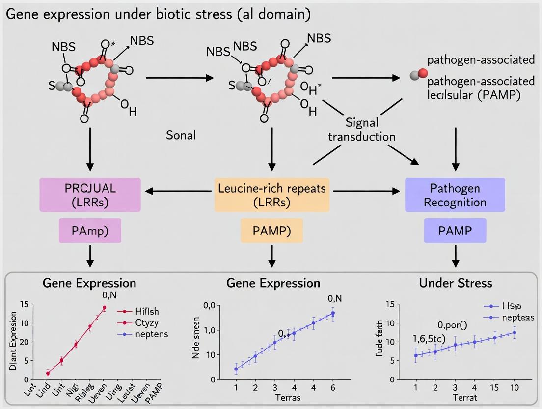
Unveiling the Plant Immune Code: NBS-LRR Gene Expression Dynamics in Biotic Stress Responses
This article provides a comprehensive review for researchers and drug development professionals on the critical role of Nucleotide-Binding Site-Leucine-Rich Repeat (NBS-LRR) genes in plant immunity.
Unveiling the Plant Immune Code: NBS-LRR Gene Expression Dynamics in Biotic Stress Responses
Abstract
This article provides a comprehensive review for researchers and drug development professionals on the critical role of Nucleotide-Binding Site-Leucine-Rich Repeat (NBS-LRR) genes in plant immunity. We explore the foundational biology of these disease resistance (R) genes, their complex transcriptional regulation during pathogen attack, and the signaling cascades they initiate. Methodologically, we detail current techniques for profiling NBS expression, including RNA-Seq and qRT-PCR, and discuss their application in transgenic approaches and marker-assisted breeding. We address common experimental challenges in quantifying these low-abundance transcripts and optimizing assays for accuracy. Finally, we compare NBS genes across plant families, validate their functions through silencing and overexpression studies, and examine their co-expression with other defense pathways. The synthesis points towards leveraging this knowledge for developing next-generation, durable crop protection strategies and novel bioactive compounds.
The Molecular Sentinels: Understanding NBS-LRR Genes and Their Role in Plant Immunity
The Nucleotide-Binding Site Leucine-Rich Repeat (NBS-LRR) gene family constitutes the largest class of plant disease resistance (R) genes, serving as the primary intracellular immune receptors for pathogen detection. Research into their expression patterns, transcriptional regulation, and functional divergence is a cornerstone of the broader thesis on plant biotic stress response. Understanding their precise definition, structural architecture, and evolutionary dynamics is fundamental to engineering durable resistance in crops and informing novel strategies for plant protection.
Gene Structure and Domain Architecture
The canonical NBS-LRR protein is defined by three core domains:
- N-terminal Domain: Typically either a coiled-coil (CC) or Toll/Interleukin-1 receptor (TIR) motif, involved in downstream signaling initiation.
- Nucleotide-Binding Site (NBS) Domain: A central, conserved ATP/GTP-binding module essential for molecular switching and activation.
- Leucine-Rich Repeat (LRR) Domain: A C-terminal, variable domain responsible for direct or indirect pathogen effector recognition.
Table 1: Core Structural Domains of NBS-LRR Proteins
| Domain | Key Motifs/Features | Proposed Function |
|---|---|---|
| TIR/CC | TIR: DDxxD, EDVID; CC: coiled-coil structure | Signaling transduction, partner interaction |
| NBS | Kinase 1a/P-loop (GxxxxGKS/T), RNBS-A, -B, -C, -D; GLPL; MHDV | Nucleotide binding/hydrolysis, regulatory switch |
| LRR | xxLxLxx (L=Leu, I, V, F) repeats | Effector recognition, specificity determinant |
Diagram: NBS-LRR Protein Domain Architecture
Classification and Phylogeny
Based on N-terminal domains and conserved motifs within the NBS domain, NBS-LRR genes are primarily classified into two major lineages: TNL (TIR-NBS-LRR) and CNL (CC-NBS-LRR), with a minor RNL (RPW8-NBS-LRR) subclade. Phylogenetic analysis of the NBS domain sequences is the standard method for classification and evolutionary inference.
Table 2: Major NBS-LRR Classes and Characteristics
| Class | N-terminal | Key NBS Motif (RNBS-D) | Prevalence | Typical Signaling Adapter |
|---|---|---|---|---|
| TNL | TIR | FLHIACKxxF | Dicots only | EDS1-PAD4-ADR1/NRG1 |
| CNL | Coiled-Coil | MHDxLxFLWL | Dicots & Monocots | NRCs / NDR1 |
| RNL | RPW8-like CC | MHDCxxFLWL | Dicots & Monocots | Often helper (e.g., NRG1, ADR1) |
Experimental Protocol: Phylogenetic Classification of NBS-LRR Genes
- Sequence Retrieval: Identify candidate sequences from genome databases using HMMER (v3.3) with Pfam profiles (PF00931 for NBS, PF00560 for TIR, PF13855 for LRR, PF05659 for RPW8).
- Multiple Sequence Alignment: Perform alignment of the NBS domain sequences using MAFFT (v7) or MUSCLE with default parameters.
- Phylogenetic Tree Construction: Build a maximum-likelihood tree using IQ-TREE (v2.2) with ModelFinder for best-fit model selection (e.g., JTT+G+I). Use 1000 ultrafast bootstrap replicates.
- Classification & Visualization: Classify clades based on topology and N-terminal identity. Annotate and visualize the tree using iTOL or FigTree.
Diagram: Workflow for NBS-LRR Phylogenetic Analysis
Evolutionary Dynamics
NBS-LRR genes exhibit rapid, lineage-specific evolution, driven by biotic stress pressures. Key mechanisms include:
- Gene Duplication: Tandem and segmental duplications create gene clusters.
- Birth-and-Death Evolution: New genes are generated by duplication; some are maintained, others degenerate into pseudogenes.
- Diversifying Selection: Positive selection acts on solvent-exposed residues in the LRR, shaping novel recognition specificities.
- Non-Homologous Recombination: Domain swapping and fusion events (e.g., between CNL and TNL loci) generate novel chimeric genes.
Table 3: Quantitative Overview of NBS-LRR Family Size in Selected Plant Genomes
| Plant Species | Genome Size (Gb) | Total NBS-LRRs* | TNLs | CNLs | RNLs/Others | Reference (Year) |
|---|---|---|---|---|---|---|
| Arabidopsis thaliana | 0.14 | ~165 | ~75 | ~90 | ~5 | TAIR (2021) |
| Oryza sativa (Rice) | 0.39 | ~500 | 0 | ~480 | ~20 | MSU RGAP (2020) |
| Zea mays (Maize) | 2.4 | ~166 | 0 | ~150 | ~16 | MaizeGDB (2023) |
| Solanum lycopersicum (Tomato) | 0.90 | ~355 | ~130 | ~225 | - | Sol Genomics (2022) |
| Note: Numbers are approximate and vary with annotation methods. |
The Scientist's Toolkit: Key Research Reagents & Materials
Table 4: Essential Research Solutions for NBS-LRR Functional Studies
| Reagent / Material | Function / Application in NBS-LRR Research |
|---|---|
| pCambia or pGreen Vectors | Binary vectors for stable plant transformation and transgenic complementation. |
| Gateway Cloning System | High-throughput cloning of full-length or domain-swapped NBS-LRR constructs. |
| Agrobacterium tumefaciens Strain GV3101 | Standard strain for transient expression (agroinfiltration) and stable transformation. |
| N. benthamiana Plants | Model plant for transient assays (e.g., cell death, co-immunoprecipitation). |
| Anti-GFP / Anti-FLAG / Anti-HA Antibodies | Detection of epitope-tagged NBS-LRR proteins in immunoblotting or Co-IP. |
| Firefly/Renilla Luciferase (LUC/REN) | Reporters for real-time measurement of immune signaling output. |
| MG132 (Proteasome Inhibitor) | To investigate NBS-LRR protein stability and turnover. |
| ATP-γ-S / ADP (Nucleotide Analogs) | To probe the role of nucleotide binding/hydrolysis in NBS-LRR activation. |
| Phytohormone Assay Kits (SA, JA, ET) | Quantify defense hormone levels upon NBS-LRR expression or activation. |
Experimental Protocol: Functional Validation via Transient Expression
Title: Agrobacterium-Mediated Transient Assay for NBS-LRR-Induced Cell Death
Methodology:
- Construct Preparation: Clone the candidate NBS-LRR gene into a binary expression vector (e.g., pBin-GFP) under a strong constitutive promoter (e.g., 35S CaMV).
- Agrobacterium Transformation: Introduce the construct into A. tumefaciens strain GV3101 (pSoup-assisted for pGreen vectors) via electroporation or freeze-thaw.
- Culture Induction: Grow single colonies in LB with appropriate antibiotics at 28°C. Pellet cells and resuspend in induction buffer (10 mM MES, pH 5.6, 10 mM MgCl₂, 150 µM acetosyringone) to an OD₆₀₀ of 0.5-1.0. Incubate for 2-4 hours at room temperature.
- Plant Infiltration: Pressure-infiltrate the bacterial suspension into leaves of 4-5 week-old N. benthamiana plants using a needleless syringe.
- Phenotyping & Analysis: Monitor infiltrated patches over 2-7 days for hypersensitive response (HR) cell death. Quantify cell death using ion conductivity measurements or trypan blue staining. For signaling analysis, co-infiltrate with reporter constructs or collect tissue for immunoblotting/RNA extraction.
Diagram: Transient Expression Assay for Functional Validation
The study of Nucleotide-Binding Site Leucine-Rich Repeat (NBS-LRR) genes forms the cornerstone of biotic stress research in plants. These genes encode intracellular immune receptors responsible for detecting pathogen-derived effectors, initiating robust defense responses. The Guard Hypothesis, proposed over two decades ago, revolutionized our understanding of this recognition by positing that NBS-LRR proteins (guards) monitor host cellular components (guardees) for perturbations caused by pathogen effectors. This framework has since expanded into broader concepts like the decoy and integrated decoy models. This whitepaper delves into the core biochemical mechanisms of pathogen recognition, framed explicitly within ongoing thesis research on the expression dynamics, functional characterization, and signaling networks of NBS domain genes under biotic stress. The elucidation of these mechanisms is not only fundamental to plant pathology but also informs novel strategies in agricultural biotechnology and inspires therapeutic approaches in mammalian innate immunity and drug development.
Core Recognition Models: From Guard to Integrated Decoy
The Guard Hypothesis
The classic Guard Hypothesis describes an indirect recognition system. Here, a plant NBS-LRR immune receptor (the guard) physically associates with a host protein (the guardee) that is a bona fide target of a pathogen virulence effector. The effector's manipulation (e.g., phosphorylation, cleavage, ubiquitination) of the guardee alters its conformation or state, which is sensed by the guard NBS-LRR. This interaction triggers a conformational change in the NBS-LRR, activating effector-triggered immunity (ETI).
The Decoy Model
An evolutionary refinement of the guard hypothesis, the Decoy Model proposes that some guardee proteins are molecular decoys. These decoys mimic real effector targets but have lost their original biochemical function. Their sole purpose is to attract effector manipulation, thereby enabling detection by the paired NBS-LRR. This allows the plant to detect effectors without the fitness cost of disrupting essential cellular pathways.
The Integrated Decoy (ID) Model
The most integrated model suggests that decoy domains are often fused directly into the NBS-LRR protein architecture itself. These integrated decoy domains (e.g., WRKY, JAZ, PBL domains) directly bind pathogen effectors, leading to autoactivation of the receptor. This represents a direct recognition mechanism but via incorporated, non-functional mimicry domains.
Table 1: Comparative Analysis of Pathogen Recognition Models
| Model | Recognition Type | Guardee/Decoy Nature | Example System | Key Evidence |
|---|---|---|---|---|
| Guard | Indirect | Functional host protein (guardee) | Arabidopsis RIN4 guarded by RPS2/RPM1 | RIN4 cleavage by AvrRpt2 (Pseudomonas) activates RPS2. |
| Decoy | Indirect | Non-functional mimic of host target | Arabidopsis PBL2 (kinase decoy) for AvrAC (Xanthomonas) | AvrAC uridylylates PBL2, activating ZAR1-RLCK complex. |
| Integrated Decoy | Direct | Domain integrated into NBS-LRR protein | Rice Pik-1 NLR with integrated HMA domain | HMA domain binds AVR-Pik (Magnaporthe) effectors directly. |
Quantitative Data on NBS-LRR Gene Expression & Polymorphism
NBS-LRR genes represent one of the largest and most dynamic gene families in plant genomes. Their expression is tightly regulated and highly responsive to stress.
Table 2: NBS-LRR Family Size and Expression Dynamics in Model Plants
| Plant Species | Approx. NBS-LRR Count | Expression Profile | Induction Fold-Change (Post-Inoculation) | Key Regulatory Mechanism |
|---|---|---|---|---|
| Arabidopsis thaliana | ~150 | Low basal; rapid, specific induction | Up to 50-fold (e.g., RPS4) | Transcriptional reprogramming via NPR1, SAR. |
| Oryza sativa (Rice) | ~500 | Tissue-specific; pathogen-responsive | 5-100 fold (e.g., Piz-t, Xa1) | Epigenetic regulation, alternative splicing. |
| Solanum lycopersicum (Tomato) | ~400 | Developmentally regulated; induced | Up to 30-fold (e.g., Mi-1.2) | Hormonal crosstalk (SA, JA, ET). |
| Zea mays (Maize) | ~125 | Low constitutive; moderate induction | 3-20 fold | Complex cis-regulatory elements. |
Table 3: Polymorphism and Diversity Metrics in NBS-LRR Genes
| Diversity Metric | Typical Value Range in NBS-LRRs | Comparison to Genome Average | Implication for Recognition |
|---|---|---|---|
| Non-synonymous/synonymous substitution rate (dN/dS) | 0.5 - >1.5 (LRR domain often >1) | Significantly higher | Positive selection for novel recognition specificities. |
| Copy Number Variation (CNV) | High frequency across accessions | Higher than housekeeping genes | Rapid adaptation to pathogen landscapes. |
| Presence/Absence Variation (PAV) | Common in cluster regions | Higher than genome average | Contributes to pan-genome and resistance spectra. |
Key Experimental Protocols in Mechanism Elucidation
Protocol: Yeast Two-Hybrid (Y2H) for Guard-Guardee-Effector Interaction Mapping
- Purpose: To identify and validate pairwise protein-protein interactions between NBS-LRRs, their guardee/decoy partners, and pathogen effectors.
- Methodology:
- Clone the coding sequence of the putative guardee/decoy into the pGADT7 (AD) vector (prey). Clone the NBS-LRR (or its specific domain like CC, TIR) into the pGBKT7 (BD) vector (bait). Clone the pathogen effector into either vector.
- Co-transform pairwise combinations (BD-bait + AD-prey) into competent yeast strain (e.g., AH109 or Y2HGold).
- Plate transformations on synthetic dropout (SD) media lacking Leu and Trp (-LW) to select for co-transformants.
- Streak positive colonies onto high-stringency SD media lacking Leu, Trp, His, and Ade (-LWAH), often with X-α-Gal for blue/white screening. Growth and color change indicate a positive interaction.
- Include controls: empty vectors, known interactors (positive), and non-interactors (negative).
- Key Reagents: pGADT7 & pGBKT7 vectors, Yeast strains (AH109), SD base, Dropout supplements, X-α-Gal, 3-AT (if needed to suppress background).
Protocol: Co-Immunoprecipitation (Co-IP) and Immunoblotting forIn PlantaComplex Validation
- Purpose: To confirm physical interactions between proteins in their native cellular environment, often post-pathogen challenge.
- Methodology:
- Generate transgenic plants or use agroinfiltration to express epitope-tagged versions (e.g., GFP, FLAG, HA, Myc) of the NBS-LRR, guardee, and/or effector.
- Harvest leaf tissue at appropriate time post-infiltration/inoculation. Grind tissue in liquid N₂ and homogenize in non-denaturing extraction buffer (e.g., with NP-40 or Triton X-100, plus protease inhibitors).
- Pre-clear lysate with control beads (e.g., protein A/G). Incubate supernatant with antibody against the bait protein's tag, followed by pull-down with protein A/G beads, or use directly with magnetic beads conjugated to the antibody.
- Wash beads extensively to remove non-specific binders. Elute proteins by boiling in SDS-PAGE loading buffer.
- Separate proteins by SDS-PAGE, transfer to PVDF membrane, and probe with antibodies against the tags of the putative interacting partners (prey).
- Key Reagents: Epitope tags (GFP, FLAG, HA), Specific antibodies, Protein A/G magnetic beads, Non-denaturing lysis buffer, Protease inhibitor cocktail, PVDF membrane, Chemiluminescent substrate.
Protocol: Quantitative RT-PCR for NBS-LRR Expression Profiling during ETI
- Purpose: To measure dynamic changes in transcript levels of specific NBS-LRR genes following pathogen recognition.
- Methodology:
- Design gene-specific primers for target NBS-LRRs and reference housekeeping genes (e.g., EF1α, ACTIN, UBIQUITIN).
- Inoculate plants with an avirulent pathogen (carrying matching Avr gene) or a virulent control. Collect tissue samples at multiple time points (e.g., 0, 2, 6, 12, 24 hours post-inoculation).
- Extract total RNA using a silica-column based kit with on-column DNase I treatment.
- Synthesize cDNA using a reverse transcriptase with oligo(dT) and/or random hexamer primers.
- Perform qPCR with SYBR Green or TaqMan chemistry in a real-time cycler. Use a standard two-step thermal cycling protocol.
- Analyze data using the comparative ΔΔCt method, normalizing target gene expression to the reference gene(s) and relative to the control condition (e.g., mock inoculation at time zero).
- Key Reagents: RNA extraction kit (e.g., RNeasy), DNase I, Reverse transcriptase, SYBR Green qPCR Master Mix, Gene-specific primers.
The Scientist's Toolkit: Key Research Reagent Solutions
Table 4: Essential Reagents for NBS-LRR and Pathogen Recognition Research
| Reagent/Material | Function/Application | Example Product/Catalog |
|---|---|---|
| Gateway Cloning System | High-throughput, recombinational cloning of NBS-LRR cDNAs into multiple expression vectors (Y2H, Co-IP, localization). | Invitrogen pDONR vectors, pEarleyGate destination vectors. |
| Agrobacterium tumefaciens Strains | Transient expression (Agroinfiltration) in Nicotiana benthamiana for protein interaction, cell death assays, and subcellular localization. | GV3101, AGL-1, EHA105 competent cells. |
| LRR Domain Consensus Prediction Software | In silico identification and structural modeling of LRR motifs for effector binding site prediction. | LRRsearch, LRRpredict, I-TASSER. |
| Anti-GFP/FLAG/HA Magnetic Beads | Efficient, high-specificity immunoprecipitation of tagged proteins from plant lysates for Co-IP assays. | ChromoTek GFP-Trap beads, Anti-FLAG M2 Magnetic Beads. |
| Plant Cell Death Assay Reagents | Quantification of hypersensitive response (HR), a hallmark of NBS-LRR activation. | Electrolyte leakage meters, Trypan Blue stain for in planta cell death visualization. |
| Pathogen Effector Libraries | Comprehensive sets of cloned effectors from major pathogens (e.g., Pseudomonas, Xanthomonas, Phytophthora) for screening against NBS-LRRs. | Custom gene synthesis libraries or publicly available repositories. |
Visualizing Signaling Pathways and Experimental Workflows
This whitepaper explores the mechanisms of transcriptional reprogramming in Nucleotide-Binding Site-Leucine-Rich Repeat (NBS-LRR) genes following biotic stress, a critical area within the broader thesis of Deciphering the Cis-Regulatory Code of NBS-LRR Genes for Enhanced Crop Resilience. NBS genes encode the largest class of plant disease resistance (R) proteins, which act as intracellular immune receptors. Their expression is tightly regulated at the transcriptional level, and biotic stressors—such as pathogens and herbivores—trigger rapid, complex changes in this regulatory landscape. Understanding this reprogramming is pivotal for developing novel strategies in plant protection and drug development targeting immune signaling pathways.
Core Signaling Pathways in NBS Gene Transcriptional Regulation
Biotic stress perception initiates signaling cascades that converge on transcription factors (TFs) to alter NBS gene expression. Two primary, interconnected pathways are central.
Diagram 1: Core Transcriptional Pathways for NBS Genes Under Biotic Stress
Key Quantitative Data on NBS Gene Expression Dynamics
Recent transcriptomic studies reveal distinct expression patterns for different NBS gene subfamilies (TNL, CNL, RNL) upon pathogen challenge.
Table 1: Temporal Expression Profiles of NBS Gene Subfamilies Post-Pathogen Inoculation
| NBS Subfamily | Key Upregulated Examples (Gene ID) | Fold-Change (Peak) | Time to Peak (Hours Post-Inoculation) | Proposed Primary Inducing Signal |
|---|---|---|---|---|
| TNL (TIR-NBS-LRR) | AtRPS4 (At5g45250) | 12-25x | 12-18 h | SA, Specific Effector Recognition |
| CNL (CC-NBS-LRR) | AtRPM1 (At3g07040) | 8-15x | 6-12 h | SA, ROS/MAPK Signaling |
| RNL (RPW8-NBS-LRR) | AtADR1 (At1g33560) | 30-50x | 24-48 h | SA, Required for TNL/CNL signaling |
| NBS (Helper, Sensor) | AtNRCs (e.g., NRC2) | 5-10x | 6-18 h | Multiple, Downstream of Sensors |
Table 2: Cis-Element Enrichment in Co-Upregulated NBS Gene Promoters
| Cis-Element Motif | Consensus Sequence | Associated Transcription Factor | Enrichment p-value (ChIP-seq/ATAC-seq) | Functional Role in Stress Response |
|---|---|---|---|---|
| W-box | (T)TGAC(C/T) | WRKY18, WRKY40, WRKY53 | < 1e-15 | Positive & Negative Regulation |
| TGA-site | TGACG | TGA2, TGA3, TGA6 | < 1e-10 | SA-Responsive, NPR1-dependent |
| G-box | CACGTG | bZIP, MYC2 | < 1e-8 | JA-Responsive, Often Repressive |
| ERE | AGGCCGCC | ERF1/2/5 | < 1e-6 | ET-Responsive |
| SARE | TTCGACCTCC | Unknown | < 1e-12 | SA-Specific Response |
Experimental Protocols for Key Methodologies
Protocol 1: Time-Course RNA-Seq for NBS Expression Profiling
Objective: Quantify genome-wide transcriptional changes in NBS genes following biotic stress.
- Plant Material & Stress: Treat Arabidopsis thaliana (Col-0) leaves with 1 mM Salicylic Acid (SA) or virulent/avirulent Pseudomonas syringae pv. tomato (Pst) strain (OD600=0.001 in 10 mM MgCl2).
- Sampling: Collect leaf tissue at 0, 1, 3, 6, 12, 24, and 48 hours post-treatment (hpt). Flash-freeze in liquid N2. Use ≥3 biological replicates.
- RNA Extraction & Library Prep: Extract total RNA using TRIzol reagent with DNase I treatment. Assess integrity (RIN > 8.0). Prepare stranded mRNA-seq libraries using Illumina TruSeq kit.
- Sequencing & Analysis: Sequence on Illumina NovaSeq platform (PE150). Align reads to TAIR10 genome with HISAT2. Count reads per gene with featureCounts. Differential expression analysis using DESeq2 (FDR < 0.05, |log2FC| > 1). Visualize NBS gene cluster dynamics with heatmaps.
Protocol 2: Chromatin Immunoprecipitation Sequencing (ChIP-seq) for TF Binding
Objective: Map genome-wide binding sites of transcription factors (e.g., WRKY, TGA) to NBS promoters.
- Crosslinking & Nuclei Isolation: Harvest SA-treated tissue (3 hpt). Crosslink with 1% formaldehyde. Quench with glycine. Isolate nuclei using Nuclei Isolation Buffer (NIB).
- Chromatin Shearing: Sonicate chromatin to ~200-500 bp fragments using a Covaris S220 sonicator.
- Immunoprecipitation: Incubate chromatin with antibody against target TF (e.g., anti-WRKY18) or IgG control. Use Protein A/G magnetic beads for pull-down.
- Library Prep & Sequencing: Reverse crosslinks, purify DNA. Prepare sequencing libraries from ChIP and Input DNA using NEBNext Ultra II kit. Sequence and map reads as in Protocol 1.
- Peak Calling & Motif Analysis: Call enriched peaks with MACS2. Annotate peaks to genomic features. Perform de novo motif discovery with MEME-ChIP on peaks proximal to NBS genes.
Diagram 2: ChIP-seq Experimental Workflow for NBS Promoter Analysis
The Scientist's Toolkit: Key Research Reagent Solutions
Table 3: Essential Reagents and Tools for NBS Transcriptional Reprogramming Research
| Item Name | Supplier/Example Catalog # | Function in Research Context |
|---|---|---|
| TRIzol Reagent | Thermo Fisher Scientific, 15596026 | Simultaneous RNA/DNA/protein isolation from plant tissue for transcriptomic studies. |
| Illumina TruSeq Stranded mRNA Library Prep Kit | Illumina, 20020594 | Preparation of strand-specific RNA-seq libraries for precise expression quantification. |
| DESeq2 R/Bioconductor Package | Bioconductor | Statistical software for differential gene expression analysis from RNA-seq count data. |
| Anti-WRKY18 / Anti-TGA2 Antibody | Agrisera, custom | Specific antibodies for Chromatin IP (ChIP) to pull down TF-bound DNA fragments. |
| Protein A/G Magnetic Beads | Pierce, 88802/88803 | Efficient capture of antibody-TF-DNA complexes during ChIP protocol. |
| NEBNext Ultra II DNA Library Prep Kit | NEB, E7645S | Preparation of high-quality sequencing libraries from ChIP-derived DNA. |
| Salicylic Acid (SA) | Sigma-Aldrich, S7401 | Key phytohormone used to induce the SA signaling pathway and NBS gene expression. |
| MG132 Proteasome Inhibitor | Sigma-Aldrich, C2211 | Used to investigate the role of proteasomal degradation in regulating NBS-related TFs. |
| Dual-Luciferase Reporter Assay System | Promega, E1910 | Quantifies promoter activity of NBS genes by measuring luciferase expression in transfected protoplasts. |
| Crispr-Cas9 Knockout Mutants (e.g., npr1-1, wrky18/40) | ABRC / TAIR | Genetic resources to dissect the role of specific signaling nodes in NBS reprogramming. |
Within the broader thesis on NBS domain gene expression in biotic stress research, this guide details the complex downstream signaling events initiated by nucleotide-binding site (NBS) domain-containing proteins, primarily Nucleotide-Binding Leucine-Rich Repeat (NLR) receptors. Upon pathogen perception, NBS proteins undergo conformational changes to form oligomeric resistosomes, which act as signaling hubs to activate robust defense programs, including transcriptional reprogramming, hormonal signaling, and localized cell death.
Core Signaling Pathways Activated by NBS Proteins
Calcium Influx and MAPK Cascade Activation
The resistosome often functions as a calcium-permeable channel. The resultant cytosolic calcium burst ([Ca²⁺]cyt) is decoded by calcium sensors like calmodulin (CaM) and CBL/CIPK networks, leading to the activation of Mitogen-Activated Protein Kinase (MAPK) cascades. Key cascades include MEKK1-MKK4/5-MPK3/6 and MEKK1-MKK1/2-MPK4, which phosphorylate downstream transcription factors.
Phytohormone Signaling Networks
NBS activation redirects hormone biosynthesis and signaling.
- Salicylic Acid (SA): A primary output. The isochorismate synthase (ICS1) pathway is induced, leading to SA accumulation. SA binds to NPR proteins, promoting the degradation of JAZ repressors and facilitating the expression of Pathogenesis-Related (PR) genes via TGA transcription factors.
- Ethylene (ET) & Jasmonic Acid (JA): Often modulated synergistically or antagonistically with SA. NBS signaling can induce ACC synthase (ACS) for ET biosynthesis and LOX, AOS for JA biosynthesis, tailoring responses to pathogen type.
- Reactive Oxygen Species (ROS) Burst: Activated directly via Respiratory Burst Oxidase Homologs (RBOHs) phosphorylated by Ca²⁺-dependent protein kinases (CDPKs) or MAPKs. ROS acts as a direct antimicrobial agent and a secondary messenger.
Transcriptional Reprogramming and Effector-Triggered Immunity (ETI)
The integrated signaling network converges on master transcriptional regulators. Key families include:
- WRKY TFs: Phosphorylated by MAPKs, bind W-box elements in promoters of defense genes.
- NPR1: The central SA receptor, changes redox state to translocate to the nucleus.
- ERF TFs: Activated by MAPK phosphorylation, bind GCC boxes in promoters of JA/ET-responsive genes.
This cascade culminates in ETI, characterized by the hypersensitive response (HR) and systemic acquired resistance (SAR).
Diagram 1: Core NBS-Activated Defense Signaling Network
Key Quantitative Data in NBS-Mediated Signaling
Table 1: Quantitative Metrics in NBS Signaling Events
| Signaling Component/Event | Measurable Parameter | Typical Experimental Range/Value | Measurement Technique |
|---|---|---|---|
| Calcium Influx | Peak [Ca²⁺]cyt increase | 1-10 µM (from ~100 nM resting) | Aequorin/GCaMP luminescence/fluorescence |
| ROS Burst | H₂O₂ accumulation rate | 1-5 µmol min⁻¹ g⁻¹ FW | Luminol-based chemiluminescence |
| MAPK Activation | MPK3/6 phosphorylation | Peak at 5-15 min post-elicitation | Immunoblot with anti-pMAPK antibody |
| SA Accumulation | Free SA in leaves | 0.5-5 µg g⁻¹ FW (can increase 10x) | HPLC or LC-MS/MS |
| Transcript Induction | PR1 gene expression | 100-1000 fold induction | qRT-PCR (relative to control) |
| HR Cell Death | Ion leakage | 40-80% conductivity increase (24hpi) | Electrolyte leakage assay |
Detailed Experimental Protocols
Protocol: Measuring NBS-Mediated Calcium Flux Using Aequorin
Purpose: To quantify the early calcium signature following NBS protein activation. Reagents: Transgenic plants expressing cytosolic aequorin, specific pathogen effector or PAMP, coelenterazine (substrate). Procedure:
- Reconstitution: Detach leaves/seedlings. Infiltrate with 5 µM coelenterazine in low-Ca²⁺ buffer. Incubate in dark for 6-12 hours.
- Elicitation: Place sample in luminometer chamber. Inject effector solution or mock.
- Measurement: Record photon counts continuously for 30-60 minutes. Use low-light photon-counting PMD.
- Calibration: At end, discharge remaining aequorin by injecting 1 M CaCl₂ in 20% ethanol. Calculate [Ca²⁺]cyt using standard formula relating log(L/Lmax) to log[Ca²⁺]. Analysis: Plot [Ca²⁺]cyt over time. Compare peak height and kinetics between genotypes/treatments.
Protocol: Assessing MAPK Activation via Immunoblot
Purpose: To detect phosphorylation/activation of MPK3/6 downstream of NBS signaling. Reagents: Plant tissue, liquid N₂, extraction buffer (HEPES, glycerol, EDTA, protease/phosphatase inhibitors), anti-p44/42 (Erk) or plant-specific anti-pMAPK antibody, anti-total MPK3/6 antibody. Procedure:
- Sample Collection: Treat plants. Harvest tissue (e.g., 100 mg) at timed intervals (0, 5, 15, 30 min) directly into liquid N₂.
- Protein Extraction: Grind tissue. Add 200 µL ice-cold extraction buffer. Centrifuge at 14,000 g, 4°C for 20 min.
- Immunoblot: Separate 20 µg supernatant protein on 10% SDS-PAGE. Transfer to PVDF membrane.
- Blocking/Probing: Block with 5% BSA. Incubate with primary anti-pMAPK antibody (1:2000) overnight at 4°C. Use HRP-conjugated secondary antibody (1:5000).
- Detection: Use ECL reagent and chemiluminescence imager.
- Reprobing: Strip membrane, reprobe with anti-total MPK3/6 antibody for loading control. Analysis: Compare band intensity of phosphorylated MAPK relative to total.
Diagram 2: Workflow for NBS Signaling Analysis
The Scientist's Toolkit: Key Research Reagent Solutions
Table 2: Essential Reagents for Investigating NBS Signaling Pathways
| Reagent/Material | Primary Function & Application | Example/Supplier Note |
|---|---|---|
| Anti-p44/42 MAPK Antibody | Detects phosphorylated/activated MPK3/6/4 homologs in immunoblots. | Cell Signaling Technology #9101; cross-reacts with plant pMAPKs. |
| GCaMP Transgenic Lines | Genetically encoded calcium indicator for live-cell imaging of [Ca²⁺]cyt flux. | Arabidopsis lines expressing GCaMP3/6 under 35S or cell-specific promoters. |
| Coelenterazine-h | Substrate for aequorin, used in reconstitution for luminescent Ca²⁺ measurement. | Thermo Fisher Scientific; dissolved in ethanol for stock. |
| L-012 (WST-8) | Highly sensitive luminol analog for detecting extracellular ROS burst. | Fujifilm Wako; used at 50-100 µM in assay buffer. |
| SA/JA/ET ELISA Kits | Quantify endogenous phytohormone levels post-NBS activation. | Numerous plant-specific kits available (e.g., MyBioSource, Agrisera). |
| Pathogen Effector Proteins | Purified recombinant proteins to specifically activate corresponding NBS receptors. | Often expressed in E. coli with His-tag, purified via Ni-NTA. |
| MAPK Inhibitors (e.g., U0126) | Chemical inhibitor of MKK1/2 activity, used to dissect MAPK role in signaling. | Used in pre-treatment controls at specified concentrations (e.g., 10 µM). |
| NLR/R-Gene Mutants | Genetic null/knockout lines to establish requirement of specific NBS protein. | Available from stock centers (e.g., ABRC, NASC) or via CRISPR lines. |
Genomic Organization and Diversity of NBS-LRR Genes Across Plant Species
Within the broader thesis investigating NBS domain gene expression in biotic stress responses, this whitepaper examines the genomic architecture and evolutionary diversification of Nucleotide-Binding Site Leucine-Rich Repeat (NBS-LRR) genes. As the largest class of plant disease resistance (R) genes, understanding their organization and diversity is foundational for research into engineered stress resistance and novel plant protection strategies.
Genomic Organization: Clustering and Distribution
NBS-LRR genes exhibit non-random genomic distributions, predominantly organized in clusters of tandem and segmentally duplicated genes. This organization facilitates rapid evolution and generation of new resistance specificities.
Table 1: Genomic Organization Metrics of NBS-LRR Genes in Model Species
| Plant Species | Total NBS-LRR Genes | % in Clusters | Avg. Cluster Size | Main Chromosomal Location(s) |
|---|---|---|---|---|
| Arabidopsis thaliana | ~165 | 75% | 2-5 | Chromosomes 1, 3, 5 |
| Oryza sativa (Rice) | ~500 | 85% | 4-15 | Chromosomes 4, 11, 12 |
| Zea mays (Maize) | ~120 | 70% | 2-7 | Chromosomes 2, 6, 10 |
| Solanum lycopersicum (Tomato) | ~350 | 90% | 3-12 | Chromosomes 4, 6, 11 |
| Glycine max (Soybean) | ~500+ | 80% | 3-10 | Multiple scaffolds |
Structural and Functional Diversity
NBS-LRR proteins are categorized into two major subfamilies based on N-terminal domains: TIR-NBS-LRR (TNL) and CC-NBS-LRR (CNL). A third, minor group (RNL) acts as helper proteins.
Table 2: Structural Diversity and Characteristics of NBS-LRR Subfamilies
| Subfamily | N-terminal Domain | Key Signaling Adapters | Presence in Monocots | Typical Resistance Spectrum |
|---|---|---|---|---|
| TNL | Toll/Interleukin-1 Receptor (TIR) | EDS1, PAD4, SAG101 | No (except some grasses) | Oomycetes, fungi, bacteria |
| CNL | Coiled-Coil (CC) | NRG1, NRC proteins | Yes | Viruses, bacteria, fungi, nematodes |
| RNL (Helper) | RPW8-like CC | ADR1, NRG1 | Yes | Broad-spectrum signaling |
Evolutionary Dynamics and Mechanisms
Diversity is driven by several evolutionary processes:
- Tandem Duplication: Primary source of intra-cluster diversity.
- Ectopic Recombination: Gene conversion and unequal crossing-over.
- Positive Selection (dN/dS >1): Acts on solvent-exposed LRR residues, shaping pathogen recognition.
- Birth-and-Death Evolution: New genes are created by duplication; some are maintained, others pseudogenize.
Table 3: Evolutionary Metrics for NBS-LRR Genes
| Evolutionary Process | Genomic Evidence | Measured Rate/Impact | Experimental Validation Method |
|---|---|---|---|
| Tandem Duplication | Clustered gene arrays | 0.05-0.2 new copies/Myr | Comparative genomics, FISH |
| Positive Selection | dN/dS ratio on LRR codons | dN/dS = 1.5 - 3.5 | PAML/SLR analysis |
| Gene Conversion | Sequence homogenization in clusters | Up to 40% of paralog pairs | Phylogenetic network analysis |
| Pseudogenization | Premature stop codons, frameshifts | 15-30% of annotated genes | PCR, RT-PCR, sequencing |
Experimental Protocols for Diversity Analysis
Protocol 4.1: Genome-Wide Identification and Annotation
Objective: To identify all NBS-LRR genes in a sequenced genome.
- HMMER Search: Use hidden Markov model profiles (PF00931 for NBS, PF00560 for LRR, PF01582 for TIR, PF05659 for CC) against the proteome (e.g.,
hmmsearch --cut_ga pfam.hmm proteome.fa). - Domain Architecture Validation: Validate candidate proteins using NCBI CD-Search or SMART for domain order and completeness.
- Manual Curation: Check for intact ORFs, remove pseudogenes (premature stops, frameshifts), and classify into TNL/CNL/RNL.
- Genomic Mapping: Map genes to chromosomes using GFF3 files and visualize with software like TBtools or custom R scripts.
Protocol 4.2: dN/dS Analysis for Positive Selection
Objective: To detect sites under positive selection in LRR regions.
- Ortholog/Paralog Alignment: Align coding sequences of orthologous gene pairs or within-species paralogs using MUSCLE or MAFFT.
- Phylogeny Reconstruction: Build a maximum-likelihood tree with IQ-TREE.
- Selection Analysis: Run the CodeML program in the PAML package. Compare site-specific models (M7 vs. M8). Sites with posterior probability >0.95 under M8 are considered under positive selection.
- Visualization: Map positively selected sites onto a protein 3D model (if available) using PyMOL.
Protocol 4.3: Expression Diversity Analysis via RNA-seq
Objective: To profile NBS-LRR expression under biotic stress.
- Treatment & RNA Extraction: Inoculate plants with pathogen/elicitor. Harvest tissue at multiple time points (0, 6, 12, 24, 48 hpi). Extract total RNA with TRIzol, check RIN >8.0.
- Library Prep & Sequencing: Prepare stranded mRNA-seq libraries (Illumina TruSeq). Sequence on NovaSeq 6000 for 150bp paired-end reads.
- Bioinformatics Pipeline:
- Read Mapping: Map clean reads to the reference genome using HISAT2.
- Read Counting: Use featureCounts to count reads mapping to each NBS-LRR gene.
- Differential Expression: Analyze with DESeq2 in R (threshold: |log2FC|>1, padj<0.05).
- Co-expression Network: Construct using WGCNA to identify regulatory modules.
Visualization of Key Concepts
Title: Evolutionary Mechanisms Driving NBS-LRR Diversity
Title: NBS-LRR Subfamily Signaling Pathways
The Scientist's Toolkit: Research Reagent Solutions
Table 4: Essential Reagents and Resources for NBS-LRR Research
| Item Name | Supplier Examples (Catalog # if typical) | Function/Application in NBS-LRR Research |
|---|---|---|
| Phytozome / EnsemblPlants | JGI / EMBL-EBI | Primary databases for genome sequences, gene models, and comparative genomics of plants. |
| Pfam HMM Profiles | Pfam Database (PF00931, PF00560, etc.) | Hidden Markov Models for identifying NBS, LRR, and other domains in protein sequences. |
| PAML (CodeML) Software | Ziheng Yang Lab | Statistical package for codon-based phylogenetic analysis and detecting positive selection (dN/dS). |
| DESeq2 R Package | Bioconductor | For differential expression analysis of RNA-seq data to identify stress-responsive NBS-LRR genes. |
| Anti-HA / Anti-Myc Tag Antibodies | Sigma, Roche, Cell Signaling | For immunoprecipitation and western blot analysis of epitope-tagged NBS-LRR proteins. |
| Gateway Cloning System | Thermo Fisher (11791020, etc.) | For high-throughput cloning of NBS-LRR genes into binary vectors for plant transformation. |
| Agrobacterium tumefaciens GV3101 | Various Biolabs | Strain for stable plant transformation or transient expression (Agroinfiltration) in leaves. |
| TRIzol Reagent | Thermo Fisher (15596026) | For high-yield, high-quality total RNA isolation from pathogen-inoculated plant tissues. |
| Luciferase Assay Kit | Promega (E1500) | For measuring activity of NBS-LRR promoter::luciferase reporters in response to elicitors. |
| Cyclopiazonic Acid (CPA) | Sigma (C1530) | Inhibitor of endoplasmic reticulum Ca2+-ATPases; used to probe calcium signaling in NBS-LRR immunity. |
From Lab to Field: Profiling NBS Expression and Engineering Resistance
This technical guide is situated within a broader thesis investigating the role of Nucleotide-Binding Site-Leucine-Rich Repeat (NBS-LRR) genes in plant biotic stress responses. NBS genes constitute one of the largest and most critical gene families for disease resistance. High-throughput profiling technologies, namely microarrays and RNA-Sequencing (RNA-Seq), are indispensable for quantifying the expression dynamics of these genes under pathogen challenge. This whitepaper provides an in-depth comparison of these platforms, detailed protocols, and analytical frameworks to advance research in this field.
Technology Comparison: RNA-Seq vs. Microarrays
Table 1: Core Comparative Analysis of RNA-Seq and Microarrays for NBS Expression Profiling
| Feature | RNA-Sequencing (RNA-Seq) | Microarrays |
|---|---|---|
| Principle | Direct sequencing of cDNA fragments (Shotgun) | Hybridization of labeled cDNA to pre-designed probes |
| Throughput & Dynamic Range | >10⁵ range; detects >6 orders of magnitude in expression | ~10³ range; limited by background and saturation |
| Resolution | Single-nucleotide; can identify SNPs, novel transcripts, and splice variants | Limited to predefined probe sequences; cannot detect novel sequences |
| Background Noise | Very low; optical noise is minimal | High; from non-specific hybridization |
| Quantitative Accuracy | High; digital counting of reads (e.g., counts per million) | Moderate; analog fluorescent intensity measurement |
| NBS Gene Suitability | Ideal for discovering novel NBS-LRR family members and paralog-specific expression | Suitable only for known, annotated NBS genes with existing probes |
| Cost per Sample (Approx.) | $500 - $2,000 (decreasing trend) | $200 - $500 |
| Primary Data Output | FASTQ files (sequence reads and quality scores) | CEL or GPR files (fluorescence intensity values) |
| Key Limitation for NBS | Complex data analysis; high computational burden | Probe cross-hybridization between highly similar NBS paralogs |
| Optimal Use Case | Discovery-phase research, non-model plants, fine-scale differential expression | High-sample-number validation studies in well-annotated model species |
Experimental Protocols
Protocol A: RNA-Seq for NBS Expression Analysis in Stressed Tissue
Objective: To profile the transcriptome, specifically NBS-LRR gene expression, in plant leaves following pathogen inoculation.
Materials: See "The Scientist's Toolkit" below.
- Sample Preparation & RNA Isolation:
- Treat plant material (e.g., Arabidopsis leaves) with pathogen or elicitor. Include mock-treated controls.
- Homogenize tissue in liquid N₂. Isolate total RNA using a silica-column based kit with on-column DNase I digestion.
- Assess RNA integrity (RIN > 8.5) using a Bioanalyzer. Quantify via fluorometry (Qubit).
- Library Preparation:
- Enrich mRNA using poly-A selection (for eukaryotic plants) or deplete ribosomal RNA.
- Fragment enriched RNA (200-300 bp) using divalent cations at elevated temperature (94°C, 5-7 min).
- Synthesize first-strand cDNA with reverse transcriptase and random hexamers, followed by second-strand synthesis.
- Perform end-repair, 3'-adenylation, and adapter ligation using a stranded library preparation kit.
- Amplify the library via 10-15 cycles of PCR with index primers to enable sample multiplexing.
- Validate library size distribution (TapeStation) and quantify (qPCR).
- Sequencing:
- Pool multiplexed libraries in equimolar ratios.
- Sequence on an Illumina NovaSeq platform to generate 150 bp paired-end reads, targeting 25-40 million reads per sample.
- Bioinformatic Analysis:
- Quality Control: Use FastQC and Trimmomatic to remove adapters and low-quality bases.
- Alignment: Map reads to the reference genome using a splice-aware aligner (e.g., HISAT2, STAR).
- Quantification: Use featureCounts or HTSeq to count reads aligning to annotated NBS-LRR and other genes.
- Differential Expression: Analyze counts with DESeq2 or edgeR in R. Identify NBS genes with significant expression changes (adjusted p-value < 0.05, log2FC > |1|).
Protocol B: Microarray Analysis for NBS Expression
Objective: To measure expression of known NBS-LRR genes across many samples in a cost-effective manner.
Materials: See "The Scientist's Toolkit" below.
- Sample & Target Preparation:
- Isolate total RNA as in Protocol A, Step 1.
- Convert RNA to cDNA using reverse transcriptase with an oligo(dT) primer incorporating a T7 promoter.
- Synthesure biotin-labeled complementary RNA (cRNA) via in vitro transcription using T7 RNA polymerase and biotin-UTP.
- Purify and fragment the labeled cRNA to 35-200 bp fragments.
- Hybridization & Washing:
- Hybridize the fragmented cRNA to a pre-designed microarray (e.g., Affymetrix GeneChip) for 16 hours at 45°C in a rotating hybridization oven.
- Perform stringent post-hybridization washes in a fluidics station according to manufacturer's protocols (e.g., non-stringent wash buffer at 25°C, followed by stringent wash buffer at 50°C).
- Scanning & Data Acquisition:
- Stain the array with streptavidin-phycoerythrin, amplify with biotinylated anti-streptavidin antibody, and re-stain.
- Scan the array using a laser confocal scanner (e.g., GeneChip Scanner 3000).
- Extract raw fluorescence intensity values (CEL files) using the scanner software.
- Data Analysis:
- Perform background correction, normalization (RMA or GCRMA), and summarization of probe-level data.
- Conduct differential expression analysis using a linear model (e.g., limma package in R). Focus analysis on predefined NBS-LRR probe sets.
Visualizations
Title: RNA-Seq Experimental and Computational Workflow
Title: Decision Flowchart: RNA-Seq vs. Microarray Selection
Title: Simplified NBS-LRR Mediated Signaling Pathway
The Scientist's Toolkit
Table 2: Essential Research Reagent Solutions for NBS Expression Profiling
| Item | Function in Experiment | Example Product / Kit |
|---|---|---|
| High-Integrity RNA Isolation Kit | Ensures pure, DNA-free total RNA with high RIN, critical for both library prep and microarray target synthesis. | Qiagen RNeasy Plant Mini Kit with on-column DNase. |
| RNA Integrity Analyzer | Accurately assesses RNA quality (RIN) to prevent wasting resources on degraded samples. | Agilent Bioanalyzer 2100 with RNA Nano Kit. |
| Stranded RNA-Seq Library Prep Kit | Converts RNA to sequencer-ready, strand-specific DNA libraries, preserving transcript origin information. | Illumina Stranded mRNA Prep. |
| Poly-A Magnetic Beads | Enriches for eukaryotic mRNA by selecting polyadenylated transcripts, reducing ribosomal RNA reads. | NEBNext Poly(A) mRNA Magnetic Isolation Module. |
| Microarray Platform & Chip | Contains immobilized probes for specific genes. Choice is species-specific. | Affymetrix GeneChip Arabidopsis ATH1 Genome Array. |
| In Vitro Transcription Labeling Kit | Produces biotin-labeled cRNA from cDNA for microarray hybridization and detection. | Affymetrix GeneChip IVT Labeling Kit. |
| Hybridization, Wash, and Stain Kit | Provides optimized buffers for the post-labeling steps of microarray processing. | Affymetrix GeneChip Hybridization, Wash, and Stain Kit. |
| NGS Alignment Software | Maps sequenced reads to a reference genome, requiring splice-awareness for eukaryotic genes. | STAR aligner or HISAT2. |
| Differential Expression Analysis Package | Statistical tool for identifying significant expression changes from count or intensity data. | DESeq2 (R/Bioconductor) for RNA-Seq; limma for microarrays. |
This guide provides a targeted framework for the quantification of Nucleotide-Binding Site Leucine-Rich Repeat (NBS-LRR) gene transcripts using reverse transcription quantitative PCR (qRT-PCR). Within a broader thesis on NBS domain gene expression in biotic stress research, accurate quantification of these transcripts is non-negotiable. These genes are central to plant innate immunity, and their expression dynamics—often low-abundance, rapidly induced, and belonging to large, highly similar gene families—present unique technical hurdles. Robust qRT-PCR data forms the critical link between observed plant stress phenotypes and the molecular mechanisms governed by the NBS-LRR resistome, enabling the validation of omics-scale discoveries and the functional characterization of candidate resistance genes.
Key Challenges & Strategic Solutions
| Challenge | Impact on qRT-PCR | Best Practice Solution |
|---|---|---|
| High Sequence Homology | Primer/probe cross-reactivity, quantifying paralogs. | Design primers spanning non-conserved regions (e.g., introns, LRR, 3'UTR). Validate with amplicon sequencing. |
| Low Basal Expression | High Cq values, increased stochastic variation. | Use high-input RNA (≥500 ng), optimized reverse transcription, and high-efficiency assays. |
| Rapid Induction Kinetics | Expression changes can be missed with poor temporal resolution. | Conduct dense time-course sampling post-inoculation (e.g., 0, 2, 6, 12, 24, 48 hpi). |
| Lack of Stable References | Normalization errors mask true expression changes. | Systematically validate 3-5 candidate reference genes under specific experimental conditions. |
| Presence of Pseudogenes | Genomic DNA amplification leads to overestimation. | Mandatory DNase I treatment, intron-spanning primer design, and -RT controls. |
Detailed Experimental Protocols
RNA Isolation & Quality Control
- Method: Use a silica-column-based kit with an on-column DNase I digestion step. For recalcitrant tissues, incorporate a CTAB-based lysis buffer to remove polysaccharides and polyphenols.
- Critical Steps: 1) Homogenize tissue in liquid N₂. 2) Perform DNase I treatment for 30 min at 25°C. 3) Elute RNA in nuclease-free water (not TE buffer).
- QC Metrics: Quantify via fluorometry (e.g., Qubit). Assess integrity using an RNA Integrity Number (RIN) > 8.0 on a Bioanalyzer or clear 28S/18S rRNA bands on gel.
Reverse Transcription
- Method: Use a two-step kit with random hexamers and oligo(dT) primers. This combination improves coverage of long transcripts and reduces 5' bias.
- Protocol: For 20 µL reaction: 500 ng – 1 µg total RNA, 1 µL 50 µM random hexamers, 1 µL 50 µM oligo(dT)₂₀, incubate at 65°C for 5 min, then chill. Add 4 µL 5x reaction buffer, 1 µL RNase inhibitor (40 U/µL), 1 µL reverse transcriptase (200 U/µL), and nuclease-free water. Incubate: 25°C for 10 min, 50°C for 50 min, 80°C for 5 min.
- Controls: Include a no-reverse transcriptase (-RT) control for each sample to monitor gDNA contamination.
qPCR Assay Design & Validation
- Primer Design: Use tools like Primer-BLAST. Target amplicons 80-150 bp. Set melting temperature (Tm) to 60 ± 1°C. Verify specificity by in silico PCR against the host genome.
- Validation: Generate a 5-point, 10-fold serial dilution of pooled cDNA (from 1:10 to 1:10,000) to create a standard curve. Run in triplicate.
- Acceptance Criteria: Amplification efficiency (E) = 90–110% (slope of -3.6 to -3.1), R² > 0.99. Confirm single-peak melt curve and a single band of correct size on an agarose gel.
qPCR Run & Analysis
- Reaction Setup: Use 10 µL reactions in a 384-well plate: 5 µL 2x SYBR Green Master Mix, 0.5 µL each primer (10 µM), 2 µL cDNA (diluted 1:10), 2 µL nuclease-free water. Run all samples in technical triplicates.
- Cycling Conditions: 95°C for 3 min; 40 cycles of 95°C for 15 sec, 60°C for 30 sec, 72°C for 30 sec (plate read); followed by melt curve analysis.
- Data Analysis: Use the comparative Cq (ΔΔCq) method. First, normalize target Cq to the geometric mean of validated reference gene Cqs (ΔCq). Then, calculate the ΔΔCq relative to the control condition (e.g., untreated, time-zero). Report as relative expression = 2^(-ΔΔCq).
Visualization of Workflow & Pathway Context
Title: NBS-LRR qRT-PCR Experimental Workflow
Title: NBS-LRR Transcriptional Activation in Immune Signaling
The Scientist's Toolkit: Research Reagent Solutions
| Item | Function & Rationale |
|---|---|
| Plant-Specific RNA Isolation Kit | Contains buffers optimized to co-purify RNA while binding plant polysaccharides/polyphenols to the column, improving yield and purity. |
| RNase Inhibitor | Protects RNA samples during handling and reverse transcription from ubiquitous RNases, critical for labile transcripts. |
| Two-Step RT-qPCR Kit | Offers flexibility for large sample sets, superior sensitivity, and the ability to optimize cDNA synthesis and qPCR separately. |
| Genomic DNA Elimination Reagent | More robust than on-column DNase I alone; provides a second, solution-phase gDNA removal step post-extraction. |
| High-Fidelity DNA Polymerase | Essential for cloning amplicons from qPCR validation runs for sequencing to confirm primer specificity. |
| Validated Reference Gene Panel | A pre-selected set of candidate genes (e.g., EF1α, ACT, UBC, PP2A) for systematic validation under specific stress conditions. |
| SYBR Green Master Mix | Cost-effective for high-throughput analysis; contains hot-start Taq polymerase, buffer, dNTPs, and the intercalating dye. |
| Optical-Grade Sealing Film | Ensures a perfect seal for 384-well plates to prevent well-to-well contamination and evaporation during cycling. |
In the context of investigating Nucleotide-Binding Site (NBS) domain gene expression in response to biotic stress, precise visualization of promoter activity is paramount. NBS domain genes, central to plant innate immunity (e.g., NBS-LRR receptors), exhibit complex, dynamic expression patterns upon pathogen perception. Promoter-β-glucuronidase (GUS) fusions serve as a foundational tool to dissect the spatial (e.g., infection sites, vascular tissues) and temporal (e.g., early vs. late response) regulation of these critical genes. This technical guide details the application of GUS reporter assays to elucidate the transcriptional control of NBS genes under stress, providing insights for engineering durable resistance.
Core Principles of Promoter-GUS Fusions
The assay hinges on creating a chimeric gene where the promoter sequence of the NBS domain gene of interest is transcriptionally fused to the coding sequence of the uidA gene, encoding GUS. Following stable transformation into a host plant, the activity of the promoter is directly reported by the accumulation of GUS enzyme. This enzyme catalyzes the hydrolysis of colorless substrates into colored or fluorescent products, allowing histochemical localization or fluorometric quantification.
Key Research Reagent Solutions
| Reagent / Material | Function in Experiment |
|---|---|
| pCAMBIA3301 Vector | Binary T-DNA vector with promoterless uidA (GUS) gene and plant selection marker (e.g., bar for phosphinothricin resistance). |
| MUG Substrate (4-Methylumbelliferyl β-D-Glucuronide) | Fluorogenic substrate for quantitative, kinetic GUS assays. Hydrolysis produces fluorescent 4-MU. |
| X-Gluc (5-Bromo-4-Chloro-3-Indolyl β-D-Glucuronide) | Chromogenic substrate for histochemical staining. Hydrolysis produces an insoluble blue indigo dye. |
| Plant Genomic DNA Isolation Kit | For isolating high-quality DNA to amplify native NBS gene promoter regions. |
| High-Fidelity DNA Polymerase | For accurate, error-free amplification of promoter sequences from genomic DNA. |
| Agrobacterium tumefaciens Strain GV3101 | Disarmed strain used for stable transformation of plant tissues via floral dip or tissue culture. |
| GUS Extraction Buffer (50 mM NaPO₄ pH 7.0, 10 mM EDTA, 0.1% Triton X-100, 10 mM β-mercaptoethanol) | Lysis buffer for extracting soluble GUS protein from plant tissues. |
| Protein Assay Dye Reagent | For normalizing fluorometric GUS activity to total protein concentration. |
Experimental Protocols
Protocol: Constructing the Promoter-GUS Fusion Vector
- Promoter Isolation: Amplify 1.5-3.0 kb of genomic sequence upstream of the NBS domain gene ATG start codon using high-fidelity polymerase. Include restriction sites compatible with the chosen binary vector (e.g., HindIII and BamHI).
- Vector Preparation: Digest the pCAMBIA3301 (or similar) binary vector with the same restriction enzymes. Purify the linearized vector via gel extraction.
- Ligation & Cloning: Ligate the isolated promoter fragment into the vector upstream of the uidA gene. Transform the ligation product into E. coli, screen colonies by PCR/restriction digest, and sequence-verify the final construct.
- Agrobacterium Transformation: Introduce the verified plasmid into Agrobacterium tumefaciens GV3101 via electroporation or freeze-thaw method.
Protocol: Histochemical GUS Staining for Spatial Expression
- Plant Material: Collect tissue from transgenic plants (and wild-type control) at specified time points post-biotic stress treatment (e.g., pathogen inoculation, elicitor application).
- Fixation: Vacuum-infiltrate tissue in chilled 90% acetone for 15-30 minutes on ice. Rinse with 50 mM sodium phosphate buffer (pH 7.2).
- Staining: Incubate tissue in X-Gluc staining solution (1 mM X-Gluc, 50 mM NaPO₄ pH 7.2, 0.5 mM potassium ferricyanide, 0.5 mM potassium ferrocyanide, 0.1% Triton X-100) at 37°C in the dark for 2-24 hours.
- Destaining: Remove chlorophyll by washing in a graded ethanol series (20%, 35%, 50%, 80%) or by incubation in 70% ethanol at 70°C.
- Imaging: Observe and photograph cleared tissue under a stereomicroscope or compound microscope.
Protocol: Fluorometric GUS Assay for Quantitative Temporal Expression
- Protein Extraction: Grind 50-100 mg of frozen plant tissue in 200-500 µL GUS extraction buffer. Centrifuge at 13,000×g for 15 min at 4°C. Keep supernatant on ice.
- Protein Quantification: Use a Bradford or similar assay to determine total protein concentration of each extract.
- Reaction Setup: For each sample, mix 10-50 µL of extract (diluted to equal protein amounts) with 1 mM MUG substrate in extraction buffer to a final volume of 500 µL. Incubate at 37°C.
- Kinetic Measurement: At time points (e.g., 0, 15, 30, 60 min), remove 100 µL of reaction mix and stop with 900 µL of 0.2 M Na₂CO₃.
- Quantification: Measure fluorescence (excitation 365 nm, emission 455 nm) using a fluorometer. Calculate GUS activity as pmol 4-MU produced per minute per µg of total protein.
Data Presentation: Quantitative GUS Activity Under Biotic Stress
Table 1: Temporal GUS Activity in PNBS-LRR::GUS Transgenic Arabidopsis Post-Pseudomonas syringae Inoculation
| Time Post-Inoculation (hpi) | Mean GUS Activity (pmol 4-MU/min/µg protein) ± SD | Fold Induction vs. Mock |
|---|---|---|
| 0 (Mock) | 12.5 ± 2.1 | 1.0 |
| 6 | 45.3 ± 5.6 | 3.6 |
| 12 | 182.7 ± 22.4 | 14.6 |
| 24 | 315.8 ± 30.1 | 25.3 |
| 48 | 89.4 ± 10.2 | 7.2 |
Table 2: Spatial GUS Staining Intensity in PNBS-LRR::GUS Plants
| Plant Tissue | Staining Intensity (0-3 scale) - Mock | Staining Intensity (0-3 scale) - Pathogen Inoculated |
|---|---|---|
| Root Apex | 1 (Weak) | 1 (Weak) |
| Mature Leaf (unwounded) | 0 (None) | 2 (Moderate) |
| Leaf Veins | 1 (Weak) | 3 (Strong) |
| Infection Site Periphery | N/A | 3 (Strong) |
| Floral Stems | 0 (None) | 1 (Weak) |
Visualizing Pathways and Workflows
Title: Promoter-GUS Reporter Assay Experimental Workflow
Title: NBS Gene Induction Pathway Visualized by GUS
Within the framework of a broader thesis on NBS (Nucleotide-Binding Site) domain gene expression in biotic stress research, this whitepaper provides a technical guide on transgenic strategies for engineering disease resistance. Focusing on the overexpression and heterologous expression of NBS-LRR (Leucine-Rich Repeat) and related defense genes, this document details current methodologies, experimental data, and protocols for developing crops with enhanced, durable resistance to pathogens.
Plant NBS-LRR genes constitute one of the largest and most critical gene families in innate immunity, encoding intracellular immune receptors that directly or indirectly recognize pathogen effectors. Transgenic manipulation of these genes—either by overexpressing endogenous alleles or expressing heterologous receptors from other species—offers a powerful avenue to engineer broad-spectrum and durable resistance, circumventing the limitations of traditional R-gene pyramiding.
Core Transgenic Strategies
Overexpression of Endogenous NBS-LRR Genes
This approach involves the constitutive or inducible overexpression of a plant's own NBS-LRR gene using strong promoters (e.g., CaMV 35S, Ubiquitin). The goal is to amplify the plant's existing defense signaling, potentially leading to a faster and stronger hypersensitive response (HR).
Heterologous Expression of Non-Host R Genes
Heterologous expression introduces an NBS-LRR gene from a non-host or wild relative into a susceptible crop plant. This can confer recognition of effectors that the crop's native immune repertoire cannot perceive, thus expanding the spectrum of resistance.
Synthetic Immune Receptor Engineering
Advanced strategies involve creating chimeric receptors by fusing novel effector recognition domains to conserved NBS-LRR signaling domains, a technique exploiting modular protein architecture.
Table 1: Efficacy of Transgenic NBS-LRR Expression in Model and Crop Plants (2020-2023)
| Target Crop | Gene Source (Gene Name) | Expression Strategy | Pathogen Tested | Reduction in Disease Severity (%) | Key Phenotype | Citation (Example) |
|---|---|---|---|---|---|---|
| Arabidopsis | Arabidopsis (RPS4) | Overexpression (35S) | P. syringae pv. tomato | 85-95 | Accelerated HR | Li et al., 2021 |
| Rice | Wild rice (O. longistaminata, Xa21) | Heterologous (Ubi) | X. oryzae pv. oryzae | 70-80 | Broad-spectrum BLB resistance | Wang et al., 2022 |
| Tomato | Pepper (Capsicum, Bs2) | Heterologous (35S) | X. gardneri | 75-85 | Specific HR to avrBs2 | Sharma et al., 2021 |
| Potato | Solanum venturii (Rpi-vnt1.1) | Heterologous (pGBM) | P. infestans | 65-75 | Late blight resistance | Jones et al., 2023 |
| Wheat | Aegilops tauschii (Sr45) | Heterologous (Ubi) | P. graminis f. sp. tritici | 60-70 | Stem rust resistance (race-specific) | Chen et al., 2022 |
| Tobacco | Synthetic (RGA5/RGA4 chimera) | Overexpression (35S) | M. oryzae (AVR-Pik) | 90-98 | Effector-triggered immunity | Narusaka et al., 2020 |
Table 2: Common Molecular and Phenotypic Assays for Validation
| Assay Category | Specific Assay | Parameter Measured | Indicator of Success |
|---|---|---|---|
| Transgene Analysis | qRT-PCR | Transcript abundance | High expression in transgenic lines. |
| Western Blot | Protein accumulation | Detection of full-length receptor. | |
| Immune Activation | Ion Leakage Assay | Electrolyte leakage | Quantification of HR strength. |
| DAB Staining | H₂O₂ accumulation | Visual detection of oxidative burst. | |
| MAPK Assay | Phosphorylation of MAPKs | Early defense signaling activation. | |
| Pathogen Resistance | Detached Leaf Assay | Lesion size/ number | Direct pathogen growth inhibition. |
| Whole Plant Inoculation | Disease score, biomass | Overall resistance in planta. |
Detailed Experimental Protocols
Protocol: Stable Transformation and Screening for NBS-LRR Overexpression inArabidopsis
Objective: Generate and identify Arabidopsis lines constitutively overexpressing an endogenous NBS-LRR gene. Materials: See "The Scientist's Toolkit" below. Workflow:
- Gene Cloning: Amplify the full-length coding sequence (CDS) of the target NBS-LRR gene from genomic DNA or cDNA. Clone into a binary vector (e.g., pBIN19 derivative) downstream of the CaMV 35S promoter and upstream of a terminator (e.g., NOS). Include a selectable marker (e.g., Bar for glufosinate resistance).
- Agrobacterium Transformation: Introduce the recombinant binary vector into A. tumefaciens strain GV3101 via electroporation.
- Floral Dip Transformation:
- Grow donor Arabidopsis (Col-0) to the stage of numerous immature floral buds.
- Resuspend a fresh culture of transformed Agrobacterium (OD₆₀₀ = ~0.8) in infiltration medium (5% sucrose, 0.05% Silwet L-77).
- Invert and submerge the aerial parts of the plant in the suspension for 30 seconds. Repeat after 7 days.
- Selection (T1 Generation): Harvest seeds (T1). Sow on soil or MS plates containing the appropriate selective agent (e.g., glufosinate). Resistant seedlings are primary transformants.
- Molecular Screening:
- Perform PCR on genomic DNA from T1 plants to confirm transgene presence.
- Use qRT-PCR on cDNA from confirmed T1 plants with gene-specific primers to assess expression levels relative to wild-type and actin control.
- Homozygous Line Selection (T3): Self T1 plants. Screen T2 progeny for 3:1 segregation resistance. Select lines showing 100% resistance in T3 for homozygous, stable lines.
Protocol: Transient Expression Assay for Heterologous R-Gene Function inNicotiana benthamiana
Objective: Rapidly validate the functionality of a heterologous NBS-LRR gene by co-expression with its cognate avirulence (Avr) effector. Materials: See "The Scientist's Toolkit." Workflow:
- Construct Preparation: Clone the candidate heterologous NBS-LRR CDS into an expression vector (e.g., pEAQ-HT). Clone the putative matching Avr effector gene into a separate vector (e.g., pGRAB).
- Agrobacterium Infiltration:
- Transform each construct into A. tumefaciens strain GV2260.
- Grow cultures, resuspend to a final OD₆₀₀ = 0.5 in MMA infiltration buffer (10 mM MES, 10 mM MgCl₂, 100 µM acetosyringone).
- Experimental Mixes: Prepare three infiltration mixes: (i) NBS-LRR strain alone, (ii) Avr strain alone, (iii) NBS-LRR and Avr strains mixed 1:1.
- Infiltration: Using a needleless syringe, infiltrate the mixes into separate patches on the abaxial side of 4-5 week-old N. benthamiana leaves.
- Phenotypic Monitoring (24-96 hpi):
- Hypersensitive Response (HR): Visually inspect for rapid, localized tissue collapse (whitening/necrosis) specifically in the co-expression zone.
- Ion Leakage Quantification: Cut leaf discs from each infiltration zone, float in distilled water, measure conductivity over time with a conductivity meter. Co-expression zones will show significantly higher ion leakage.
- Biochemical Assays: Harvest tissue from zones for Western blot (protein expression confirmation) or DAB staining (H₂O₂ detection).
Signaling Pathways and Workflows
Diagram 1: NBS-LRR Activation and Defense Signaling Pathway
Diagram 2: Stable Transgenic Line Development Workflow
The Scientist's Toolkit: Key Research Reagent Solutions
Table 3: Essential Materials for Transgenic Resistance Research
| Category | Item / Reagent | Function & Rationale |
|---|---|---|
| Cloning & Vectors | High-Fidelity DNA Polymerase (e.g., Phusion) | Error-free amplification of NBS-LRR CDS for cloning. |
| Gateway or Golden Gate Modular Binary Vectors | Enables rapid, standardized assembly of expression constructs (Promoter-Gene-Terminator). | |
| pEAQ-HT or pGRAB Vectors | Optimized for high-level transient protein expression in N. benthamiana. | |
| Transformation | Agrobacterium tumefaciens GV3101/GV2260 | Standard disarmed strains for stable (GV3101) and transient (GV2260) transformation. |
| Silwet L-77 | Surfactant critical for efficient Agrobacterium infiltration during floral dip or transient assays. | |
| Acetosyringone | Phenolic compound that induces Agrobacterium vir gene expression, enhancing T-DNA transfer. | |
| Selection & Screening | Herbicides/ Antibiotics (e.g., Glufosinate, Kanamycin) | Selective agents for plants transformed with corresponding resistance markers (Bar, nptII). |
| TIANGEN Plant DNA/RNA Kits | Reliable extraction of high-quality nucleic acids from diverse plant tissues. | |
| SYBR Green qPCR Master Mix | For sensitive, quantitative analysis of transgene expression levels (qRT-PCR). | |
| Phenotypic Analysis | Pathogen Isolates (Wild-type & Avr mutants) | Essential for challenge inoculations to assess specificity and spectrum of resistance. |
| Conductivity Meter | Quantifies ion leakage as a precise, numerical measure of HR cell death. | |
| 3,3'-Diaminobenzidine (DAB) | Histochemical stain that polymerizes in the presence of H₂O₂, visualizing oxidative burst. | |
| Protein Analysis | Anti-GFP or Anti-Myc Tag Antibodies | Common for detecting tagged transgenic NBS-LRR proteins via Western blot or microscopy. |
| Phospho-p44/42 MAPK (Erk1/2) Antibody | Detects activation of conserved defense-related MAP kinases. |
This whitepaper details a technical framework for integrating Nucleotide-Binding Site (NBS) gene polymorphisms into Marker-Assisted Selection (MAS) programs. It is situated within a broader thesis investigating the expression dynamics of NBS domain genes in response to biotic stress. NBS-LRR genes constitute a major plant disease resistance (R-gene) family. Polymorphisms within these genes, particularly in the NBS domain, are directly linked to specific pathogen recognition capabilities. By moving beyond correlative markers to causative functional polymorphisms, breeders can achieve precise, durable resistance stacking in elite crop varieties, accelerating development cycles and enhancing food security.
Core NBS Gene Polymorphisms and Their Functional Impact
Polymorphisms in NBS genes are critical for generating novel resistance specificities. Key polymorphism classes are summarized in the table below.
Table 1: Functional Classes of NBS Gene Polymorphisms for MAS
| Polymorphism Type | Genomic Location | Molecular Consequence | Impact on Resistance Phenotype | Suitability for MAS |
|---|---|---|---|---|
| Non-Synonymous SNPs (nsSNPs) | Exon (especially P-loop, RNBS-A, RNBS-D motifs) | Alters amino acid sequence of NBS domain; affects ATP-binding/hydrolysis or protein conformation. | Can broaden, narrow, or abolish recognition specificity; often quantitative. | High (Causative, requires functional validation) |
| Presence/Absence Variations (PAVs) | Entire gene or large exonic segments | Complete gain or loss of a specific NBS-LRR gene copy. | Binary effect: presence confers potential recognition, absence results in susceptibility. | Very High (Easy to score, strong effect) |
| Indels (In-frame) | Exon (between motifs) | Insertion/Deletion of amino acids, altering domain spacing/geometry. | Modulates signaling intensity or recognition spectrum. | Moderate to High |
| Variable Tandem Repeats | LRR domain (adjacent to NBS) | Changes in copy number of LRR sub-motifs. | Directly alters pathogen effector binding affinity and specificity. | High |
| Promoter Polymorphisms | Cis-regulatory regions | Modifies expression level (constitutive or inducible). | Alters timing and magnitude of defense response; quantitative resistance. | High (for expression-based resistance) |
Experimental Protocols for NBS Polymorphism Discovery & Validation
Protocol: Targeted NBS Gene Enrichment and High-Throughput Sequencing
Objective: To capture and sequence the repertoire of NBS-encoding genes from multiple plant genotypes for polymorphism discovery.
- DNA Extraction: Isolate high-molecular-weight genomic DNA (>50 kb) from leaf tissue of parental and mapping population lines using a CTAB-based protocol with RNase A treatment.
- Probe Design: Synthesize biotinylated RNA or DNA probes complementary to conserved NBS domain motifs (e.g., P-loop, Kinase-2, GLPL). Probes should be 80-120 nt in length.
- Solution-Based Hybrid Capture:
- Fragment 1 µg of genomic DNA to 200-300 bp via sonication.
- Prepare Illumina sequencing libraries with unique dual-index adapters.
- Hybridize the library with the biotinylated NBS probes for 16-24 hours at 65°C in a hybridization buffer.
- Capture probe-bound fragments using streptavidin-coated magnetic beads.
- Wash stringently to remove non-specifically bound DNA.
- Elute and PCR-amplify the enriched NBS library.
- Sequencing & Analysis: Sequence on an Illumina NovaSeq platform (PE 150 bp). Map reads to a reference genome using BWA-MEM. Call SNPs and Indels using GATK HaplotypeCaller. Identify PAVs using CNVnator or read-depth analysis.
Protocol: Functional Validation via Allelic Replacement and Pathogen Assay
Objective: To establish causality between a specific NBS polymorphism and a resistance phenotype.
- Vector Construction (CRISPR-Cas9 mediated Allelic Replacement):
- Design two sgRNAs flanking the polymorphic region of the target NBS gene in the susceptible recipient line.
- Clone sgRNAs into a plant Cas9 expression vector (e.g., pHEE401E).
- Synthesize a donor template containing the resistant allele sequence from the donor line, flanked by ~1 kb homology arms.
- Plant Transformation & Selection:
- Transform the susceptible genotype via Agrobacterium-mediated transformation.
- Regenerate plants on selection media.
- Screen T0 plants via PCR and sequencing for precise homologous recombination.
- Phenotyping:
- Challenge T1 homozygous edited lines and controls with the target pathogen.
- For fungal/bacterial pathogens: use standardized inoculum, measure disease index, lesion size, and perform pathogen quantification (CFU/g tissue).
- For viruses: use mechanical inoculation or vector transmission, assess symptom severity via visual scoring and viral titer via qRT-PCR.
- Biochemical Confirmation:
- Perform immunoblotting to confirm protein expression of the new allele.
- Conduct an in vitro ATP-binding/GTPase assay using recombinant NBS domains to assess the functional impact of nsSNPs.
Integration into MAS Breeding Workflows
A systematic pipeline for deploying NBS polymorphisms in MAS is visualized below.
Diagram Title: MAS Pipeline for NBS Polymorphisms
NBS-Mediated Resistance Signaling Pathway
Understanding the signaling context is essential for predicting polymorphism effects. The core pathway is diagrammed below.
Diagram Title: NBS-LRR Signaling & Polymorphism Impact
The Scientist's Toolkit: Research Reagent Solutions
Table 2: Essential Reagents for NBS Polymorphism Research and MAS Integration
| Reagent / Material | Supplier Examples | Function in NBS-MAS Workflow |
|---|---|---|
| NBS Domain Conserved Motif Probes (Biotinylated) | Integrated DNA Technologies (IDT), Agilent | For targeted sequence capture to enrich NBS gene family members from complex genomes prior to sequencing. |
| KASP (Competitive Allele-Specific PCR) Assay Primers | LGC Biosearch Technologies, Thermo Fisher | For high-throughput, low-cost genotyping of validated SNP polymorphisms in breeding populations. |
| Plant CRISPR-Cas9 Allelic Replacement Vector System (e.g., pHEE401E) | Addgene, personal constructs | For functional validation of polymorphisms via precise gene editing and allelic exchange in susceptible backgrounds. |
| Recombinant NBS Domain Protein (Wild-type & Mutant) | Expressed in E. coli or wheat germ system | For in vitro biochemical assays (ATPase, GTPase) to quantify the functional impact of nsSNPs. |
| Pathogen-Specific Antibodies / ELISA Kits | Agdia, APS Biocontrol | For accurate quantification of pathogen load during phenotyping assays for resistance validation. |
| High-Fidelity DNA Polymerase for Amplicon Sequencing (e.g., Q5, Phusion) | New England Biolabs, Thermo Fisher | For error-free amplification of NBS gene loci from multiple genotypes prior to Sanger or next-generation sequencing. |
| Next-Generation Sequencing Library Prep Kit for Low-Input DNA | Illumina, NuGEN | For preparing sequencing libraries from enriched NBS DNA or small population pools for bulked segregant analysis. |
| Fluorescent dsDNA Binding Dye for HRM Analysis (e.g., EvaGreen) | Bio-Rad, Biotium | For high-resolution melt curve analysis to detect SNPs and indels in NBS amplicons during initial screening. |
Navigating Experimental Challenges in NBS-LRR Expression Studies
This technical guide, framed within a broader thesis on Nucleotide-Binding Site Leucine-Rich Repeat (NBS-LRR) domain gene expression in biotic stress research, details advanced methodologies to overcome the central challenge of low-abundance transcript and protein detection. Effective study of these critical plant immune components requires precise enrichment and ultra-sensitive detection. This whitepear provides researchers and drug development professionals with current, actionable protocols and analytical frameworks.
NBS-LRR genes, encoding the largest class of plant disease resistance (R) proteins, are often expressed at constitutively low levels or transiently induced during pathogen attack. Their low basal abundance, coupled with high sequence homology among family members, complicates expression profiling, protein-protein interaction studies, and functional characterization. This necessitates a multi-faceted approach combining physical or molecular enrichment with state-of-the-art detection technologies.
Enrichment Strategies for Transcripts and Proteins
Transcriptomic Enrichment
Pre-enrichment of target transcripts reduces background and increases sequencing depth.
Protocol 2.1.1: Targeted RNA-Seq via Hybrid Capture
- Principle: Biotinylated DNA oligonucleotides (baits) complementary to NBS-LRR gene sequences are used to pull down target RNAs from a total RNA library before sequencing.
- Detailed Methodology:
- Library Preparation: Generate double-stranded cDNA from total RNA (e.g., using SMARTer technology). Ligate sequencing adapters.
- Probe Design: Design 80-120mer biotinylated DNA probes tiling across all known/predicted NBS-LRR coding sequences in the organism of interest. Include conserved domain regions and variable LRR regions.
- Hybridization: Denature the library (95°C, 10 min) and incubate with probe pool in hybridization buffer (e.g., SureSelect Hybridization Buffer) at 65°C for 16-24 hours.
- Capture: Add streptavidin-coated magnetic beads to bind biotinylated probe:target complexes. Wash stringently (e.g., with Wash Buffer 1 & 2 from Agilent) to remove non-specific hybrids.
- Elution & Amplification: Elute captured DNA with NaOH, neutralize, and PCR-amplify for 10-14 cycles.
- Sequencing: Perform high-depth sequencing on Illumina platforms.
Table 1: Comparison of Transcript Enrichment Methods
| Method | Principle | Enrichment Factor | Key Advantage | Key Limitation |
|---|---|---|---|---|
| Hybrid Capture | Solution hybridization with biotinylated DNA baits | 100-10,000x | High multiplexing capability; custom panels | Requires prior sequence knowledge |
| Ampliseq (PCR-based) | Multiplex PCR from cDNA | Up to 1,000,000x | Fast, requires low input | Primer design critical; bias risk |
| Ribo-depletion | Removal of ribosomal RNA | ~100x (relative to rRNA) | Broad transcriptome view | Does not enrich specific gene families |
| 3’ mRNA Seq | Poly-A selection | ~100x (relative to non-polyA) | Standard for coding RNA | Cannot enrich specific families within mRNA |
Proteomic Enrichment
Low-abundance NBS-LRR proteins require isolation from dominant cellular proteins.
Protocol 2.2.1: Immunoaffinity Purification (IP) for NBS-LRR Proteins
- Principle: Antibodies specific to a conserved NBS domain epitope (or a tagged version of the protein) are used to isolate the target protein and its interacting partners.
- Detailed Methodology:
- Sample Preparation: Grind frozen plant tissue under liquid N2. Homogenize in non-denaturing IP lysis buffer (e.g., 50 mM Tris-HCl pH 7.5, 150 mM NaCl, 0.5% NP-40, 1x protease inhibitor cocktail, 1 mM PMSF). Centrifuge at 15,000g for 15 min at 4°C.
- Antibody Coupling: Incubate 2-5 µg of anti-NBS domain monoclonal antibody or anti-tag antibody with 50 µL of protein A/G magnetic beads for 1 hour at RT with rotation. Wash beads twice with PBS.
- Immunoprecipitation: Incubate clarified lysate (1-2 mg total protein) with antibody-coupled beads for 2-4 hours at 4°C with rotation.
- Washing: Wash beads 4-5 times with ice-cold lysis buffer.
- Elution: Elute proteins using low-pH glycine buffer (pH 2.5-3.0) or competitive elution with tag peptide. Neutralize immediately with Tris buffer.
Sensitive Detection and Quantification Methods
Nucleic Acid Detection
Digital PCR (dPCR): For absolute quantification of rare NBS-LRR splice variants or low-fold changes.
- Protocol: Partition a cDNA sample into 20,000 nanoliter-sized droplets or microwells. Perform end-point PCR in each partition. Count positive (fluorescent) vs. negative partitions. Apply Poisson statistics to calculate absolute copy number/µL without a standard curve.
Table 2: Sensitivity Metrics for Nucleic Acid Detection
| Method | Limit of Detection (LoD) | Dynamic Range | Best For |
|---|---|---|---|
| qRT-PCR (TaqMan) | ~10 copies/reaction | 7-8 logs | Validating expression of specific isoforms |
| Digital PCR | 1-3 copies/reaction | 5 logs | Absolute quantitation of rare transcripts |
| RNA-Seq (Standard) | ~0.1 TPM | 4-5 logs | Discovery |
| Targeted RNA-Seq | ~0.001 TPM | >5 logs | Profiling full NBS family |
Protein Detection
Single Molecule Array (Simoa): Enables detection of proteins at sub-femtogram/mL levels.
- Protocol: (Conceptual) Antibody-coated paramagnetic beads capture NBS-LRR protein from a lysate. A biotinylated detector antibody and β-galactosidase (β-Gal) conjugated streptavidin form a complex. Beads are sealed in femtoliter wells. If a bead carries the target protein, the enzyme converts substrate to a fluorescent product, creating a localized high concentration detectable via fluorescence imaging.
Integrated Workflow for NBS-LRR Analysis
A recommended pipeline for comprehensive analysis from tissue to data.
Diagram 1: Integrated NBS-LRR Analysis Workflow
Diagram 2: NBS-LRR Activation in Biotic Stress Signaling
The Scientist's Toolkit: Research Reagent Solutions
Table 3: Essential Materials for Low-Abundance NBS-LRR Research
| Item | Function & Rationale | Example Product/Catalog |
|---|---|---|
| RNase Inhibitor | Prevents degradation of low-copy-number mRNA during extraction. Critical for preserving integrity. | Protector RNase Inhibitor (Roche) |
| Magnetic Beads (Streptavidin) | For hybrid capture or IP. Enable rapid, stringent wash steps to reduce non-specific background. | Dynabeads MyOne Streptavidin C1 |
| Target-Specific Biotinylated Probe Pool | Enriches the entire NBS-LRR family from complex cDNA libraries for sequencing. | Custom xGen Lockdown Probes (IDT) |
| Conserved Domain Antibody | Immunoprecipitates multiple NBS-LRR proteins via shared epitope for pooled analysis. | Anti-NBS Domain (Arabidopsis) (Agrisera) |
| Crosslinker (for ChIP) | Stabilizes transient transcription factor-DNA interactions for studying NBS-LRR regulation. | DSG (Disuccinimidyl glutarate) |
| Phosphatase/Protease Inhibitor Cocktail | Preserves post-translational modification states of signaling proteins during lysis. | PhosSTOP/cOmplete (Roche) |
| Single Molecule Assay Kit | Ultrasensitive quantitation of key immune signaling proteins or cytokines. | Simoa Planar Array Kit (Quanterix) |
| High-Fidelity PCR Enzyme | Essential for accurate amplification of rare targets and library prep for sequencing. | KAPA HiFi HotStart ReadyMix |
| Size Selection Beads | Cleanup and size selection of libraries post-enrichment to remove adapter dimers. | SPRIselect Beads (Beckman Coulter) |
| MS-Grade Trypsin | For digesting low-concentration IP eluates into peptides for LC-MS/MS identification. | Trypsin Gold, Mass Spec Grade (Promega) |
Advancing NBS-LRR research in biotic stress demands a conscious pipeline integrating targeted enrichment with frontier detection technologies. The protocols and tools detailed here provide a framework to transform low-abundance targets from technical obstacles into quantifiable, biologically interpretable data, directly supporting the development of novel disease-resistant plant varieties and informed therapeutic strategies.
Addressing Gene Family Redundancy and High Sequence Homology in Assay Design
The Nucleotide-Binding Site Leucine-Rich Repeat (NBS-LRR) gene family represents a critical frontline in plant innate immunity against biotic stress. In Arabidopsis thaliana, this family comprises over 200 members, while in crops like rice and soybean, the number often exceeds 500. This expansion, driven by tandem duplication and polyploidization, results in significant sequence homology (often >80% amino acid identity within subclades) and functional redundancy. For researchers investigating biotic stress responses, this creates a formidable barrier to designing assays that can accurately quantify expression, silence, or edit specific paralogs without cross-reactivity. This whitepaper provides a technical guide to navigating these challenges, framed within the broader thesis of elucidating the coordinated expression dynamics of NBS domain genes under pathogen attack.
Quantitative Scope of the Problem
The scale of redundancy and homology in plant NBS-LRR genes is substantial. The following table summarizes key quantitative data:
Table 1: Scale of Redundancy & Homology in Model Plant NBS-LRR Families
| Plant Species | Estimated NBS-LRR Genes | Major Clades/Subfamilies | Avg. Intra-Clade Amino Acid Identity | Common Genomic Organization |
|---|---|---|---|---|
| Arabidopsis thaliana | ~200 | TIR-NBS-LRR (TNL), CC-NBS-LRR (CNL) | 75-95% | Clustered tandems |
| Oryza sativa (Rice) | ~500 | CNL, RNL, NL | 80-98% | Dense clusters |
| Glycine max (Soybean) | >700 | TNL, CNL | 70-90% | Large clustered arrays |
| Solanum lycopersicum (Tomato) | ~300 | CNL, TNL | 78-96% | Clusters and singletons |
Core Strategies for Specific Assay Design
In SilicoIdentification of Unique Target Regions
The first step is a comprehensive bioinformatic analysis to identify minimally homologous regions suitable for specific probe or primer design.
Experimental Protocol: Bioinformatic Pipeline for Unique Target Identification
- Gene Family Compilation: Retrieve all NBS-LRR sequences for your organism from databases (e.g., TAIR, RGAP, Phytozome).
- Multiple Sequence Alignment (MSA): Perform a rigorous MSA using tools like Clustal Omega or MAFFT.
- Homology Heatmap & Phylogeny: Generate a percent identity matrix and a phylogenetic tree to visualize clades and redundancy.
- Variable Region Identification: Use tools like Geneious or custom Python/R scripts to scan the MSA for regions of maximal sequence divergence. Ideal targets often reside in:
- The 3' or 5' Untranslated Regions (UTRs).
- Hypervariable loops within the LRR domain.
- Intronic sequences (for genomic DNA assays).
- The less conserved N-terminal domain (TIR or CC).
- Specificity Validation In Silico: Perform a BLASTN/BLASTP search of the candidate probe/primer sequence against the host genome. Require at least 2-3 mismatches, preferably consecutive or at the 3' end, for all non-target paralogs.
Quantitative PCR (qPCR) Assay Design
qPCR remains the gold standard for expression analysis but is highly susceptible to cross-amplification.
Experimental Protocol: Design and Validation of Gene-Specific qPCR Assays
- Primer Design Rules: Design primers (18-22 bp) spanning an exon-exon junction to preclude genomic DNA amplification. Target the unique regions identified in Section 3.1. Maintain a Tm of 60 ± 1°C.
- Inclusive Amplification Control (for Redundancy Studies): Design a second primer set that targets a conserved region (e.g., the P-loop of the NBS domain) to amplify all paralogs in a clade. This estimates total clade expression.
- Stringent Specificity Testing:
- Amplify from Genomic DNA: Use gDNA as a template containing all paralogous genes. Run the qPCR with a melting curve analysis. A single, sharp peak indicates specificity. Multiple peaks indicate cross-amplification.
- Cross-Amplification Test: Clone representative paralogs (3-5 from the same clade) into a standard vector. Perform qPCR using each plasmid as a template. Calculate the amplification efficiency for each non-target; a ΔCq > 10 compared to the true target is typically required for confidence.
- Data Normalization: Use multiple, stable reference genes (e.g., EF1α, UBQ5) validated for the specific biotic stress condition.
Diagram 1: qPCR Primer Design Strategy for Homologous Genes
RNAIn SituHybridization and Single-Cell RNA-Seq
For spatial expression resolution, RNA in situ hybridization (RISH) is key, but homology necessitates careful probe design.
Experimental Protocol: RISH Probe Design for Homologous Genes
- Long Probe Design (~500 bp): Generate DIG-labeled RNA probes by in vitro transcription from a cloned template corresponding to the most unique region of the target gene (often the 3' UTR).
- Stringent Washes: Optimize post-hybridization wash stringency (often using low-concentration RNAse A and/or elevated temperature in the wash buffer) to dissociate imperfectly matched probes.
- Parallel Control: Always run a parallel assay with a conserved NBS domain probe to visualize total clade expression and confirm the unique probe's specificity by differential signal.
CRISPR-Cas9 Editing for Functional Validation
Knocking out specific members of a redundant family requires sgRNAs with absolute specificity.
Experimental Protocol: Design of Paralog-Specific sgRNAs
- Target Site Identification: Use CRISPR design tools (e.g., CRISPR-P, CHOPCHOP) to scan the unique regions of your target gene for suitable NGG PAM sites.
- Off-Target Analysis: Perform a genome-wide search allowing for up to 3-4 mismatches, with special emphasis on the seed region (8-12 bp proximal to PAM). Exclude any sgRNA with a perfectly matched or 1-mismatch off-target site in another NBS-LRR gene.
- Validation Strategy: Always design sgRNAs for 2-3 independent target sites within the same gene. Phenotypic convergence from multiple guides confirms true gene function rather than off-target effects.
- Multiplexing for Redundancy: To overcome functional redundancy, design a cocktail of sgRNAs targeting multiple paralogs within the same clade, using a tRNA or crRNA array system.
The Scientist's Toolkit: Research Reagent Solutions
Table 2: Essential Reagents & Tools for Assay Design Against Redundant Gene Families
| Reagent/Tool | Function & Rationale |
|---|---|
| High-Fidelity DNA Polymerase (e.g., Q5, Phusion) | Critical for error-free amplification of specific targets from complex, homologous gene templates. |
| Locked Nucleic Acid (LNA) or Bridged Nucleic Acid (BNA) Probes | Incorporation into qPCR probes or FISH probes increases binding affinity (Tm) and discrimination power, allowing shorter, more specific sequences to be used. |
| CRISPR-Cas9 Nickase (Cas9n) or High-Fidelity Cas9 (e.g., SpCas9-HF1) | Reduces off-target cleavage events dramatically compared to wild-type SpCas9, essential when targeting sequences with homologous relatives. |
| TILLING or EMS-Mutagenized Populations | Provides an alternative reverse-genetics resource to identify natural, single-nucleotide polymorphisms in target genes that can break homology and be used for specific PCR-based assays. |
| Long-Range PCR Kit | Necessary for amplifying and cloning large, repetitive genomic segments of NBS-LRR genes, including promoter regions, for reporter construct creation. |
| In Vitro Transcription Kit (DIG/ Fluorescein labeled) | For generating strand-specific RNA probes for high-sensitivity in situ hybridization with minimized background. |
| Universal ProbeLibrary (UPL) or TaqMan MGB Probes | Hydrolysis probes with proprietary chemistries that are shorter and can be designed for higher specificity in SNP-dense divergent regions. |
Integrated Workflow for Expression and Functional Analysis
Diagram 2: Integrated Workflow for NBS-LRR Gene Study Under Biotic Stress
Successfully addressing gene family redundancy in NBS domain research requires a multi-layered, validation-heavy approach. By integrating rigorous in silico design with empirical specificity testing and employing a combination of quantitative (qPCR), spatial (RISH), and functional (CRISPR) assays, researchers can dissect the individual and collective contributions of homologous genes to the plant biotic stress response. This precise toolkit moves the field beyond the limitation of redundancy, enabling the functional characterization essential for translating basic knowledge into strategies for crop improvement.
Optimizing RNA Extraction from Challenging, Stressed Plant Tissues
Within the framework of a doctoral thesis investigating Nucleotide-Binding Site-Leucine-Rich Repeat (NBS-LRR) gene expression dynamics under biotic stress, the integrity of extracted RNA is paramount. Challenged plant tissues—such as those infected with pathogens, undergoing senescence, or rich in secondary metabolites—present significant obstacles to RNA extraction. These tissues often contain high levels of polysaccharides, polyphenols, RNases, and other compounds that co-precipitate with nucleic acids or degrade RNA. Obtaining high-quality, intact RNA from such samples is the critical first step for downstream applications like RT-qPCR, RNA-Seq, and microarray analysis, which are essential for elucidating NBS domain gene regulatory networks.
Key Challenges & Underlying Biochemistry
The primary challenges stem from the plant's stress response physiology, directly relevant to biotic stress studies.
- Polyphenols & Quinones: Oxidize to form covalent bonds with RNA, turning samples brown and rendering RNA unusable.
- Polysaccharides: Co-precipitate with RNA, forming viscous, gel-like pellets that inhibit enzymatic reactions in cDNA synthesis and PCR.
- Endogenous RNases: Activity often increases in stressed, damaged, or senescing tissues.
- Secondary Metabolites: Alkaloids, terpenes, and tannins interfere with lysis and precipitation steps.
- Acidic Vacuolar Contents: Released upon cell lysis, lowering pH and promoting RNA hydrolysis.
Comparative Analysis of Key Methods & Reagents
The table below summarizes the efficacy, yield, and suitability of common methods for difficult tissues within a biotic stress research pipeline.
Table 1: Comparison of RNA Extraction Methods for Challenging Plant Tissues
| Method | Core Principle | Best For Tissues High In: | Average Yield (μg/g FW)* | A260/A280 (Quality) | Suitability for NBS Gene RT-qPCR |
|---|---|---|---|---|---|
| CTAB-Based | Selective precipitation of polysaccharides with CTAB in high-salt buffer. | Polysaccharides, Polyphenols (e.g., tubers, woody plants). | 50-150 | 1.8-2.0 (Good) | Excellent, but may require extra DNase I treatment. |
| Hot Acid Phenol | Phenol-chloroform extraction at low pH (4.5-5.0) to partition RNA to aqueous phase. | RNases, General contaminants (e.g., fungal-infected leaves). | 30-100 | 1.9-2.1 (Very Good) | Excellent, provides high-purity RNA. |
| Commercial Silica-Column Kits (Modified) | Lysis with optimized, often proprietary buffers followed by binding/elution from silica. | Most challenges, with pre-lysis modifications. | 20-80 | 1.9-2.1 (Very Good) | Excellent, fast, and consistent for high-throughput. |
| Lithium Chloride Precipitation | Selective precipitation of RNA with LiCl, leaving contaminants in solution. | Polysaccharides. | 40-120 | 1.7-2.0 (Variable) | Good, but may co-precipitate some contaminants. |
FW = Fresh Weight; Yields are highly tissue-dependent.
Detailed Optimized Protocol: CTAB-Hot Acid Phenol Hybrid Method
This in-house protocol combines the strengths of CTAB and acidic pH, ideal for NBS-LRR studies in heavily stressed tissues (e.g., Phytophthora-infected roots, wounded leaves).
Reagents & Solutions:
- 2% CTAB Buffer: 2% CTAB, 2% PVP-40, 100 mM Tris-HCl (pH 8.0), 25 mM EDTA, 2.0 M NaCl, 0.5 g/L Spermidine. Add 2% β-mercaptoethanol just before use.
- Acid Phenol:Chloroform:IAA: Phenol equilibrated to pH 4.5-5.0.
- DNase I Buffer: 10 mM Tris-HCl (pH 8.0), 2.5 mM MgCl₂, 0.5 mM CaCl₂.
- 3M Sodium Acetate (pH 5.2)
- Absolute Ethanol & 70% Ethanol
- RNase-free Water
Procedure:
- Pre-chill mortar, pestle, and liquid nitrogen.
- Grinding: Flash-freeze 100 mg tissue in LN₂. Grind to a fine powder. Keep frozen.
- Lysis: Transfer powder to a pre-warmed (65°C) 2 ml tube containing 1 ml hot CTAB buffer. Vortex vigorously. Incubate at 65°C for 10 min with occasional mixing.
- Deproteination & Decontamination: Cool to room temp. Add 1 volume of Acid Phenol:Chloroform:IAA (pH 4.5). Vortex thoroughly for 2 min. Centrifuge at 12,000 x g, 15 min, 4°C.
- RNA Partitioning: Transfer the upper aqueous phase to a new tube. Repeat step 4.
- Precipitation: Transfer final aqueous phase. Add 1/10 volume 3M NaOAc (pH 5.2) and 2.5 volumes ice-cold 100% ethanol. Mix. Precipitate at -80°C for 1 hour or overnight.
- Pellet Washing: Centrifuge at 16,000 x g, 30 min, 4°C. Wash pellet twice with 70% ethanol. Air-dry briefly.
- DNase Treatment: Resuspend pellet in 50 µl RNase-free water. Add 5.7 µl 10x DNase I buffer and 3 µl DNase I (RNase-free). Incubate at 37°C for 30 min.
- Purification: Add RNase-free water to 100 µl. Add 1/10 vol NaOAc and 2.5 vols ethanol. Re-precipitate at -80°C for 30 min. Wash pellet with 70% ethanol. Resuspend in 30 µl RNase-free water.
- QC: Assess concentration (Nanodrop), integrity (Bioanalyzer/TapeStation RIN >7.0), and purity (A260/A280 ~2.0, A260/A230 >2.0).
The Scientist's Toolkit: Research Reagent Solutions
Table 2: Essential Reagents for RNA Extraction from Stressed Plant Tissues
| Item | Function & Rationale |
|---|---|
| Polyvinylpyrrolidone (PVP-40) | Binds polyphenols via hydrogen bonds, preventing oxidation and complexation with RNA. Critical for phenolic-rich tissues. |
| β-Mercaptoethanol (or DTT) | Strong reducing agent. Denatures RNases and prevents polyphenol oxidation by scavenging oxygen. |
| Cetyltrimethylammonium bromide (CTAB) | Ionic detergent. Effective at denaturing proteins and, in high-salt buffer, selectively precipitating polysaccharides. |
| Spermidine (Triamine) | Binds to and helps precipitate polysaccharides and nucleic acids, improving yield from difficult lysates. |
| Acidified Phenol (pH ~4.5) | At acidic pH, RNA partitions to the aqueous phase, while DNA remains in the interphase/organic phase, providing initial DNA removal. |
| 8-Hydroxyquinoline | Added to phenol as an antioxidant and partial RNase inhibitor. Also chelates metal ions. |
| LiCl (8M) | Selectively precipitates RNA from an aqueous solution, leaving many polysaccharides and some DNA in supernatant. |
| RNase-free DNase I | Essential for complete genomic DNA removal prior to sensitive gene expression assays like RT-qPCR for NBS genes. |
| RNA-stabilizing Agents (e.g., RNAlater) | Penetrate tissue to rapidly inactivate RNases in situ at collection. Vital for field sampling or pausing an experiment. |
Visualization of Workflow & Biological Context
Diagram 1: RNA Extraction & Downstream Analysis Workflow
Diagram 2: Biotic Stress Path to RNA Extraction Challenges
Selecting Appropriate Reference Genes for Normalization Under Biotic Stress
Within the context of a broader thesis on Nucleotide-Binding Site-Leucine-Rich Repeat (NBS-LRR) domain gene expression under biotic stress, accurate gene expression normalization is paramount. Reference genes, often called housekeeping genes, are essential internal controls in quantitative real-time PCR (qRT-PCR) to correct for non-biological variations. Their expression must be stable across different experimental conditions, including pathogen or pest challenge. Selecting inappropriate reference genes is a major source of error, leading to misinterpretation of the expression dynamics of NBS-LRR genes, which are central to plant defense signaling.
The Criticality of Reference Gene Validation in Biotic Stress Studies
Under biotic stress, the plant's transcriptional landscape undergoes massive reprogramming. Traditional housekeeping genes (e.g., ACTIN, GAPDH, TUBULIN) are often involved in cytoskeletal dynamics and primary metabolism, processes that can be altered during immune responses. Using non-validated reference genes can artificially inflate, suppress, or obscure the true expression patterns of target NBS-LRR genes, compromising downstream analyses like defense pathway modeling or biomarker discovery.
Candidate Reference Genes and Evaluation Criteria
Common candidate genes span various functional classes to increase the likelihood of identifying stable ones. Key evaluation involves both a priori knowledge and empirical stability measurement using dedicated algorithms.
Table 1: Common Candidate Reference Genes for Plant Biotic Stress Studies
| Gene Symbol | Full Name | Primary Function | Potential Pitfall Under Biotic Stress |
|---|---|---|---|
| ACT | Actin | Cytoskeleton structural protein | Expression altered during cellular restructuring for defense. |
| EF1α | Elongation Factor 1-alpha | Protein translation | Generally stable, but may vary in high metabolic shifts. |
| UBQ | Polyubiquitin | Protein degradation | Ubiquitin-proteasome system is heavily involved in immune signaling. |
| GAPDH | Glyceraldehyde-3-phosphate dehydrogenase | Glycolysis | Central metabolism is stress-responsive. |
| PP2A | Protein Phosphatase 2A subunit | Reversible protein phosphorylation | Signaling component, potentially regulated. |
| TIP41 | TIP41-like protein | Vesicular trafficking/ signaling | Often shows high stability in genome-wide studies. |
| SAND | SAND family protein | Chromatin remodeling | Suggested as stable in model plants like Arabidopsis. |
| CYP | Cyclophilin | Peptidyl-prolyl cis-trans isomerase | Foldase activity, often stable. |
Experimental Protocol for Reference Gene Selection
Sample Preparation and RNA Extraction
- Biotic Stress Induction: Inoculate experimental plants (e.g., Arabidopsis, tomato, rice) with the pathogen of interest (e.g., Pseudomonas syringae, Fusarium oxysporum) or herbivores using standardized methods (e.g., spray inoculation, root dipping, needle infiltration). Include appropriate mock-inoculated controls.
- Time-Course Sampling: Collect tissue from at least three biological replicates at multiple time points post-inoculation (e.g., 0, 6, 24, 48, 72 hours).
- RNA Extraction: Use a reliable kit (e.g., Qiagen RNeasy Plant Mini Kit) with on-column DNase I digestion to eliminate genomic DNA contamination. Verify RNA integrity via agarose gel electrophoresis (sharp 18S and 28S rRNA bands) and quantify using a spectrophotometer (e.g., NanoDrop; acceptable A260/A280 ratio: ~2.0).
cDNA Synthesis and qRT-PCR
- Reverse Transcription: Synthesize first-strand cDNA from 1 µg of total RNA using a reverse transcriptase kit (e.g., High-Capacity cDNA Reverse Transcription Kit, Applied Biosystems) with oligo(dT) and/or random primers.
- Primer Design: Design gene-specific primers for 8-12 candidate reference genes and target NBS-LRR genes. Amplicon length should be 80-200 bp. Validate primer efficiency via a standard curve (5-point, 10-fold dilutions). Acceptable efficiency range is 90-110%, with correlation coefficient (R²) > 0.990.
- qRT-PCR Run: Perform reactions in triplicate (technical replicates) using a SYBR Green master mix on a real-time PCR system. Standard cycling conditions: 95°C for 10 min, followed by 40 cycles of 95°C for 15 sec and 60°C for 1 min. Include a no-template control (NTC) for each primer pair.
Data Analysis and Stability Ranking
- Cq Value Extraction: Obtain quantification cycle (Cq) values using a consistent threshold.
- Stability Analysis: Input Cq values into specialized algorithms:
- geNorm: Calculates a stability measure (M) for each gene; stepwise exclusion of the least stable gene; determines the optimal number of reference genes via pairwise variation (Vn/Vn+1). A V value < 0.15 suggests n genes are sufficient.
- NormFinder: Estimates intra- and inter-group variation, providing a stability value; considers sample subgroups.
- BestKeeper: Uses pairwise correlations based on raw Cq values and calculates standard deviations.
- Comprehensive Ranking: Use a consensus ranking from at least two algorithms to select the top 2-3 most stable genes.
Table 2: Example Stability Ranking Output (Hypothetical Data: Tomato - Phytophthora infestans)
| Gene | geNorm (M-value) | Rank | NormFinder (Stability Value) | Rank | BestKeeper (Std Dev [± Cq]) | Rank | Final Consensus Rank |
|---|---|---|---|---|---|---|---|
| TIP41 | 0.125 | 1 | 0.098 | 1 | 0.15 | 2 | 1 |
| PP2A | 0.130 | 2 | 0.105 | 2 | 0.14 | 1 | 2 |
| EF1α | 0.215 | 3 | 0.210 | 3 | 0.28 | 3 | 3 |
| UBQ | 0.450 | 5 | 0.401 | 5 | 0.55 | 6 | 5 |
| ACT | 0.420 | 4 | 0.395 | 4 | 0.45 | 4 | 4 |
| GAPDH | 0.680 | 6 | 0.750 | 6 | 0.80 | 5 | 6 |
Signaling Pathways and Experimental Workflow
Diagram 1: Ref Gene Selection Workflow
Diagram 2: Biotic Stress Signaling & Gene Impact
The Scientist's Toolkit: Research Reagent Solutions
Table 3: Essential Reagents and Kits for Reference Gene Validation Studies
| Item Name (Example) | Function & Application in Protocol | Critical Consideration |
|---|---|---|
| RNA Stabilization Reagent (e.g., RNAlater) | Immediately stabilizes RNA in harvested tissue, preventing degradation. | Crucial for field samples or time-course experiments with rapid transcriptional changes. |
| Plant-Specific RNA Extraction Kit (e.g., RNeasy Plant Mini Kit) | Isolates high-quality, DNA-free total RNA from polysaccharide/polyphenol-rich plant tissues. | Must include DNase I step. Yield and purity are non-negotiable for qRT-PCR. |
| High-Capacity cDNA Reverse Transcription Kit | Converts RNA to stable cDNA with high efficiency and reproducibility. | Use a kit with both random hexamers and oligo(dT) for comprehensive coverage. |
| SYBR Green qPCR Master Mix (e.g., PowerUp SYBR) | Provides all components (enzyme, buffer, dye) for robust, sensitive qPCR amplification. | Must be optimized for the specific real-time PCR instrument in use. Verify lack of primer-dimer formation. |
| Validated Primer Pairs | Gene-specific oligonucleotides for amplifying candidate reference and target genes. | Primer efficiency validation (90-110%) is mandatory. Pre-designed, validated panels are available for some model species. |
| Digital PCR System (Optional but Advanced) | Provides absolute quantification without a standard curve; excellent for low-abundance targets or rare splice variants. | Useful for ultimate validation of reference gene copy number stability under stress. |
For NBS-LRR gene expression studies under biotic stress, reference gene selection is not a preliminary step but a core component of experimental rigor. Best practices mandate:
- Never assume stability: Always empirically validate reference genes for the specific plant species, tissue, and biotic stressor.
- Use multiple candidates: Screen 8-12 genes from different functional pathways.
- Employ statistical algorithms: Rely on geNorm, NormFinder, or comparable tools for objective ranking.
- Normalize with multiple genes: Use the geometric mean of the 2-3 most stable genes for optimal normalization.
- Validate post-hoc: Confirm that the use of selected reference genes yields expected expression patterns for a control gene with known stress-responsive behavior. Adherence to this protocol ensures the accuracy of NBS-LRR expression data, forming a reliable foundation for understanding plant immune signaling networks and identifying potential targets for engineered resistance.
Within the study of Nucleotide-Binding Site-Leucine-Rich Repeat (NBS-LRR) genes in plant biotic stress responses, a fundamental challenge is distinguishing whether the observed expression of a candidate gene is constitutive (basal, always present) or induced (upregulated specifically by pathogen perception). Misinterpretation can lead to incorrect conclusions about gene function in resistance pathways. This guide details the critical experimental frameworks, timing, and controls required to make this essential distinction, focusing on technical rigor for researchers and drug development professionals investigating plant immunity.
Core Conceptual Framework and Experimental Variables
The expression profile of an NBS gene is defined by measuring its transcript abundance under controlled conditions. Two primary variables are manipulated:
- Time: The duration between the application of a biotic stress (e.g., pathogen inoculation, elicitor treatment) and sample collection.
- Treatment: The condition of the experimental organism (e.g., mock-inoculated, pathogen-inoculated, mutant backgrounds, chemical inhibitor pre-treatment).
A constitutive expression pattern shows no significant change in transcript levels over time following stress treatment compared to the mock control. An induced pattern shows a statistically significant increase in transcript levels at one or more time points post-treatment.
Quantitative Data: Key Temporal Expression Profiles
Data from model studies on NBS-LRR genes (e.g., Arabidopsis R genes, rice blast resistance genes) typically reveal distinct kinetic patterns.
Table 1: Characteristic Expression Kinetics of NBS-LRR Genes Post-Pathogen Challenge
| Expression Pattern | Description | Typical Time to First Detectable Induction | Example Peak Expression Timeframe | Relative Fold-Change (Pathogen vs. Mock) |
|---|---|---|---|---|
| Constitutive / Basal | Steady-state level, unaffected by pathogen perception. | Not Applicable (N/A) | N/A | ~1x (No significant change) |
| Rapidly Induced | Early upregulation, often part of immediate signaling cascades. | 0.5 - 3 Hours Post-Inoculation (HPI) | 6 - 12 HPI | 5x to 50x |
| Delayed / Sustained Induction | Later upregulation, often associated with sustained defense. | 6 - 12 HPI | 24 - 72 HPI | 3x to 20x |
Table 2: Essential Experimental Controls for Expression Studies
| Control Type | Purpose | Recommended Implementation |
|---|---|---|
| Untreated/Mock Control | Baseline for constitutive expression. | Apply inoculation buffer without pathogen. Sample at identical time points. |
| Time-Zero Control | Defines starting transcript level. | Harvest tissue immediately before treatment application. |
| Housekeeping Gene | Normalizes for RNA input & integrity. | Use validated, stable genes (e.g., EF1α, UBQ5, ACT2). Test for stability under your conditions. |
| Positive Control Gene | Validates the efficacy of the induction treatment. | Include a known pathogen-responsive gene (e.g., PR1) in assays. |
| Genotype Control | Assesses genetic specificity of response. | Include susceptible or mutant (e.g., npr1, eds1) genotypes. |
Detailed Experimental Protocols
Protocol 1: Time-Course Analysis for Expression Kinetics Objective: To delineate the expression profile of a target NBS gene.
- Plant Growth & Pathogen Preparation: Grow plants under standardized conditions. Prepare a standardized inoculum of the relevant pathogen (e.g., Pseudomonas syringae at OD600=0.001 in MgCl2) or a defined elicitor (e.g., flg22 at 100 nM).
- Treatment & Time Points: Divide plants into two groups: (A) Pathogen/Elicitor-treated and (B) Mock-treated. Apply treatment via infiltration, spraying, or dipping. Design a time course series: e.g., 0, 1, 3, 6, 12, 24, 48, and 72 hours post-inoculation (HPI).
- Tissue Harvest & RNA Extraction: At each time point, harvest tissue from both groups (≥3 biological replicates). Immediately freeze in liquid N2. Extract total RNA using a column-based kit with on-column DNase I treatment.
- Reverse Transcription & qPCR: Synthesize cDNA from 1 µg of total RNA using oligo(dT) or random hexamer primers. Perform quantitative PCR (qPCR) using gene-specific primers for the target NBS gene, a housekeeping gene, and a positive control gene (PR1). Use a SYBR Green master mix with a robust thermal cycling protocol.
- Data Analysis: Calculate ∆Ct values relative to the housekeeper. Normalize ∆Ct of treated samples to the average ∆Ct of mock samples at the corresponding time point or to the time-zero control to calculate ∆∆Ct and fold-change (2^-∆∆Ct). Perform statistical analysis (e.g., Student's t-test) between treated and mock at each time point.
Protocol 2: Using Inhibitors to Probe Signaling Requirements Objective: To determine if induction requires specific signaling pathways (e.g., transcriptional activation, protein synthesis).
- Pre-treatment: Prior to pathogen/elicitor challenge, pre-treat one set of plants with an inhibitor (e.g., Actinomycin D for transcriptional inhibition, Cycloheximide for translational inhibition) and a control set with the inhibitor solvent (e.g., DMSO).
- Challenge & Sampling: Inoculate all plants with the pathogen. Harvest tissue at the predetermined peak induction time (from Protocol 1).
- Expression Analysis: Proceed with RNA extraction and qPCR as in Protocol 1.
- Interpretation: If induction is blocked by Actinomycin D, it suggests de novo transcription is required. Unchanged expression in the presence of Cycloheximide may suggest the transcript is short-lived or its induction does not require new protein synthesis.
Visualization of Key Concepts and Workflows
Title: Signaling Pathway from Pathogen Perception to NBS Gene Expression
Title: Experimental Workflow for Distinguishing Expression Patterns
The Scientist's Toolkit: Research Reagent Solutions
Table 3: Essential Reagents for NBS Gene Expression Studies
| Reagent / Material | Function & Application | Key Considerations |
|---|---|---|
| Column-based RNA Extraction Kit (e.g., Qiagen RNeasy) | High-quality, genomic DNA-free total RNA isolation. Essential for sensitive downstream qPCR. | Ensure includes DNase I step. High RIN (>7) indicates integrity. |
| Reverse Transcription Kit with Random Hexamers/Oligo(dT) | cDNA synthesis from RNA template. Random hexamers give broader representation. | Use a kit with high efficiency and RNase inhibitor. |
| SYBR Green qPCR Master Mix | Fluorescent detection of amplified cDNA during qPCR. Enables melt curve analysis for specificity. | Choose a mix with robust performance and low background. |
| Validated Housekeeping Gene Primers | Internal control for normalization of qPCR data. | Must be empirically verified for stable expression under all experimental conditions. |
| Chemical Inhibitors (Actinomycin D, Cycloheximide) | Probe mechanistic requirements for gene induction (transcription, translation). | Use optimal, non-lethal concentrations determined in dose-response assays. |
| Defined Elicitors (e.g., flg22, chitin) | Standardized, reproducible induction of PTI responses, simplifying initial kinetic studies. | Purity and concentration are critical for consistency. |
| Patharium-Qualified Plant Growth Chamber | Provides consistent, controllable environmental conditions to minimize expression variability. | Control light, humidity, temperature, and photoperiod precisely. |
Validation, Cross-Species Comparison, and Network Integration of NBS Genes
Functional validation of candidate genes is a cornerstone of modern plant molecular biology, particularly within the context of Nucleotide-Binding Site-Leucine-Rich Repeat (NBS-LRR) gene family research in biotic stress responses. This technical guide details three pivotal approaches—Virus-Induced Gene Silencing (VIGS), CRISPR-Cas9 knockouts, and Complementation Assays—framed within a thesis investigating the role of specific NBS domain genes in mediating defense against pathogens. These methodologies enable researchers to move from in silico identification to conclusive demonstration of gene function.
Virus-Induced Gene Silencing (VIGS)
VIGS is a rapid, transient post-transcriptional gene silencing technique used to knock down gene expression. It is particularly valuable for high-throughput functional screening in plants that are recalcitrant to stable transformation.
Core Protocol: TRV-Based VIGS inNicotiana benthamiana
Principle: The Tobacco Rattle Virus (TRV) vector system is engineered to carry a fragment of the target NBS gene. Upon infection, plant RNAi machinery processes the viral RNA, generating siRNAs that target the corresponding endogenous mRNA for degradation.
Detailed Methodology:
- Target Sequence Selection (~300-500 bp): Design primers to amplify a unique, non-conserved fragment of the target NBS gene to avoid off-target silencing of homologous genes.
- Vector Construction: Clone the PCR product into the TRV2 vector (e.g., pTRV2) using Gateway cloning or restriction enzyme/ligation. The construct is transformed into Agrobacterium tumefaciens strain GV3101.
- Agroinfiltration:
- Grow Agrobacterium cultures containing pTRV1 (viral RNA1) and pTRV2-target (viral RNA2 + insert) to OD₆₀₀ ~1.0.
- Pellet cells and resuspend in infiltration buffer (10 mM MES, 10 mM MgCl₂, 200 µM acetosyringone, pH 5.6).
- Mix the pTRV1 and pTRV2-target cultures 1:1.
- Using a needleless syringe, infiltrate the mixed culture into the abaxial side of 2-3 fully expanded leaves of 2-3 week-old N. benthamiana plants.
- Phenotypic Analysis: After 2-3 weeks, challenge silenced plants with the pathogen of interest (e.g., Phytophthora infestans for late blight studies). Monitor disease symptoms, pathogen biomass, and defense marker gene expression (e.g., PR1, LOX) compared to empty vector (TRV2-EV) controls.
- Validation of Silencing: Confirm knockdown efficiency via qRT-PCR on tissue sampled just prior to pathogen challenge.
Table 1: Representative VIGS Data for NBS-LRR Gene Knockdown
| Target Gene (Species) | Pathogen Challenge | Silencing Efficiency (% Reduction) | Observed Phenotype Change vs. Control | Key Measured Parameter (e.g., Lesion Size, Pathogen Biomass) |
|---|---|---|---|---|
| NBS-LRR1 (Solanum lycopersicum) | Pseudomonas syringae pv. tomato | 70-85% | Enhanced susceptibility | 2.5-fold increase in bacterial CFU/g tissue |
| RNL-type NBS (Nicotiana benthamiana) | Colletotrichum orbiculare | 60-75% | Compromised hypersensitive response (HR) | HR lesion formation reduced by 80% |
| TNL Gene Cluster (Arabidopsis thaliana) | Hyaloperonospora arabidopsidis | 50-65% | Partial loss of resistance | Disease index increased from 2 to 7 (scale 0-9) |
CRISPR-Cas9-Mediated Knockouts
CRISPR-Cas9 enables targeted, heritable gene knockout, allowing for the generation of stable mutant lines to study the non-redundant function of NBS genes.
Core Protocol: Multiplexed Knockout in Diploid Plants
Principle: The Cas9 endonuclease is guided by a single-guide RNA (sgRNA) to a specific genomic locus, creating a double-strand break (DSB). Repair via error-prone non-homologous end joining (NHEJ) leads to insertion/deletion (indel) mutations and gene disruption.
Detailed Methodology:
- sgRNA Design: Identify 20-nt target sequences immediately preceding a 5'-NGG PAM in the first few exons of the target NBS gene. Use two sgRNAs per gene to delete a larger fragment for more reliable knockouts. Tools like CRISPR-P or CHOPCHOP are recommended.
- Vector Assembly: Clone sgRNA expression cassettes (under Pol III promoters like AtU6) into a plant CRISPR-Cas9 binary vector (e.g., pHEE401E for Arabidopsis, pYLCRISPR/Cas9 for rice) harboring a codon-optimized Cas9 driven by a Pol II promoter (e.g., 35S, ZmUbi).
- Plant Transformation: Transform the construct into plants using Agrobacterium-mediated transformation (floral dip for Arabidopsis, tissue culture for crops).
- Mutant Screening (T1 Generation):
- Select transgenic plants on appropriate antibiotics/herbicides.
- Extract genomic DNA from leaf tissue.
- PCR-amplify the target region and subject products to restriction enzyme digestion (if the sgRNA site is destroyed) or High-Resolution Melt (HRM) analysis to identify heteroduplexes.
- Sanger sequence PCR products from putative mutants to characterize indel patterns.
- Homozygous Line Selection (T2/T3): Self T1 plants. Screen progeny for Cas9-free, homozygous mutant lines by PCR and sequencing. These lines are used for downstream biotic stress assays.
Diagram 1: CRISPR-Cas9 knockout workflow for plants.
Complementation Assays
Complementation assays provide definitive proof of gene function by restoring the wild-type phenotype in a mutant background, confirming that the observed phenotype is due to the disrupted gene.
Core Protocol: Stable Genetic Complementation
Principle: The wild-type genomic DNA or cDNA of the target NBS gene, including its native promoter and terminator, is introduced into a homozygous CRISPR knockout or natural mutant line.
Detailed Methodology:
- Complementation Construct: Clone the full genomic region (promoter, coding sequence, introns, terminator) of the target NBS gene into a binary vector (e.g., pCAMBIA1300). For cDNA complementation, use the native promoter to ensure correct spatial/temporal expression.
- Plant Transformation: Transform the complementation construct into the homozygous mutant plant via Agrobacterium.
- Transgenic Line Selection: Select independent transgenic lines (T1) on appropriate selection media.
- Functional Validation:
- Confirm transgene integration and expression via PCR and qRT-PCR.
- Challenge the complemented lines (T2/T3 homozygous for the transgene) with the relevant pathogen.
- The key outcome is the restoration of the wild-type resistant phenotype, demonstrating that the introduced gene is both necessary and sufficient for the function.
Integrated Validation Pathway
Diagram 2: Gene function validation cascade from screening to proof.
The Scientist's Toolkit: Research Reagent Solutions
Table 2: Essential Materials for Functional Validation in Plant Biotic Stress Research
| Reagent / Solution | Supplier Examples | Function in Experiment |
|---|---|---|
| TRV VIGS Vectors (pTRV1, pTRV2) | TAIR, Addgene | RNA virus-based vectors for inducing post-transcriptional gene silencing. |
| CRISPR-Cas9 Binary Vectors (e.g., pHEE401E, pYLCRISPR/Cas9) | Addgene, CGIR | All-in-one plasmids for expressing Cas9 and sgRNAs in plants. |
| Gateway Cloning Kit | Thermo Fisher | Efficient, site-specific recombination system for rapid vector construction. |
| Agrobacterium tumefaciens Strain GV3101 | Lab stocks, CICC | Disarmed strain for efficient transformation of binary vectors into plants. |
| Acetosyringone | Sigma-Aldrich | Phenolic compound that induces Agrobacterium vir genes during infiltration. |
| Infiltration Buffer (MES, MgCl₂) | Prepared in lab | Optimized buffer for Agrobacterium delivery into plant tissue. |
| Plant Selection Antibiotics (e.g., Kanamycin, Hygromycin) | Thermo Fisher, GoldBio | For selecting transgenic plants carrying resistance markers on vectors. |
| High-Fidelity DNA Polymerase (e.g., Phusion, KAPA) | NEB, Roche | For accurate amplification of gene fragments and vector components. |
| qRT-PCR Mix with SYBR Green | Bio-Rad, Thermo Fisher | For quantitative assessment of gene silencing and transgene expression. |
| Pathogen Spores/Cultures (e.g., P. infestans, P. syringae) | Lab stocks, DSMZ | For controlled biotic stress challenges post-genetic manipulation. |
This whitepaper provides a technical guide to comparative genomic analyses of Nucleotide-Binding Site Leucine-Rich Repeat (NBS-LRR) genes, central to plant innate immunity, within key crop families (e.g., Poaceae, Fabaceae, Solanaceae). The content is framed within a broader thesis investigating NBS domain gene expression and regulation in response to biotic stress. Understanding the syntenic relationships and diversification patterns of these resistance (R) genes across lineages is critical for elucidating evolutionary mechanisms of pathogen recognition and for engineering durable disease resistance in crops.
Core Concepts: Synteny and NBS Gene Diversification
Synteny refers to the conserved order of genes on chromosomes across different species, stemming from a common ancestor. Comparative synteny analysis identifies orthologous NBS gene loci, distinguishing them from lineage-specific duplications (paralogs).
Diversification Mechanisms driving NBS gene evolution include:
- Tandem Duplication: The primary mechanism for NBS gene family expansion, creating clusters of closely related paralogs.
- Ectopic Recombination: Non-allelic homologous recombination leading to sequence exchange and new specificities.
- Birth-and-Death Evolution: Continuous gene duplication, followed by diversification or pseudogenization.
- Positive Selection: Particularly in the LRR domain, favoring amino acid changes that alter pathogen recognition.
Current Data Synthesis: NBS Genes in Major Crop Families
Data gathered from recent genome assemblies and comparative studies are summarized below.
Table 1: NBS Gene Repertoire in Model Crops
| Crop Species (Family) | Genome Size (Gb) | Total Predicted NBS Genes | Genes in Tandem Clusters | Major Chromosomal Locations | Key Reference (Year) |
|---|---|---|---|---|---|
| Oryza sativa (Poaceae) | ~0.43 | ~500 | ~70% | Chr 11, Chr 4 | Shang et al. (2022) |
| Zea mays (Poaceae) | ~2.3 | ~150 | ~50% | Chr 2, Chr 10 | Wang et al. (2023) |
| Glycine max (Fabaceae) | ~1.1 | ~319 | ~65% | Chr 16, Chr 11 | Kang et al. (2022) |
| Solanum lycopersicum (Solanaceae) | ~0.9 | ~191 | ~75% | Chr 11, Chr 5 | Kim et al. (2023) |
| Solanum tuberosum (Solanaceae) | ~0.84 | ~438 | ~80% | Chr 11, Chr 5 | Lin et al. (2024) |
Table 2: Conserved Syntenic Blocks Harboring NBS Genes
| Syntenic Block ID | Conserved Across Families | Characteristic NBS Subfamily | Inferred Evolutionary Event |
|---|---|---|---|
| SB-01 | Poaceae, Fabaceae | TNL (CC-TNLS in Poaceae) | Ancient pre-diversification locus |
| SB-02 | Solanaceae, Rosaceae | CNL | Speciation-era tandem expansion |
| SB-03 | Poaceae only | RNL (RPW8-like) | Lineage-specific conservation |
Detailed Experimental Protocols
Protocol: Identification and Annotation of NBS Genes from Genome Assemblies
1. Data Retrieval:
- Download high-quality, chromosome-level genome assemblies (FASTA) and annotation files (GFF3) from Phytozome, NCBI, or Ensembl Plants.
2. NBS Domain HMM Search:
- Use HMMER (v3.3) with Pfam profiles for NBS (NB-ARC, PF00931) and LRR (PF00560, PF07723, PF07725, PF12799, PF13306, PF13516, PF13855) domains.
- Command:
hmmsearch --cpu 8 --domtblout output.domtbl NB-ARC.hmm protein.fasta - Extract sequences with significant E-values (e.g., < 1e-5). Combine overlapping hits from the same gene model.
3. Classification and Subfamily Assignment:
- Align NBS domains using MAFFT (v7). Construct a maximum-likelihood phylogenetic tree (IQ-TREE2, ModelFinder).
- Classify genes into CNL (Coiled-Coil-NBS-LRR), TNL (TIR-NBS-LRR), RNL (RPW8-NBS-LRR), and NL (NBS-LRR only) subfamilies based on clading with known reference sequences and presence of upstream domains (via MEME/InterProScan).
Protocol: Comparative Synteny Analysis of NBS Loci
1. Whole-Genome Alignment:
- Use MCScanX or JCVI utility libraries. First, perform all-vs-all protein sequence alignment using BLASTP.
- Generate a collinearity file from BLAST results and genome GFF annotations.
2. Synteny Network Construction:
- Identify homologous blocks with a minimum of 5 gene pairs. Use
python -m jcvi.compara.catalog orthologfor pairwise comparisons. - Visualize with
jcvi.graphics.karyotypeto generate synteny maps, highlighting NBS gene positions.
3. Microsynteny Analysis:
- For focal NBS clusters, extract gene orders and orientations from the GFF files for 2-5 species.
- Use tools like Genomicus Plants or custom Python/R scripts with Bioconductor (GenomicRanges) to visualize fine-scale conservation.
Protocol: Expression Profiling Under Biotic Stress (Thesis Context)
1. Plant Material and Stress Treatment:
- Grow plants under controlled conditions. Inoculate with a defined pathogen (e.g., Pseudomonas syringae pv. tomato DC3000 for tomato) or treat with a defined elicitor (e.g., flg22). Mock-inoculate controls.
- Harvest tissue (e.g., leaves) at multiple time points (e.g., 0, 6, 12, 24, 48 hours post-inoculation) with biological replicates (n>=3).
2. RNA-seq Library Preparation and Analysis:
- Extract total RNA (TRIzol method), check quality (RIN > 8.0). Prepare stranded mRNA-seq libraries (Illumina TruSeq).
- Sequence on Illumina platform (e.g., 2x150 bp, 30M reads/sample).
- Map reads to the reference genome using HISAT2 or STAR. Count reads per gene with featureCounts.
- Perform differential expression analysis (e.g., DESeq2 in R), comparing stress vs. mock at each time point. Focus on NBS gene set.
3. Integration with Genomic Data:
- Correlate expression patterns (e.g., induction, repression) with NBS gene location (singleton vs. cluster), subfamily, and syntenic conservation status.
Visualization Diagrams
Diagram 1: NBS Gene Evolution and Analysis Workflow
Diagram 2: NBS-LRR Signaling in Biotic Stress Response
The Scientist's Toolkit: Research Reagent Solutions
Table 3: Essential Reagents and Resources for NBS Gene Research
| Item/Category | Specific Example/Supplier | Function in Research |
|---|---|---|
| Reference Genomes | Phytozome v13, Ensembl Plants | High-quality assemblies for synteny and gene prediction. |
| HMM Profiles | Pfam (NB-ARC PF00931), custom HMMs | Sensitive identification of NBS and LRR domains in protein sequences. |
| Comparative Genomics Software | JCVI (v1.x), MCScanX, OrthoFinder | Detection of syntenic blocks and ortholog groups across species. |
| Phylogenetic Analysis Suite | IQ-TREE2, MEGA11, RAxML | Constructing robust trees for NBS gene classification and evolution. |
| Positive Selection Detection | PAML (codeml), HyPhy (FUBAR, MEME) | Identifying sites under diversifying selection in NBS/LRR regions. |
| Plant Growth/PATHOGEN Strains | ABRC, NCPPB, lab-maintained isolates | Standardized biological material for controlled stress assays. |
| RNA-seq Library Prep Kit | Illumina TruSeq Stranded mRNA | High-quality, strand-specific cDNA libraries for expression profiling. |
| qPCR Validation Reagents | SYBR Green Master Mix, gene-specific primers | Validation of RNA-seq results for key NBS gene candidates. |
| Cloning & Transformation | Gateway LR Clonase II, Agrobacterium GV3101 | Functional validation via overexpression or silencing in plants. |
Nucleotide-Binding Site Leucine-Rich Repeat (NBS-LRR) genes constitute the largest family of plant disease resistance (R) genes. Their expression is a critical determinant of a plant's ability to perceive pathogens and initiate defense signaling cascades. Within the broader thesis of NBS domain gene expression in biotic stress research, a key question arises: what are the genetic determinants underlying the natural variation in NBS gene expression among individuals? Expression Quantitative Trait Locus (eQTL) mapping provides a powerful statistical genetics framework to address this. By correlating genomic loci (single nucleotide polymorphisms - SNPs) with variation in transcript levels (typically measured via RNA-seq), eQTL mapping identifies cis- (local) and trans- (distant) regulatory variants controlling NBS expression. This guide details the protocols, analysis, and interpretation for mapping eQTLs for NBS genes.
Core Experimental Workflow for NBS eQTL Mapping
The following workflow is essential for a robust eQTL study focused on NBS genes.
Diagram Title: eQTL Mapping Workflow for NBS Genes
Detailed Experimental Protocols
Population Design & Stress Treatment
- Recombinant Inbred Line (RIL) Population: A common choice for plants (e.g., Arabidopsis, rice). Cross two parental lines with divergent NBS expression profiles and biotic stress responses. Advance to ~F8 generation to achieve homozygous lines.
- Biotic Stress Protocol: To capture stress-responsive eQTLs, treat plants with a defined pathogen or elicitor.
- Material: RIL seedlings at identical developmental stage.
- Treatment Group: Inoculate with a standardized concentration of pathogen (e.g., Pseudomonas syringae at 1x10^8 CFU/mL) or spray with 100 µM flg22 peptide.
- Control Group: Mock treatment (e.g., MgCl2 buffer).
- Harvest: Collect leaf tissue from both groups at multiple timepoints (e.g., 0, 6, 24 hours post-inoculation) into liquid N₂. Minimum 3 biological replicates per line/treatment.
RNA-seq for Expression Phenotyping
- Total RNA Extraction: Use a kit with on-column DNase digestion (e.g., Qiagen RNeasy Plant Mini Kit). Assess integrity (RIN > 8.0, Agilent Bioanalyzer).
- Library Preparation: Use a stranded mRNA-seq library prep kit (e.g., Illumina TruSeq Stranded mRNA). Poly-A selection is standard.
- Sequencing: Aim for a minimum depth of 20-30 million paired-end (150bp) reads per sample.
- Bioinformatics Pipeline:
- Quality Control: FastQC, Trimmomatic.
- Alignment: Hisat2 or STAR to the reference genome.
- Quantification: FeatureCounts or HTSeq-count, using a custom GTF file annotating all NBS-LRR genes (from Pfam domain search).
- Normalization: Generate TPM or FPKM values. For eQTL mapping, use normalized count data (e.g., from DESeq2 or edgeR's variance stabilizing transformation).
Genotyping & Variant Calling
- DNA Sequencing: Whole-genome resequencing (10-15x coverage) of all RILs and parents, or high-density SNP array.
- Variant Calling:
- Align reads with BWA-MEM.
- Follow GATK best practices: MarkDuplicates, HaplotypeCaller.
- Filter SNPs (QUAL > 30, depth > 10).
- Genotype Matrix: Create a harmonized SNP matrix (0,1,2 for homozygous ref, heterozygous, homozygous alt) for all RILs.
Statistical eQTL Mapping
Use the MatrixEQTL (R package) for efficient computation.
Significance Threshold: Apply a False Discovery Rate (FDR) correction (Benjamini-Hochberg) across all tests. An FDR < 0.05 is standard.
Key Data Outputs and Interpretation
Table 1: Example eQTL Results for Hypothetical NBS Gene RGA5
| NBS Gene | Lead SNP | Chr | Position | eQTL Type | Effect Size (β) | p-value | FDR | Putative Regulatory Gene |
|---|---|---|---|---|---|---|---|---|
| RGA5 | rs_10234 | 1 | 15,234,567 | cis | 2.1 | 3.2e-10 | 0.001 | (Promoter variant of RGA5) |
| RGA5 | rs_59821 | 4 | 72,189,456 | trans | -1.4 | 8.7e-08 | 0.012 | WRKY22 (Transcription Factor) |
| RPM1 | rs_33455 | 2 | 33,456,789 | cis | 1.8 | 1.4e-09 | 0.002 | (Enhancer variant) |
| RPS2 | rs_77890 | 5 | 10,123,456 | trans | 0.9 | 2.1e-06 | 0.043 | EDS1 (Signaling Component) |
- cis-eQTL: The SNP is located within 1 Mb upstream/downstream of the NBS gene's transcription start site. Likely affects its own regulation (promoter, enhancer variants).
- trans-eQTL: The SNP is distant, often on a different chromosome. Implicates a separate regulatory gene (e.g., a transcription factor) that controls the NBS gene's expression.
The Scientist's Toolkit: Key Research Reagents & Solutions
Table 2: Essential Reagents for NBS eQTL Mapping Experiments
| Item | Function/Description | Example Product/Catalog |
|---|---|---|
| High-Quality RNA Extraction Kit | Isolate intact, DNA-free total RNA from plant tissue, often with challenging polysaccharide/polyphenol content. | Qiagen RNeasy Plant Mini Kit, Zymo Research Quick-RNA Plant Kit |
| Stranded mRNA-seq Library Prep Kit | For construction of strand-specific Illumina sequencing libraries from poly-A+ mRNA. | Illumina TruSeq Stranded mRNA, NEBNext Ultra II Directional RNA Library Prep |
| Whole Genome Amplification & Seq Kit | For low-input DNA from RILs for high-coverage resequencing. | Illumina DNA Prep, Nextera DNA Flex Library Prep |
| Pathogen/Elicitor | Standardized biotic stress agent to induce NBS gene expression. | Pseudomonas syringae pv. tomato DC3000, synthetic flg22 peptide (Genscript) |
| DNase I (RNase-free) | Critical for removal of genomic DNA contamination during RNA purification. | Qiagen RNase-Free DNase Set, Thermo Fisher DNase I (RNase-free) |
| Reverse Transcriptase | For cDNA synthesis during qRT-PCR validation of eQTLs. | Thermo Scientific Maxima H Minus Reverse Transcriptase |
| Gel Shift Assay Kit | For validating SNP effects on transcription factor binding (EMSA). | Thermo Scientific LightShift Chemiluminescent EMSA Kit |
| Dual-Luciferase Reporter Assay System | For functional validation of cis-regulatory haplotype activity in planta. | Promega Dual-Luciferase Reporter Assay System |
Pathway to Functional Validation
Identifying an eQTL is an association; functional validation establishes causality.
Diagram Title: Functional Validation Pathways for cis and trans eQTLs
This whitepaper, framed within a broader thesis on Nucleotide-Binding Site-Leucine-Rich Repeat (NBS-LRR) gene expression under biotic stress, details a methodological framework for constructing and interpreting co-expression networks that integrate NBS genes with Pathogenesis-Related (PR) proteins and core phytohormone signaling pathways. The analysis aims to elucidate coordinated defense mechanisms in plants, providing insights for developing novel plant protection strategies.
Core Conceptual Framework and Signaling Pathway Integration
Plant innate immunity involves a complex interplay between receptors (including many NBS-LRR proteins), downstream signaling cascades, and effector outputs like PR proteins. Phytohormones such as salicylic acid (SA), jasmonic acid (JA), and ethylene (ET) orchestrate these responses, often in an antagonistic or synergistic manner. A co-expression network analysis reveals modules of genes that are functionally linked and co-regulated during stress.
Diagram: Integrated Plant Immune Signaling Pathway
Experimental Protocol for Co-Expression Network Construction
3.1. Data Acquisition and Preprocessing
- Source: Download relevant RNA-Seq datasets from public repositories (e.g., NCBI SRA, ArrayExpress). Focus on experiments involving pathogen infection (bacterial, fungal, oomycete) or hormone treatment in model plants (e.g., Arabidopsis thaliana, Oryza sativa).
- Inclusion Criteria: Select studies with multiple time points post-inoculation/treatment and biological replicates.
- Processing: Use a standardized pipeline (e.g., FastQC, Trimmomatic, HISAT2/StringTie or STAR/RSEM, or Kallisto for alignment and quantification). Merge fragments per kilobase per million (FPKM) or transcripts per million (TPM) values from all samples into a master expression matrix.
3.2. Network Inference and Module Detection
- Tool: Utilize Weighted Gene Co-Expression Network Analysis (WGCNA) in R.
- Steps:
- Filter lowly expressed genes. Perform variance stabilization.
- Choose a soft-thresholding power (β) to achieve a scale-free network topology (scale-free R² > 0.85).
- Construct an adjacency matrix and transform it into a Topological Overlap Matrix (TOM).
- Perform hierarchical clustering on the TOM-based dissimilarity matrix.
- Use dynamic tree cutting to identify modules of highly co-expressed genes, each assigned a unique color label.
- Calculate module eigengenes (MEs) – the first principal component of a module – representing its expression profile.
- Correlate MEs with sample traits (e.g., time post-infection, hormone level, disease severity score) to identify biologically relevant modules.
3.3. Integration with Known Genes
- Create a reference list of known NBS-LRR genes (from databases like UniProt or TAIR), PR proteins (e.g., PR-1, PR-2, PR-5), and markers for SA (e.g., PR1, ICS1), JA (e.g., LOX2, VSP2), and ET (e.g., ERF1) pathways.
- Map these genes onto the co-expression modules to determine their network placement.
3.4. Functional Enrichment and Visualization
- Perform Gene Ontology (GO) and Kyoto Encyclopedia of Genes and Genomes (KEGG) pathway enrichment analysis for genes within key modules.
- Visualize the network using Cytoscape, focusing on intramodular connectivity and hub genes.
Diagram: Co-Expression Network Analysis Workflow
Key Research Reagent Solutions
| Reagent / Material | Function in Experiment |
|---|---|
| TRIzol Reagent | Total RNA isolation from plant tissue, preserving integrity for RNA-Seq. |
| Illumina TruSeq Stranded mRNA Kit | Library preparation for next-generation sequencing, includes poly-A selection. |
| RNase Inhibitor (e.g., RiboLock) | Prevents RNA degradation during cDNA synthesis and library construction. |
| SYBR Green qPCR Master Mix | Validating RNA-Seq results via quantitative PCR of hub NBS, PR, or hormone genes. |
| WGCNA R Package | Primary software tool for constructing weighted co-expression networks. |
| Cytoscape Software | Open-source platform for visualizing complex molecular interaction networks. |
| Phytohormone Standards (SA, JA, MeJA, ACC) | For exogenous treatment experiments or as analytical standards for LC-MS/MS quantification. |
| Pathogen Strains (e.g., Pseudomonas syringae pv. tomato DC3000) | Standardized biotic stress elicitor for infection assays. |
Exemplar Quantitative Data from Network Analysis
Table 1: Example Module-Trait Correlations from a Simulated Analysis of *Arabidopsis Infected with Hyaloperonospora arabidopsidis (Oomycete).*
| Module Color (Label) | Number of Genes | Correlation with SA Level (24 hpi) | Correlation with JA Level (24 hpi) | Enriched Functional Terms (Top Hit) | Key Hub Genes Identified |
|---|---|---|---|---|---|
| Salmon | 1450 | 0.92 (p=3e-12) | -0.15 (p=0.32) | Defense Response, SA-mediated Signaling | AT3G52430 (PR1), AT1G64280 (NBS-LRR) |
| Turquoise | 3200 | 0.08 (p=0.55) | 0.88 (p=8e-10) | Response to JA, Woundin g | AT5G44420 (PDF1.2), AT1G32640 (MYC2) |
| Brown | 850 | 0.75 (p=2e-06) | 0.41 (p=0.08) | Cell Death, Hypersensitive Response | AT4G11170 (NBS-LRR), AT1G19250 (FRK1) |
| Yellow | 620 | 0.31 (p=0.12) | 0.71 (p=5e-05) | Ethylene Biosynthesis, Terpenoid Metabolism | AT1G05010 (ERF1), AT3G25810 (ACS6) |
Table 2: Distribution of Known Gene Families Across Key Modules in the Exemplar Network.
| Gene Family | Total Genes in Reference List | Genes Found in Salmon Module | Genes Found in Turquoise Module | Genes Found in Other/Unaffiliated Modules |
|---|---|---|---|---|
| NBS-LRR | 150 | 38 | 12 | 100 |
| PR Proteins | 18 | 11 (PR-1, -2, -5) | 4 (PR-3, -4, -12) | 3 |
| SA Pathway Markers | 8 | 7 | 0 | 1 |
| JA Pathway Markers | 10 | 1 | 8 | 1 |
The integration of NBS genes with PR proteins and phytohormone pathways via co-expression network analysis reveals distinct defense sub-networks. As exemplified in the tables, a strong SA-associated module (Salmon) is enriched for specific NBS-LRRs and PR proteins, indicating coordinated transcriptional regulation. In contrast, JA/ET-responsive modules contain different sets of genes. Hub genes within these modules (e.g., specific NBS-LRRs with high intramodular connectivity) are prime candidates for functional validation through mutagenesis or overexpression. This systems biology approach, central to a thesis on NBS gene expression, moves beyond single-gene studies to map the regulatory landscape of plant immunity, identifying critical nodes for potential therapeutic or agricultural intervention.
Within the broader thesis on Nucleotide-Binding Site-Leucine-Rich Repeat (NBS-LRR) domain gene expression in biotic stress research, this whitepaper provides a technical benchmark. The aim is to compare the efficacy, sustainability, and molecular mechanisms of plant resistance mediated by endogenous NBS-LRR genes against exogenous chemical pesticides and biological control agents. This comparison is critical for developing integrated pest management (IPM) strategies and guiding future drug/biological development.
Core Mechanisms and Signaling Pathways
NBS-LRR Mediated Resistance Pathway
NBS-LRR proteins are intracellular immune receptors that recognize pathogen effector proteins, initiating Effector-Triggered Immunity (ETI). This leads to a hypersensitive response (HR) and systemic acquired resistance (SAR).
Diagram 1: NBS-LRR Signaling Cascade in ETI
Comparative Intervention Pathways
Diagram 2: Comparative Stress Intervention Workflow
Quantitative Performance Benchmarking
Table 1: Efficacy & Durability Comparison Across Control Strategies
| Performance Metric | NBS-Based Resistance (e.g., R-gene Introgressed) | Chemical Pesticides (e.g., Strobilurin) | Biological Controls (e.g., Bacillus spp.) |
|---|---|---|---|
| Initial Efficacy (% Disease Suppression) | 85-98% (Strain-Specific) | 90-99% (Broad-Spectrum) | 40-80% (Variable) |
| Onset of Action | 24-72 hrs (Post-Recognition) | 1-24 hrs | 48-120 hrs |
| Duration of Protection | Whole Season (SAR) / Plant Life (R-gene) | 7-21 days | 10-30 days (Persistence Dependent) |
| Risk of Resistance Evolution | High (Pathogen Effector Mutation) | High (Target-Site Mutation) | Low to Moderate |
| Impact on Non-Target Organisms | Negligible | High | Low to Moderate |
| Residual Environmental Impact | None | Moderate to High | Negligible to Low |
Table 2: Molecular & Phenotypic Response Metrics
| Characteristic | NBS-Based Response | Chemical Interference | Biological Interference |
|---|---|---|---|
| Key Gene Expression Markers | PR1, PR2, NPR1, EDS1, PAD4 | Detoxification Genes (e.g., CYP450s) | ISR Markers (e.g., MYC2, VSP2) |
| ROS Burst Intensity (nmol/g FW) | High (100-200) | Variable (Often High: 50-150) | Low to Moderate (20-60) |
| Phytohormone Profile | SA ↑↑, JA ↓ | Often Disrupted | JA/ET ↑ (ISR) |
| Fitness Cost (Yield Impact) | Can be Negative (-5 to 15%) | Can be Negative (-5 to 10%) | Typically Neutral or Positive |
Detailed Experimental Protocols for Benchmarking
Protocol A: Quantifying NBS-LRR-Mediated Resistance
Objective: Measure the efficacy of an NBS-based resistance via pathogen challenge and transcriptomic analysis.
- Plant Material: Use near-isogenic lines (NILs) differing only at the target NBS-LRR locus (R-gene) and a susceptible wild-type.
- Pathogen Inoculation: Prepare a calibrated spore suspension (e.g., 1x10⁵ spores/mL) of the avirulent pathogen strain. Pressure-infiltrate into leaves of 4-week-old plants.
- Phenotypic Scoring: At 0, 24, 48, and 72 hours post-inoculation (hpi):
- Assess HR lesions via trypan blue staining.
- Quantify pathogen biomass using qPCR with pathogen-specific primers (e.g., EF1α for fungi).
- Gene Expression Analysis: At 12 and 24 hpi, harvest tissue for RNA-seq or RT-qPCR. Key targets: PR1, PR2, and the introgressed NBS-LRR gene itself.
Protocol B: Benchmarking Against Chemical Foliar Application
Objective: Compare the direct effect of a commercial fungicide.
- Chemical Application: Prepare recommended field concentration (e.g., 0.1% v/v) of systemic fungicide (e.g., Azoxystrobin). Apply as a fine mist to runoff on susceptible plants.
- Challenge & Assessment: 24 hours post-application, inoculate with the virulent pathogen strain. Monitor disease progression (lesion size/number) daily for 7 days. Compare to untreated, inoculated controls and NBS-resistant lines.
Protocol C: Benchmarking Against Biological Control Agents (BCAs)
Objective: Evaluate the protective effect of a rhizosphere-colonizing BCA.
- Bacterial Inoculation: Drench soil of susceptible plants with a suspension of Bacillus subtilis (1x10⁸ CFU/mL) 7 days before pathogen challenge.
- Pathogen Challenge & Analysis: Inoculate foliar pathogen as in Protocol A. Assess disease symptoms and collect root/leaf samples for:
- Quantification of BCA population (CFU counting on selective media).
- Expression analysis of Induced Systemic Resistance (ISR) markers (MYC2, VSP2) via RT-qPCR.
The Scientist's Toolkit: Essential Research Reagents
Table 3: Key Reagent Solutions for NBS-Biotic Stress Research
| Reagent / Material | Function in Experiment | Example Product / Specification |
|---|---|---|
| Near-Isogenic Lines (NILs) | Isolates the effect of a single NBS-LRR gene for clean phenotypic and molecular comparison. | Tomato NILs for Mi-1.2 (nematode/aphid resistance). |
| Pathogen Isogenic Lines | Pairs of avirulent (Avr+) and virulent (Avr-) strains to study gene-for-gene specificity. | Pseudomonas syringae pv. tomato DC3000 (AvrRpt2+ vs. AvrRpt2-). |
| Salicylic Acid (SA) ELISA Kit | Quantifies endogenous SA levels, a central phytohormone in NBS-mediated SAR. | High-sensitivity plant SA ELISA (Detection limit: 10 ng/g FW). |
| ROS Detection Dye (e.g., H2DCFDA) | Visualizes and quantifies the oxidative burst, an early event in ETI. | Cell-permeable fluorogenic probe for microscopy/plate readers. |
| PR1 & NBS-LRR TaqMan Assays | Gold-standard for precise, specific quantification of key gene expression markers via RT-qPCR. | Gene-specific primers & FAM-labeled probes. |
| Selective Media for BCAs | Allows for the selective growth and quantification of applied biological control agents from complex plant microbiomes. | Bacillus selection media with antibiotics (e.g., Polymyxin B). |
| Trypan Blue Stain | Visualizes and distinguishes dead (stained) cells in the hypersensitive response from live tissue. | 0.05% solution in lactophenol for leaf clearing and staining. |
Conclusion
The study of NBS-LRR gene expression provides a fundamental window into the sophisticated architecture of plant innate immunity. As detailed across foundational, methodological, troubleshooting, and validation intents, these genes are not static entities but dynamically regulated hubs integrating pathogen perception with robust defense responses. For biomedical and clinical researchers, understanding these plant-based molecular recognition systems offers analogies for pattern recognition in mammalian immunity and inspires novel strategies for engineering durable resistance. Future directions must focus on translating expression profiles into predictive models of resistance, leveraging synthetic biology to design novel NBS receptors with expanded recognition spectra, and exploring the potential of NBS-derived signaling components as targets for novel, sustainable bioactive compounds. The continued integration of omics technologies and advanced gene editing will be pivotal in harnessing the NBS arsenal to safeguard global food security and health.