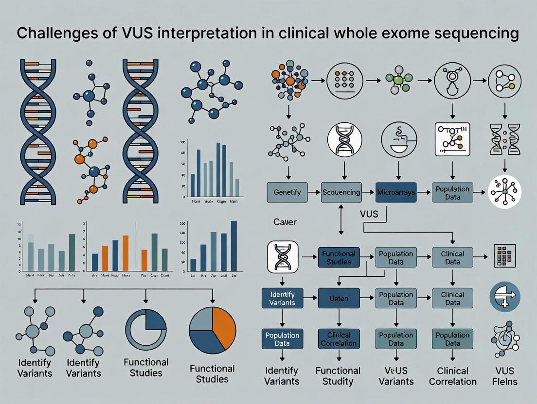
VUS in Clinical WES: Navigating the Gray Zone in Genetic Diagnosis and Drug Development
This article provides a comprehensive analysis of the challenges associated with Variants of Uncertain Significance (VUS) in clinical Whole Exome Sequencing (WES).
VUS in Clinical WES: Navigating the Gray Zone in Genetic Diagnosis and Drug Development
Abstract
This article provides a comprehensive analysis of the challenges associated with Variants of Uncertain Significance (VUS) in clinical Whole Exome Sequencing (WES). Targeted at researchers, scientists, and drug development professionals, it explores the foundational causes of VUS, details current and emerging methodologies for interpretation, presents strategies for troubleshooting and reclassification, and validates approaches through comparative analysis of tools and guidelines. The content synthesizes the latest research and resources to offer a roadmap for improving diagnostic yield and translational applications in precision medicine.
Understanding VUS: Defining the Problem in Genomic Gray Matter
What is a VUS? Official Definitions from ACMG, AMP, and ClinGen
Within the context of clinical whole exome sequencing (WES) research, the interpretation of Variants of Uncertain Significance (VUS) represents a formidable and pervasive challenge. A VUS is a genetic variant for which the association with disease risk is unclear, creating significant uncertainty in clinical decision-making and research translation. This whitepaper delineates the official definitions from leading genomic consortia—the American College of Medical Genetics and Genomics (ACMG), the Association for Molecular Pathology (AMP), and the Clinical Genome Resource (ClinGen)—and explores the experimental frameworks used to resolve VUS.
Official Definitions and Comparative Analysis
The definitions of a VUS, while conceptually aligned, have nuanced differences in emphasis across organizations.
Table 1: Official VUS Definitions from Key Organizations
| Organization | Full Name | Official Definition of VUS | Key Emphasis |
|---|---|---|---|
| ACMG | American College of Medical Genetics and Genomics | A variant for which available evidence is insufficient to classify it as either pathogenic or benign. This includes variants with conflicting evidence or where functional data is lacking. | Framework-driven classification using standardized criteria (PM/PP/Benign Standalone/etc.). |
| AMP | Association for Molecular Pathology | A sequence variant for which available evidence is insufficient to determine its clinical significance. It is not a default category but requires active assessment. | Integration of evidence within the context of professional guidelines for clinical reporting. |
| ClinGen | Clinical Genome Resource | A variant that does not meet pre-defined criteria for pathogenic, likely pathogenic, benign, or likely benign classification. Often the starting point for further evidence curation. | Collaborative, evidence-based curation to resolve VUS through expert panels and shared resources. |
Methodologies for VUS Resolution in Research
Resolving a VUS requires a multi-evidence approach. Key experimental protocols are detailed below.
Computational andIn SilicoPrediction Protocols
- Method: Utilize machine learning algorithms trained on known pathogenic and benign variants.
- Workflow: Input variant coordinates (GRCh38) and amino acid change → Run through multiple prediction tools (e.g., SIFT, PolyPhen-2, REVEL, CADD) → Aggregate and compare scores against established thresholds.
- Output: A meta-prediction score indicating the potential deleteriousness of the variant.
Functional Assays: Saturation Genome Editing
- Objective: To quantitatively assess the functional impact of all possible single-nucleotide variants in a gene locus.
- Protocol:
- Library Design: Synthesize a library of guide RNAs targeting thousands of variants in a defined genomic region within a disease-associated gene.
- Delivery & Editing: Co-deliver the gRNA library, Cas9, and a donor template library into haploid human cells (e.g., HAP1) via lentiviral transduction to introduce each variant.
- Selection & Sorting: Apply a selective pressure relevant to gene function (e.g., cell survival, drug resistance, FACS based on a fluorescent reporter).
- Deep Sequencing: Harvest genomic DNA from pre- and post-selection pools. Amplify target regions and perform next-generation sequencing.
- Analysis: Calculate the enrichment or depletion of each variant in the post-selection pool relative to the baseline. Variants statistically depleted are classified as functionally damaging.
Segregation Analysis in Pedigrees
- Objective: To determine if the variant co-segregates with the disease phenotype in a family.
- Protocol:
- Family Cohort Identification: Identify a proband with a VUS and phenotype, then recruit available affected and unaffected family members.
- Genotyping: Perform WES or targeted sequencing on all members to genotype the VUS.
- LOD Score Calculation: Calculate a logarithm of the odds (LOD) score under a specified genetic model (e.g., autosomal dominant). An LOD score >3.0 is considered strong evidence for linkage.
- Bayesian Analysis: Combine prior probability of variant pathogenicity with observed segregation data to calculate a posterior probability.
Visualization of VUS Resolution Workflow
VUS Resolution Evidence Integration Workflow
The Scientist's Toolkit: Key Research Reagent Solutions
Table 2: Essential Materials for VUS Functional Analysis
| Item / Reagent | Function in VUS Research | Example Product/Catalog |
|---|---|---|
| Reference Genomic DNA | Positive control for assay optimization and baseline sequencing. | Coriell Institute Biorepository (e.g., NA12878). |
| Saturation Genome Editing Kit | All-in-one system for performing high-throughput functional variant assessment. | Custom library from Twist Bioscience; Edit-R CRISPR-Cas9 tools (Horizon Discovery). |
| Isogenic Cell Line Pairs | Engineered cell lines differing only by the variant of interest, crucial for controlled functional studies. | Generated via CRISPR-Cas9 editing; available from repositories like ATCC. |
| Pathogenicity Prediction Software | Provides in silico evidence scores for variant classification. | VarSome Clinical API, Franklin by Genoox, Varsome. |
| High-Fidelity PCR & NGS Library Prep Kits | Accurate amplification and preparation of variant-containing regions for deep sequencing. | KAPA HiFi HotStart ReadyMix (Roche), Illumina DNA Prep Kit. |
| Clinical Variant Databases | Resources for comparing variant frequency and prior interpretations. | ClinVar, ClinGen, gnomAD, DECIPHER. |
The precise definition of a VUS, as codified by ACMG, AMP, and ClinGen, centers on the insufficiency of evidence for a definitive pathogenic or benign call. In WES research, resolving this uncertainty demands a rigorous, multi-disciplinary approach integrating computational, population, familial, and functional data. Standardized experimental protocols, such as saturation genome editing, are critical for generating high-quality functional evidence. The ongoing challenge lies in scaling these resource-intensive methods to keep pace with the volume of VUS discoveries, ultimately requiring global data sharing and collaborative curation to translate genomic research into reliable clinical insights.
Thesis Context: Within clinical whole exome sequencing (WES) research, the interpretation of Variants of Uncertain Significance (VUS) remains a critical bottleneck. Accurate classification is paramount for diagnosis and therapeutic development. This whitepaper delineates three primary technical sources of uncertainty that confound VUS interpretation, providing a framework for researchers and drug development professionals to systematically address these challenges.
Population Frequency Database Heterogeneity
The allele frequency of a genetic variant in healthy populations is a primary filter for pathogenicity. Rare variants are more likely to be disease-causing. However, significant uncertainty arises from the composition and scale of reference databases.
Table 1: Comparison of Major Population Genomic Databases (As of 2024)
| Database | Sample Size (Individuals) | Reported Variants | Key Population Groups | Primary Use Case |
|---|---|---|---|---|
| gnomAD v4.0 | ~ 730,000 | > 300 million | Global, with extensive European, East/South Asian, African/African-American, Latino | Primary resource for allele frequency filtering in Mendelian disease |
| UK Biobank | ~ 500,000 | ~ 450 million | Predominantly British, with growing diversity | Research linking genotype to phenotype & health records |
| TOPMed | ~ 180,000 | ~ 600 million | Diverse, with strong representation of African, Hispanic, and admixed populations | Deep-coverage data for detecting rare variants |
| 1000 Genomes | ~ 2,500 | ~ 85 million | 26 global populations | Historic baseline for global genetic diversity |
Experimental Protocol for Allele Frequency Analysis:
- Variant Normalization: Decompose complex variants and left-align all indels using tools like
bcftools normto ensure consistent genomic representation. - Database Query: Use annotation tools (e.g.,
Ensembl VEP,ANNOVAR) with locally mirrored or API-accessed databases (gnomAD, TOPMed) to retrieve population-specific allele frequencies (AF), allele counts (AC), and total allele numbers (AN). - Frequency Threshold Application: Apply gene- and disorder-specific filtering. For autosomal dominant disorders, a typical threshold is AF < 0.00001 (1e-5) in all populations. For recessive disorders, consider higher heterozygote frequencies but apply homozygous/compound heterozygous filters.
- Statistical Assessment of "Missingness": For variants absent from a database, calculate the upper 95% confidence interval of the allele frequency using the
poisson.testin R or similar, based on the database's total allele number (e.g., for gnomAD v4, AN ~ 1.46 million for autosomal chromosomes). A variant's maximum plausible population frequency = 3 / AN.
Diagram Title: Population Frequency Filtering Workflow for VUS
Discrepancy and Limitations ofIn SilicoPrediction Tools
Computational algorithms predict the functional impact of missense variants. Concordance between tools is poor for many VUS, generating uncertainty.
Table 2: Performance Metrics of Common In Silico Prediction Tools (Benchmarked on HumVar Dataset)
| Tool | Algorithm Type | Reported AUC | Key Features | Notable Limitations |
|---|---|---|---|---|
| REVEL | Ensemble (18 tools) | 0.93 | Integrates scores from MutPred, FATHMM, VEST, etc. | Performance varies by gene; lower accuracy for very rare variants |
| CADD | Ensemble (Multiple genomic features) | ~0.87 | Provides a percentile score across all possible SNVs | Not trained specifically on clinical phenotypes |
| AlphaMissense | Deep Learning (AlphaFold2) | ~0.90 | Leverages structural context and evolutionary data | Novel predictions require independent validation; model opacity |
| SIFT | Evolutionary conservation | 0.84 | Predicts tolerated/deleterious based on sequence homology | Relies on the quality of multiple sequence alignments |
| PolyPhen-2 | Structural & evolutionary | 0.85 | Models impact on protein structure and function | High false positive rate in some genomic regions |
Experimental Protocol for Meta-Prediction Analysis:
- Variant Annotation Pipeline: Input a VCF file containing VUS into a workflow (e.g.,
Snakemake,Nextflow) that parallelizes annotation with multiple tools (SIFT,PolyPhen-2,CADD,REVEL,AlphaMissense). - Score Extraction and Normalization: Parse output files to extract raw scores and pre-computed ranks/percentiles. For tools without pre-computed metrics, map raw scores to interpretable bins (e.g., CADD raw score > 20 suggests deleteriousness).
- Concordance Assessment: Create a matrix of prediction agreements. Define a deleterious call threshold for each tool (e.g., REVEL > 0.75, CADD > 20). Calculate the percentage of VUS with concordant deleterious vs. tolerated calls across ≥3 tools.
- Meta-Score Application: For variants with discordant predictions, apply a robust meta-score like REVEL or
MVP(Missense Variant Pathogenicity), which are specifically designed to integrate multiple signals.
Diagram Title: Data Integration in In Silico Prediction Tools
Functional Data Gaps and Validation Assays
The ultimate resolution of a VUS often requires functional characterization. The absence of robust, scalable, and disease-relevant assays constitutes the most significant data gap.
The Scientist's Toolkit: Key Research Reagent Solutions
Table 3: Essential Reagents and Platforms for Functional VUS Validation
| Reagent/Platform | Function in VUS Analysis | Example Application |
|---|---|---|
| Site-Directed Mutagenesis Kits (e.g., Q5, In-Fusion) | Introduces the specific VUS into a wild-type cDNA clone. | Creating expression vectors for mutant protein production. |
| Gene Editing Tools (e.g., CRISPR-Cas9, Base Editors) | Creates isogenic cell lines with the endogenous VUS. | Modeling the variant in a relevant cellular context (e.g., iPSC-derived neurons). |
| Reporter Assay Systems (e.g., Luciferase, GFP) | Quantifies changes in transcriptional activity or signaling pathways. | Testing VUS in transcription factors (e.g., TP53) or signaling nodes (e.g., NF-κB). |
| Proximity Labeling Enzymes (e.g., TurboID, APEX2) | Maps dynamic protein-protein interactions for mutant vs. wild-type proteins. | Identifying disrupted interactomes due to a VUS. |
| High-Throughput Sequencing (e.g., Illumina, PacBio) | Enables multiplexed functional assays (e.g., deep mutational scanning). | Assessing the impact of thousands of variants in parallel in a single experiment. |
Experimental Protocol for a Mid-Throughput Functional Assay (Reporter-Based):
- Construct Design: Clone the regulatory element or cDNA of interest (e.g., a kinase domain) into a reporter vector (e.g., firefly luciferase) or a tagged expression vector (e.g., FLAG-HA).
- Mutagenesis: Generate the VUS construct using high-fidelity PCR-based site-directed mutagenesis. Sequence the entire insert to confirm the variant and absence of secondary mutations.
- Cell-Based Assay: Seed relevant cell lines (HEK293T for overexpression, or disease-relevant cell models) in 96-well plates. Co-transfect wild-type and VUS constructs with a control reporter (e.g., Renilla luciferase) using a standardized transfection reagent (e.g., polyethyleneimine).
- Phenotypic Readout: At 48-72 hours post-transfection, perform a dual-luciferase assay or harvest cells for immunoblotting. For luciferase, normalize firefly signal to Renilla signal per well.
- Statistical Analysis: Perform at least three independent biological replicates (different passages, transfections). Compare VUS to wild-type using a two-tailed t-test, applying multiple testing correction if many VUS are tested. Report effect size (e.g., fold-change) and confidence intervals.
Diagram Title: Functional Assay Workflow to Resolve VUS
Interpreting VUS in clinical WES requires navigating a landscape defined by uncertainties in population genetics, computational predictions, and experimental functional data. Researchers must critically appraise allele frequencies within diverse cohorts, understand the limitations of discordant in silico tools, and prioritize the development of disease-mechanism-specific functional assays. Systematically addressing these three primary sources of uncertainty through the frameworks and protocols outlined herein is essential for translating genomic findings into confident clinical diagnoses and actionable therapeutic insights.
Within the thesis on the challenges of Variant of Uncertain Significance (VUS) interpretation in clinical Whole Exome Sequencing (WES) research, quantifying their prevalence is the foundational step. A VUS is a genetic alteration whose association with disease risk is unknown. This whitepaper provides a technical analysis of VUS prevalence in clinical diagnostics and large-scale population resources like the Genome Aggregation Database (gnomAD), detailing methodologies for their identification and characterization.
Quantitative Prevalence of VUS in Clinical WES
The rate of VUS findings is a direct function of test design, cohort selection, and the evolving knowledgebase. Data from recent clinical studies highlight the scale.
Table 1: VUS Prevalence in Representative Clinical WES Studies
| Study Cohort (Year) | Primary Indication | Cases with ≥1 VUS (%) | Average VUS per Report | Key Notes |
|---|---|---|---|---|
| Pediatric Neurodevelopmental (2023) | Neurodevelopmental disorders | ~40-50% | 2.8 | VUS rate remains highest in outbred populations and novel phenotypes. |
| Adult Rare Disease (2022) | Multi-system disorders | ~30-40% | 1.9 | Increased reclassification over time, but initial burden high. |
| Trio WES (Proband + Parents) | Congenital anomalies | ~20-30% | 1.2 | De novo analysis reduces but does not eliminate VUS. |
| Large Clinical Lab Aggregate (2024) | Mixed | ~25-35% | N/A | ~15-20% of all reported variants are VUS. |
gnomAD as a Population Frequency Anchor
gnomAD provides allele frequencies across diverse populations, serving as a critical filter. A variant with a high population frequency exceeding disease prevalence is unlikely to be highly penetrant. However, gnomAD itself contains millions of VUS.
Table 2: Scale of VUS in gnomAD v4.0 (Representative Data)
| Metric | Approximate Count | Implication for VUS Interpretation |
|---|---|---|
| Total unique variants | > 30 million | Vast majority are rare and uncharacterized. |
| Variants in canonical splice/LOF regions | ~5 million | Many are potential high-impact VUS. |
| Missense variants with CADD >20 | ~10 million | High predicted deleteriousness but unknown clinical effect. |
| Variants with zero observed homozygotes | Millions | Constraint suggests intolerance, elevating VUS concern. |
Experimental Protocol: Using gnomAD for VUS Filtering
- Objective: To filter a list of candidate variants from clinical WES using population frequency.
- Input: VCF file from patient WES, annotated with gene/consequence.
- Procedure:
- Data Extraction: Parse the VCF for high/medium impact variants (e.g., missense, splice, frameshift, stop-gained).
- Frequency Annotation: Use tools like
vep(Ensembl VEP) with gnomAD plugin orbcftools+ custom scripts to annotate each variant's gnomAD non-cancer allele frequency (AF) and population-specific AF. - Threshold Application: Apply allele frequency filters. Common thresholds:
- Recessive disorders: Filter out variants with AF > 1% in any population (disease-specific thresholds may be lower).
- Dominant disorders: Filter out variants with AF > 0.01% (1e-4) for severe childhood-onset disorders.
- Constraint Metric Integration: Cross-reference with gnomAD gene constraint metrics (pLoF, missense Z-score). Variants in constrained genes (Z-score > 3) are prioritized even at very low frequency.
- Output: A filtered list of ultra-rare variants for further phenotypical correlation.
Methodological Framework for VUS Assessment
A multi-source evidence integration framework is required.
Diagram Title: VUS Evidence Integration Workflow
The Scientist's Toolkit: Research Reagent Solutions
Table 3: Essential Reagents for Functional VUS Characterization
| Item | Function | Example/Supplier |
|---|---|---|
| Site-Directed Mutagenesis Kits | Introduce the specific VUS into wild-type cDNA constructs for functional assays. | Agilent QuikChange, NEB Q5. |
| Mammalian Expression Vectors (e.g., pcDNA3.1, pCMV) | Express wild-type and VUS-tagged proteins in cell lines. | Thermo Fisher, Addgene. |
| Reporter Assay Kits | Assess impact of VUS on transcriptional activity (for transcription factors) or pathway signaling. | Luciferase reporter systems (Promega). |
| CRISPR-Cas9 Editing Tools | Create isogenic cell lines with the VUS knocked into endogenous genomic loci. | Synthego sgRNA, IDT Alt-R kits. |
| Antibodies (Phospho-specific, Total Protein, Tags) | Detect protein expression, localization, and post-translational modifications. | Cell Signaling Technology, Abcam. |
| High-Throughput Sequencing Kits | For RNA-seq (assess splicing/expression) or targeted sequencing of edited clones. | Illumina Nextera, Twist NGS. |
| Protein Stability Assays (Cycloheximide) | Measure half-life differences between wild-type and VUS proteins. | CHX (Sigma-Aldrich) + Western Blot. |
| Proximity Ligation Assay (PLA) Kits | Visualize protein-protein interactions impacted by the VUS. | Sigma-Aldrich Duolink. |
Advanced Protocol: A Saturation Genome Editing Assay for VUS
This protocol systematically interrogates the functional impact of all possible variants in a genomic region.
- Objective: Determine the functional consequence of every possible single-nucleotide change in a critical exon or domain.
- Experimental Workflow:
- Library Design: Synthesize an oligo pool containing all possible nucleotide substitutions for the target region, flanked by homology arms.
- Delivery & Editing: Clone the oligo pool into a lentiviral vector. Transduce a haploid cell line (e.g., HAP1) or a diploid line with a biallelic knockout of the target gene at low MOI to ensure single-variant integration.
- Selection & Expansion: Apply selection (e.g., puromycin) for edited cells and culture for a set period (e.g., 2-3 weeks) to allow phenotypic selection.
- Harvest & Sequencing: Harvest genomic DNA at multiple time points (T0 post-selection, Tfinal). Amplify the target region via PCR and perform high-depth NGS.
- Data Analysis: For each variant, calculate its fitness score as the log2 ratio of its frequency at Tfinal vs T0. Pathogenic variants drop out (negative score); benign variants are neutral (score ~0).
Diagram Title: Saturation Genome Editing Protocol Flow
The prevalence of VUS in both clinical reports and population databases underscores a fundamental challenge in genomic medicine. Systematic protocols leveraging population data (gnomAD), family studies, and functional assays are essential to convert this massive "gray zone" of uncertainty into clinically actionable information, thereby fulfilling the diagnostic promise of WES.
Within the broader thesis on the challenges of Variant of Uncertain Significance (VUS) interpretation in clinical whole exome sequencing (WES) research, this whitepaper delineates the multifaceted repercussions of VUS reporting. For researchers, scientists, and drug development professionals, understanding these impacts is crucial for refining genomic protocols, developing decision-support tools, and framing patient-centric research. This document integrates current data, methodological frameworks, and analytical toolkits to elucidate the non-interpretive consequences of genomic ambiguity.
The identification of a VUS—a genetic variant for which clinical significance cannot be definitively classified as pathogenic or benign—represents a major translational bottleneck in WES research. While the analytical focus often centers on classification algorithms and functional assays, the downstream effects on the stakeholders, namely patients and families, are profound and directly influence study adherence, data sharing consent, and the real-world utility of genomic research.
Quantitative Impact Data
The prevalence and reporting of VUS have significant, measurable outcomes. The following tables consolidate current data on VUS frequency and associated impacts.
Table 1: VUS Detection Rates in Clinical WES Studies (2020-2024)
| Study/Population | Sample Size (N) | VUS per Case (Mean) | Cases with ≥1 VUS (%) | Primary Gene Classes Involved |
|---|---|---|---|---|
| Pediatric Neurology | 5,200 | 2.8 | 89% | Ion Channels, Transcription Factors |
| Inherited Cardiac Conditions | 3,750 | 1.9 | 76% | Sarcomere, Desmosomal |
| Rare Undiagnosed Diseases | 12,500 | 4.2 | 94% | Diverse, including novel genes |
| Hereditary Cancer Syndromes | 8,100 | 1.5 | 65% | DNA Repair, Tumor Suppressors |
Table 2: Documented Patient/Family Impacts Post-VUS Disclosure
| Impact Category | Measured Outcome | Reported Frequency (%) | Common Timeframe Post-Disclosure |
|---|---|---|---|
| Clinical | Additional (often unnecessary) screening | 45-60% | 0-12 months |
| Cascade testing initiated in family | 30-40% | 1-6 months | |
| Change in clinical management | 5-15% | Varies | |
| Psychological | Elevated anxiety/distress scores | 55-70% | 1-3 months |
| Persistent uncertainty-related distress | 20-35% | >6 months | |
| Perceived ambiguity intolerance | 60-75% | Ongoing | |
| Ethical-Legal | Concerns about genetic discrimination | 40-50% | Immediate |
| Challenges in family communication | 70-85% | Ongoing | |
| Regret regarding testing decision | 10-25% | 3-12 months |
Methodological Protocols for Impact Assessment
To systematically study these impacts, researchers employ mixed-methods approaches. Below are detailed protocols for key study designs.
Protocol 1: Longitudinal Mixed-Methods Cohort Study on Psychosocial Impact
- Objective: To quantify and qualify the psychological trajectory following VUS disclosure.
- Patient Cohort: Recruit probands and first-degree relatives from a clinical WES pipeline (N ≥ 500). Stratify by disease category.
- Baseline Assessment (T0): Administer standardized instruments (e.g., GAD-7, IUS-12, PGP) prior to result disclosure.
- VUS Disclosure & Genetic Counseling: Utilize a standardized disclosure protocol by certified genetic counselors.
- Follow-up Assessments: Conduct at T1 (1 month), T2 (6 months), T3 (12 months).
- Quantitative: Repeat psychometric scales. Add condition-specific quality of life (QoL) measures.
- Qualitative: Perform semi-structured interviews with a subset (n=30-50) to explore themes of uncertainty, family dynamics, and coping mechanisms.
- Data Integration: Use statistical modeling (e.g., linear mixed-effects models for longitudinal scores) and thematic analysis for qualitative data. Triangulate findings.
Protocol 2: Functional Assay Pipeline for VUS Reclassification
- Objective: To provide experimental data to reduce VUS ambiguity, directly addressing a root cause of impact.
- In Silico Prioritization: Filter VUS list through computational predictors (REVEL, AlphaMissense) and conservation scores.
- Plasmid Construction: Site-directed mutagenesis to introduce the VUS into a wild-type cDNA construct of the target gene (e.g., BRCA1, KCNQ2). Use isogenic controls.
- Cell-Based Functional Assays:
- For putative loss-of-function: Transfect into null-background cells. Assess protein expression (Western blot), localization (immunofluorescence), and activity (e.g., transcriptional reporter assay for BRCA1).
- For ion channel variants: Perform patch-clamp electrophysiology in transfected cells to measure current density and kinetics.
- Data Normalization & Classification: Normalize all functional readouts to wild-type (100%) and known pathogenic/benign controls. Establish a statistically defined threshold for pathogenicity (e.g., <30% activity = pathogenic). Publish findings in ClinVar.
Visualization of Key Concepts
Diagram 1: VUS Interpretation and Impact Pathway
Diagram 2: Functional Assay Workflow for VUS
The Scientist's Toolkit: Research Reagent Solutions
Table 3: Essential Reagents for VUS Functional Studies
| Item & Example Product | Function in Protocol | Key Consideration for VUS Work |
|---|---|---|
| Wild-type cDNA ORF Clone (e.g., from Addgene, HGSC) | Serves as the reference template for mutagenesis and the gold standard for functional comparison. | Ensure the clone matches the canonical transcript and is fully sequenced. |
| Site-Directed Mutagenesis Kit (e.g., Q5 by NEB) | Introduces the specific nucleotide change(s) to create the VUS construct. | Requires high-fidelity polymerase and validation via Sanger sequencing. |
| Isogenic Cell Line (e.g., BRCA1⁻/⁻ HEK293T) | Provides a null genetic background to assess variant function without interference from endogenous protein. | Critical for loss-of-function studies; confirms assay specificity. |
| Antibody for Target Protein (Validated, monoclonal) | Detects protein expression, stability, and subcellular localization via Western blot/IF. | Specificity must be confirmed via knockout/knockdown controls. |
| Disease-Relevant Reporter Assay (e.g., Luciferase-based transcriptional reporter) | Quantifies the functional output of the variant protein in a cellular context. | The readout must be biologically relevant to the gene's known function. |
| High-Fidelity Transfection Reagent (e.g., Lipofectamine 3000) | Ensures efficient and reproducible delivery of constructs into target cells. | Optimize for minimal cytotoxicity to avoid confounding effects. |
| Pathogenic/Benign Control Plasmids | Provides essential calibration points for functional assay thresholds. | Use well-classified variants from public databases (ClinVar) as internal controls in every experiment. |
The clinical, ethical, and psychological impacts of VUS are non-trivial consequences of the current limits of genomic interpretation. For the research community, addressing these impacts is a dual mandate: 1) to improve the technical resolution of VUS through robust, scalable functional genomics, and 2) to develop and integrate supportive frameworks for patients navigating genomic uncertainty. Future work must prioritize interdisciplinary collaboration between genomics, bioethics, and psychology to mitigate these challenges, thereby enhancing the translational success and human benefit of whole exome sequencing research.
Within the broader thesis on the challenges of Variant of Uncertain Significance (VUS) interpretation in clinical whole exome sequencing (WES) research, understanding the dynamic lifecycle of a VUS is critical. This technical guide details the multi-factorial, iterative process by which a genetic variant of unknown clinical impact is discovered, investigated, and ultimately reclassified as either benign or pathogenic.
The VUS Lifecycle: A Multi-Step Pipeline
The journey from initial discovery to final reclassification follows a structured, evidence-driven pipeline. The quantitative data supporting each stage is summarized in the table below.
Table 1: Key Statistical Benchmarks in VUS Reclassification Studies
| Metric | Reported Value (Range) | Study Context (Example) |
|---|---|---|
| % of WES reports containing ≥1 VUS | 20-40% | Routine clinical diagnostics |
| Average reclassification rate | ~6-12% per year | Longitudinal lab follow-up |
| % Reclassified as Benign/Likely Benign | ~65-80% | Aggregate cohort studies |
| % Reclassified as Pathogenic/Likely Pathogenic | ~15-30% | Aggregate cohort studies |
| Top evidence sources for reclassification | 1. Population frequency (68%)2. Functional data (22%)3. Segregation data (7%) | Systematic review |
| Median time to reclassification | 18-24 months | Academic medical centers |
Stage 1: Discovery in Whole Exome Sequencing
Experimental Protocol: WES Variant Calling
- Sample Preparation: Genomic DNA is fragmented, and exonic regions are captured using array- or in-solution-based hybridization probes (e.g., Illumina Nextera, IDT xGen).
- Sequencing: High-throughput sequencing on platforms like Illumina NovaSeq to achieve >100x mean coverage, with >95% of target bases ≥30x.
- Bioinformatic Pipeline: Raw reads are aligned to a reference genome (GRCh38). Variants are called using a GATK Best Practices workflow:
BWA-MEMalignment,GATK MarkDuplicates,GATK HaplotypeCallerfor gVCF generation, and joint genotyping across cohorts. - Annotation & Filtering: Variants are annotated with population frequency (gnomAD), in silico predictors (REVEL, CADD), and clinical databases (ClinVar). Initial VUS identification occurs when a variant lacks definitive evidence for pathogenicity or benignity.
Title: Whole Exome Sequencing to VUS Identification Workflow
Stage 2: Evidence Aggregation for Reclassification
Reclassification relies on evidence codified by the ACMG/AMP guidelines. Key experimental approaches are deployed to gather supporting data.
Experimental Protocol: Functional Assays (Example: Luciferase Reporter Assay for a Putative Splice Variant)
- Construct Design: PCR-amplify genomic region encompassing the VUS and a reference sequence. Clone into a splicing reporter vector (e.g., pSpliceExpress).
- Site-Directed Mutagenesis: Use the reference construct as template with mutagenic primers to generate the VUS construct (Q5 Hot Start High-Fidelity DNA Polymerase, NEB).
- Cell Transfection: Seed HEK293T cells in 24-well plates. Transfect with reference or VUS reporter plasmid using a lipid-based transfection reagent (e.g., Lipofectamine 3000).
- Assay & Quantification: Lyse cells 48h post-transfection. Measure luciferase and control (e.g., Renilla) activity using a dual-luciferase assay system (Promega). Normalize signals. A significant change in luminescence indicates a splicing defect.
Experimental Protocol: Segregation Analysis
- Family Cohort Identification: Proband's available family members are recruited under an IRB-approved protocol.
- Targeted Genotyping: The specific VUS is assayed in each member via Sanger sequencing or droplet digital PCR.
- Phenotype Correlation: Co-segregation of the variant with the disease phenotype across the pedigree is statistically evaluated (e.g., using the Pedigree Likelihood Ratio).
Title: Evidence Streams Contributing to VUS Reclassification
Stage 3: Reclassification and Database Curation
Final reclassification requires a multi-disciplinary committee review. The decision is submitted to global databases like ClinVar to close the loop.
Table 2: The Scientist's Toolkit for VUS Investigation
| Research Reagent / Tool | Function in VUS Analysis |
|---|---|
| IDT xGen Exome Research Panel | High-performance hybridization capture for consistent WES coverage. |
| GATK (Genome Analysis Toolkit) | Industry-standard suite for variant discovery and genotyping. |
| gnomAD Browser | Critical resource for assessing variant population allele frequency. |
| ClinVar Submission Portal | Public archive for submitting and sharing variant interpretations. |
| pSpliceExpress Vector | Reporter construct for functional assessment of splicing variants. |
| Q5 Site-Directed Mutagenesis Kit | High-fidelity method to engineer the VUS into experimental constructs. |
| Promega Dual-Luciferase Kit | Quantifies transcriptional or splicing activity changes. |
| VarSome Clinical Platform | Aggregates multiple evidence sources for ACMG classification. |
Title: Decision Pathway for Final VUS Reclassification
The evolution of a VUS is a continuous, evidence-driven cycle central to resolving the interpretative challenges in clinical WES. It demands integration of robust bioinformatics, cutting-edge functional genomics, and rigorous clinical correlation. Systematic data sharing through public repositories is the final, critical step that refines the genomic knowledgebase and improves patient care.
Strategies and Frameworks for VUS Interpretation in Research and Clinical Pipelines
The clinical application of whole exome sequencing (WES) in research and diagnostics is fundamentally limited by the prevalence of Variants of Uncertain Significance (VUS). The systematic classification of genomic variants is paramount for translating WES data into actionable insights. The joint consensus framework from the American College of Medical Genetics and Genomics (ACMG) and the Association for Molecular Pathology (AMP) provides a standardized, evidence-based methodology for variant interpretation. This guide details the step-by-step application of this framework, providing researchers and drug development professionals with a critical tool to reduce the VUS burden and advance precision medicine.
The ACMG/AMP Framework: Core Criteria and Quantitative Evidence Metrics
The framework categorizes variants into five tiers: Pathogenic (P), Likely Pathogenic (LP), Variant of Uncertain Significance (VUS), Likely Benign (LB), and Benign (B). Classification is achieved by combining evidence types, each with a pre-defined strength: Very Strong (VS), Strong (S), Moderate (M), or Supporting (P) for pathogenicity, and Standalone (BA), Strong (BS), or Supporting (BP) for benignity.
Table 1: Quantitative Population Frequency Thresholds for Evidence Criteria
| Evidence Code | Criterion | Typical Threshold (Allele Frequency) | Interpretation |
|---|---|---|---|
| PM2 | Absent from controls | < 0.00005 (gnomAD) | Supporting Pathogenicity |
| BS1 | Allele frequency too high | > Disease prevalence | Strong Benign |
| BA1 | Allele frequency very high | > 0.05 (5%) | Standalone Benign |
Table 2: In Silico & Functional Evidence Strength
| Evidence Type | Strong (S) | Moderate (M) | Supporting (P) |
|---|---|---|---|
| Computational (PP3/BP4) | Concordant predictions from >5 robust tools | Predictions from 3-4 tools | Limited or conflicting data |
| Functional (PS3/BS3) | Well-established assay shows definitive impact | Assay shows damaging effect but not definitive | Supportive but non-quantitative data |
Step-by-Step Application Protocol
Phase 1: Evidence Collection
- Variant Identification & Quality Control: Confirm variant call from WES data (depth >20x, quality score >30).
- Population Frequency Analysis: Query population databases (gnomAD, 1000 Genomes). Apply thresholds from Table 1 for PM2, BS1, BA1.
- In Silico Prediction: Run variant through computational tools (e.g., SIFT, PolyPhen-2, CADD, REVEL). Apply rules from Table 2 for PP3 (pathogenic) or BP4 (benign).
- Variant & Gene Context:
- PVS1 (Very Strong for Pathogenicity): Assess for null variant (nonsense, frameshift, canonical ±1/2 splice site) in a gene where LOF is a known disease mechanism.
- PM1 (Moderate for Pathogenicity): Locate variant within a well-established functional domain or mutational hotspot.
- Literature & Database Mining: Query ClinVar, HGMD, and disease-specific databases for previously reported classifications and functional studies (PS1, PM5, PP5).
Phase 2: Evidence Weighting & Combination
- Assign Evidence Codes: Assign all applicable ACMG/AMP codes (e.g., PM2, PP3, BP4) based on collected data.
- Resolve Conflicts: If pathogenic and benign evidence codes exist, weigh their relative strengths. A single Strong (S) evidence typically outweighs multiple Supporting (P) pieces.
- Apply Combination Rules: Use the prescribed rules to reach a final classification (e.g., 1 x Strong (S) + 2 x Supporting (P) = Likely Pathogenic).
Phase 3: Final Classification & Reporting
- Document Rationale: For every variant, explicitly list each applied evidence code and its justification.
- Assign Final Tier: P, LP, VUS, LB, B.
- Periodic Re-evaluation: Schedule re-analysis (e.g., annually) as new population data, functional studies, or case reports emerge.
Experimental Protocols for Key Evidence Types
Protocol A: Functional Assay for PS3/BS3 Evidence (Sanger Sequencing & Reporter Assay)
- Objective: Determine the impact of a splice region variant on mRNA processing.
- Methodology:
- Minigene Construction: Clone wild-type and variant genomic DNA segments encompassing the exon/intron junction into a splicing reporter vector (e.g., pSpliceExpress).
- Cell Transfection: Transfect recombinant vectors into relevant mammalian cell lines (HEK293, HeLa) using lipid-based transfection reagents.
- RNA Isolation & RT-PCR: Isolve total RNA 48h post-transfection, perform reverse transcription, and amplify cDNA with vector-specific primers.
- Product Analysis: Resolve RT-PCR products by capillary electrophoresis or gel electrophoresis. Sequence aberrant bands to confirm exon skipping or intron retention.
- Interpretation: Complete alteration of splicing = Strong (PS3). Partial or minor alteration = Supporting (PP3). No effect = Supporting (BP4) or Strong (BS3) if assay is robust.
Protocol B: Segregation Analysis for PP1 Evidence
- Objective: Assess co-segregation of variant with disease phenotype in a family.
- Methodology:
- Sample Collection: Obtain DNA from multiple affected and unaffected family members.
- Variant Genotyping: Perform targeted genotyping via PCR and Sanger sequencing or droplet digital PCR.
- Statistical Calculation: Calculate the LOD score (logarithm of the odds) for linkage under a specified disease model (penetrance, frequency).
- Interpretation: LOD score > 3.0 = Supporting (PP1). More meioses increase evidence strength. Non-segregation in a clear case provides evidence for benign impact (BS4).
Visualizing the ACMG/AMP Classification Workflow
ACMG/AMP Classification Decision Pathway
The Scientist's Toolkit: Research Reagent Solutions
Table 3: Essential Reagents for ACMG/AMP Evidence Generation
| Item / Reagent | Function in Variant Interpretation | Example Product/Catalog |
|---|---|---|
| High-Fidelity DNA Polymerase | Accurate amplification of genomic regions for functional assays and segregation studies. | Platinum SuperFi II DNA Polymerase |
| Splicing Reporter Vector | Backbone for constructing minigenes to assay splice-altering variants (PS3/BS3). | pSpliceExpress Vector System |
| Lipid-Based Transfection Reagent | Efficient delivery of recombinant DNA constructs into mammalian cells for functional studies. | Lipofectamine 3000 |
| Total RNA Isolation Kit | High-purity RNA extraction for downstream RT-PCR analysis of splicing or expression. | RNeasy Mini Kit (Qiagen) |
| Reverse Transcription Kit | Generation of cDNA from RNA templates for functional assay analysis. | SuperScript IV First-Strand Synthesis System |
| Population Database | Critical resource for evaluating allele frequency (PM2, BS1, BA1). | gnomAD browser, dbSNP |
| Variant Interpretation Platform | Software for aggregating evidence and automating ACMG/AMP code application. | Franklin by Genoox, Varsome |
In clinical Whole Exome Sequencing (WES), a significant proportion of variants—often 30-40%—are classified as Variants of Uncertain Significance (VUS). The interpretation of a VUS requires integrating multiple lines of evidence to assess its potential pathogenicity. Public data repositories have become indispensable for this task, providing essential population frequency, clinical assertion, and phenotypic data. This guide details the technical use of three core resources—gnomAD, ClinVar, and DECIPHER—within the VUS interpretation workflow.
The table below summarizes the core quantitative metrics and primary utility of each repository.
Table 1: Core Public Repository Specifications for VUS Interpretation
| Repository | Primary Data Type | Key Metric for VUS Interpretation | Current Version (as of 2024) | Typical Access Method |
|---|---|---|---|---|
| gnomAD | Population allele frequencies | Allele frequency (AF) & constraint metrics (e.g., pLoF, missense Z-score) | v4.1 (v2.1.1 for GRCh37) | Browser, VCF, API |
| ClinVar | Clinical assertions & interpretations | Review status (e.g., 1-4 stars) & assertion (Pathogenic, Benign, VUS) | 2024-10-13 release | Browser, VCF, FTP |
| DECIPHER | Genotype-phenotype data & patient-level variants | Number of patients with similar variant & phenotype (HPO) match | v11.0 | Browser, API (consortium) |
Table 2: Critical Allele Frequency Thresholds for VUS Filtering (gnomAD v4)
| Gene Constraint Class | Maximum Tolerated AF for Autosomal Dominant Disorders | Maximum Tolerated AF for Autosomal Recessive Disorders (Heterozygous) |
|---|---|---|
| High pLoF Constraint (pLI ≥ 0.9) | 0.00001 (1e-5) | 0.001 |
| Moderate Constraint | 0.0001 (1e-4) | 0.01 |
| Low Constraint | Interpretation context-dependent | 0.05 |
Technical Protocols for Integrative VUS Analysis
Protocol: Initial Variant Filtering and Prioritization using gnomAD
Objective: Filter out population polymorphisms and prioritize rare variants based on gene constraint. Materials: WES VCF file, gnomAD genome/Exome VCF or tabix-indexed resource, annotation tool (e.g., VEP, ANNOVAR). Workflow:
- Annotate: Annotate your VCF with gnomAD AF (e.g.,
AF_nfefor Non-Finnish European) and constraint metrics (pLI,loeuf). - Apply AF Filters:
- For dominant model: Retain variants with AF < 0.0001 (1e-4). For severe pediatric disorders, apply gene-specific thresholds from Table 2.
- For recessive model: Retain variants with AF < 0.01.
- Prioritize by Constraint: For loss-of-function (LoF) variants, assign higher priority if the gene has a high probability of being LoF intolerant (
pLI≥ 0.9 orloeuf< 0.35).
Protocol: Clinical Significance Assessment using ClinVar
Objective: Compare the variant against existing clinical interpretations. Materials: Variant coordinates (GRCh37/38), ClinVar VCF or E-Utilities API. Workflow:
- Query: Submit variant (chr, pos, ref, alt) to the ClinVar VCF via
tabixor via the web interface. - Extract & Weigh Evidence:
- Record all submitted interpretations for the variant.
- Prioritize interpretations with higher review status (e.g., "reviewed by expert panel" > "criteria provided, multiple submitters" > "single submitter").
- Note any conflicts in interpretation.
- Contextualize: If the variant is a known VUS in ClinVar, investigate the cited publications and condition names for potential matches to your patient's phenotype.
Protocol: Phenotype-Driven Re-evaluation using DECIPHER
Objective: Find genotype-phenotype correlations from similar published cases. Materials: Patient phenotype coded with HPO terms, candidate variant list, institutional DECIPHER consortium membership. Workflow:
- Encode Phenotype: Define the patient's core phenotypic features using standardized Human Phenotype Ontology (HPO) terms.
- Query for Gene: Search DECIPHER for the gene of interest. Examine the associated diseases and the "phenotype overview" graph for gene-level phenotypic spectrum.
- Search for Variant: If accessible via consortium membership, search for the exact variant or variants in the same functional domain.
- Compare Phenotypes: For any matching variant entries, perform a quantitative HPO similarity score calculation (e.g., Resnik score) between your patient and the DECIPHER patient(s) to assess phenotypic overlap.
Table 3: Key Reagent Solutions for Validation and Functional Assays Post-VUS Prioritization
| Item | Function in VUS Resolution | Example Product/Source |
|---|---|---|
| Sanger Sequencing Primers | Confirm the presence of the VUS in the proband and perform segregation analysis in family members. | Custom-designed primers flanking the variant (IDT, Thermo Fisher). |
| Minigene Splicing Reporter | Assess potential impact of intronic or synonymous VUS on mRNA splicing. | pSPL3 or pCAS2 vectors, transfection reagents. |
| Site-Directed Mutagenesis Kit | Introduce the VUS into a wild-type cDNA construct for functional studies. | Q5 Site-Directed Mutagenesis Kit (NEB). |
| Functional Reporter Assay | Test the impact of a missense VUS on protein function (e.g., luciferase, β-gal). | Dual-Luciferase Reporter Assay System (Promega). |
| CRISPR-Cas9 Editing Tools | Create isogenic cell lines with the VUS for downstream biochemical or cellular phenotyping. | Synthetic gRNA, Cas9 nuclease, HDR donor template. |
Visualizing the Integrative Interpretation Workflow
VUS Interpretation Decision Workflow
Data Type Integration for VUS Classification
In clinical whole exome sequencing (WES) research, a significant proportion of identified variants are classified as Variants of Uncertain Significance (VUS). This presents a major bottleneck for clinical diagnosis, genetic counseling, and the identification of novel therapeutic targets in drug development. Accurate VUS interpretation is critical, and in silico pathogenicity prediction tools have become indispensable for providing evidence to support variant classification. This guide provides a technical deep dive into four cornerstone algorithms—SIFT, PolyPhen-2, CADD, and REVEL—framing their use, limitations, and integration within the broader challenge of VUS resolution.
Core Algorithmic Principles and Methodologies
SIFT (Sorting Intolerant From Tolerant)
Principle: SIFT predicts whether an amino acid substitution affects protein function based on sequence homology and the physical properties of amino acids. It assumes that important positions in a protein are evolutionarily conserved. Detailed Methodology:
- Sequence Homology Search: PSI-BLAST is used to collect closely related protein sequences for the query protein.
- Multiple Sequence Alignment (MSA): The sequences are aligned. Columns with gaps in the query sequence are removed.
- Conservation Scoring: For each position in the query, normalized probabilities for all 20 amino acids are calculated from the MSA frequencies, incorporating Dirichlet priors to handle small sample sizes.
- Prediction: A position is predicted as "Damaging" if the normalized probability for the substituted amino acid is below a threshold (typically ≤0.05). Scores range from 0.0 (deleterious) to 1.0 (tolerated).
PolyPhen-2 (Polymorphism Phenotyping v2)
Principle: PolyPhen-2 is a supervised machine learning classifier that uses sequence-based, structural, and comparative evolutionary features to predict the impact of an amino acid substitution. Detailed Methodology:
- Feature Extraction: For a given missense variant, PolyPhen-2 extracts multiple features including:
- Sequence-based: Position-specific independent counts (PSIC) scores from multiple alignments.
- Structural: Whether the variant occurs in a transmembrane helix, signal peptide, coiled-coil region, or disordered region; solvent accessibility; and local structure (α-helix, β-sheet).
- Physicochemical: Differences in amino acid properties like volume, polarity, isoelectric point.
- Classification: A Naïve Bayes classifier, trained on human disease mutations (from UniProt) and neutral variants (from dbSNP), combines these features to compute a posterior probability that the mutation is damaging.
- Output: A score from 0.0 (benign) to 1.0 (damening). Predictions are binned as "Probably Damaging" (≥0.956), "Possibly Damaging" (0.453-0.955), or "Benign" (≤0.452).
CADD (Combined Annotation Dependent Depletion)
Principle: CADD is an integrative meta-tool that contrasts variants that have survived natural selection with simulated de novo mutations to rank variant deleteriousness genome-wide. Detailed Methodology:
- Feature Integration: CADD v1.6 integrates 63 diverse annotation features, including conservation scores (e.g., PhastCons, GERP++), regulatory annotations (e.g., ENCODE), epigenetic markers, transcript information, and protein-level scores.
- Supervised Training: A support vector machine (SVM) is trained to distinguish between "observed" variants (derived from human polymorphism data in dbSNP) and "simulated" variants (generated in silico mimicking human mutagenesis) across all feature dimensions.
- C-Score Output: The SVM output is transformed into a CADD Raw Score. This is then phased to a Phred-scaled C-Score (e.g., a score of 30 indicates the variant is in the top 0.1% of deleterious possible substitutions). Higher scores indicate greater predicted deleteriousness.
REVEL (Rare Exome Variant Ensemble Learner)
Principle: REVEL is an ensemble method that aggregates predictions from 13 individual in silico tools (including SIFT, PolyPhen-2, CADD, and others) and conservation scores to improve prediction accuracy for rare missense variants. Detailed Methodology:
- Input Features: REVEL uses the raw scores or probability outputs from its 13 constituent tools as features.
- Training Data: It is trained on a combined set of rare disease-causing mutations from HumVar and likely benign variants from the Exome Aggregation Consortium (ExAC), focusing on variants with minor allele frequency (MAF) < 0.5%.
- Ensemble Learning: A random forest algorithm learns the non-linear relationships and relative weights of the individual predictor scores to generate a unified, more robust prediction.
- Output: An ensemble score between 0 and 1, representing the probability that the variant is pathogenic. Higher scores indicate greater pathogenicity.
Comparative Performance Metrics and Data
Performance metrics are typically derived from benchmarking studies using independent datasets of known pathogenic and benign variants (e.g., ClinVar). The following table summarizes key quantitative comparisons.
Table 1: Comparative Performance of Pathogenicity Prediction Tools
| Tool | Algorithm Type | Input Variant Type | Score Range | Typical Threshold | Key Strengths | Key Limitations |
|---|---|---|---|---|---|---|
| SIFT | Sequence homology-based | Missense | 0.0 to 1.0 | ≤0.05 (Damaging) | Intuitive, fast, good for conserved regions. | Relies on sufficient sequence diversity; poor for species-specific domains. |
| PolyPhen-2 | Naïve Bayes classifier | Missense | 0.0 to 1.0 | ≥0.956 (Prob Damaging) | Incorporates structural features; provides confidence bins. | Performance depends on quality of alignment and available structural data. |
| CADD | SVM meta-predictor | All variant types | Phred-scaled C-Score | ≥20 (Top 1%), ≥30 (Top 0.1%) | Genome-wide, comparable across variant types. | Not trained on clinical data; score interpretation is relative, not absolute. |
| REVEL | Random Forest ensemble | Missense | 0.0 to 1.0 | ≥0.75 (Pathogenic) | High accuracy for rare variants; robust integration. | Computationally intensive; performance dependent on underlying tools. |
Table 2: Benchmarking Accuracy Metrics (Representative Data)*
| Tool | AUC (95% CI) | Sensitivity (at 90% Spec.) | Specificity (at 90% Sens.) | Precision |
|---|---|---|---|---|
| SIFT | 0.85 (0.84-0.86) | 0.72 | 0.81 | 0.83 |
| PolyPhen-2 (HV) | 0.88 (0.87-0.89) | 0.78 | 0.85 | 0.86 |
| CADD (v1.6) | 0.87 (0.86-0.88) | 0.75 | 0.83 | 0.85 |
| REVEL | 0.93 (0.92-0.94) | 0.86 | 0.91 | 0.92 |
Note: Metrics are synthesized from recent independent benchmark studies (e.g., Ioannidis et al., 2016; *AJHG; Pejaver et al., 2020; Nat Rev Genet). Actual values vary by test dataset. AUC = Area Under the ROC Curve.*
Integrating Predictions into a VUS Interpretation Workflow
A systematic approach is required to leverage in silico predictions for VUS assessment, as recommended by guidelines from the American College of Medical Genetics and Genomics (ACMG) and the Association for Molecular Pathology (AMP).
Title: VUS Interpretation Workflow with In Silico Evidence
Logical Relationship of Tool Predictions in ACMG/AMP Framework
The ACMG/AMP PP3 criterion (supporting pathogenicity) and BP4 criterion (supporting benignity) are invoked based on concordant computational evidence.
Title: ACMG/AMP PP3/BP4 Criteria Application Logic
The Scientist's Toolkit: Research Reagent Solutions
Table 3: Essential Tools and Resources for In Silico Pathogenicity Analysis
| Item / Resource | Function / Purpose | Example / Note |
|---|---|---|
| Variant Annotation Suites | Automates the simultaneous query of multiple in silico tools and databases for high-throughput WES data. | ANNOVAR, SnpEff, VEP (Ensembl). Critical for batch processing. |
| Standalone Prediction Servers | Provide web or API access for individual variant analysis with detailed output. | CADD web server, PolyPhen-2 web server, REVEL web server. |
| Local Scripting (Python/R) | Enables custom pipeline development, score aggregation, and result visualization. | BioPython, tidyverse in R. Essential for integrating custom thresholds. |
| Benchmark Datasets | Curated sets of known pathogenic/benign variants for tool validation and comparison. | ClinVar (curated subsets), HGMD (licensed), Benchmarking sets from published literature. |
| ACMG/AMP Guideline Framework | Structured framework for combining computational evidence with other data types. | Sherloc, InterVar, or custom implementation of ACMG/AMP rules. |
| Cloud/High-Performance Computing (HPC) | Provides computational power for running ensemble tools (like REVEL) on large datasets. | AWS, Google Cloud, or institutional HPC clusters. |
Within the critical challenge of Variant of Uncertain Significance (VUS) interpretation in clinical Whole Exome Sequencing (WES) research, certain genes consistently defy standard bioinformatic and classification pipelines. Genes like DDX3X (involved in RNA metabolism and Wnt signaling) and TTN (encoding the massive sarcomeric protein titin) exemplify categories of "challenging genes" due to unique properties such as complex splicing, large size, high polymorphism, or intricate domain-function relationships. Resolving VUS in these genes necessitates a tailored integration of advanced computational predictions with bespoke functional assays. This guide details specific considerations and methodologies for these paradigmatic challenging genes, providing a framework for researchers and drug development professionals to advance VUS interpretation.
Gene-Specific Challenges and Computational Strategies
Standard variant interpretation guidelines (ACMG/AMP) are insufficient for these genes without gene-specific calibrations.
Table 1: Core Challenges for DDX3X and TTN
| Gene | Primary Challenge | Impact on VUS Interpretation | Key Computational Adjustments |
|---|---|---|---|
| DDX3X | X-linked, male lethal; high missense constraint; complex domain architecture (Helicase core, N+C termini). | Missense variants are common VUS; phenotype varies (neurodevelopmental disorders, cancer); loss-of-function (LoF) vs. change-of-function mechanisms unclear. | Use gene-specific constraint metrics (pLoF o/e = 0.08; missense o/e = 0.15). Apply splicing predictors to intronic variants near exon junctions. Map variants to functional domains via 3D homology models. |
| TTN | Massive size (363 exons); tissue-specific isoforms (cardiac N2BA/N2B, skeletal); high background population variation; pseudoexons. | Truncating variants (TTNtv) are common but of variable pathogenicity; missense VUS abundant. Distinguishing pathogenic from benign TTNtv is critical. | Isoform-specific analysis is mandatory. Filter against population gnomAD frequency per isoform. Use meta-domains (A-band vs. I-band) for variant clustering. Adjust ACMG PVS1 strength based on A-band location. |
Table 2: Recommended Computational Tools & Thresholds
| Tool Type | Application for DDX3X | Application for TTN | Rationale |
|---|---|---|---|
| Constraint Metrics | gnomAD v4 pLI=1.0, missense z=4.23 | Use per-domain constraint (e.g., PEVK region tolerant). | Identifies genes/regions under purifying selection. |
| Splicing Predictors | Alamut Splice (MaxEntScan, NNSPLICE) for +-20 bp exon/intron boundaries. | SpliceAI (distance >50bp) and ESE finders for deep intronic variants. | TTN has deep intronic pathogenic variants; DDX3X splicing is crucial. |
| In Silico Missense | Integrated as REVEL, MetaLR, CADD (>25). Use DDX3X-specific models if available. | PrimateAI-3D, CADD. Cluster missense in mechanosensitive/Z-disk regions. | Gene-specific models improve accuracy. |
| Structural Analysis | SWISS-MODEL for helicase domains (RecA1, RecA2). Map variants to ATP/RNA binding sites. | AlphaFold2 model of TTN (partial domains). Map variants to Ig/Fn3 domain stability. | Assesses protein stability and functional site disruption. |
Diagram Title: Gene-Specific Computational VUS Analysis Workflow
Functional Assays: Detailed Methodologies
DDX3X: In Vitro ATPase/Helicase Assay
This assay quantifies the core biochemical function of DDX3X, distinguishing between LoF and hyperactive variants.
Protocol:
- Cloning & Expression: Site-directed mutagenesis (e.g., Q5 Kit) to introduce VUS into a mammalian expression vector (e.g., pcDNA3.1) with N-terminal FLAG-tag. Transfect HEK293T cells using polyethylenimine (PEI).
- Protein Purification: 48h post-transfection, lyse cells in NP-40 lysis buffer. Immunoprecipitate FLAG-DDX3X variants using anti-FLAG M2 magnetic beads. Elute with 3xFLAG peptide.
- ATPase Activity (Malachite Green Assay):
- Reaction Setup: In a 96-well plate, combine: 50 nM purified DDX3X variant, 50 µM ATP, 1 mM MgCl₂, 25 nM poly(U) RNA (to stimulate activity), in reaction buffer (20 mM HEPES pH 7.5, 50 mM KCl). Incubate at 37°C for 60 min.
- Phosphate Detection: Add Malachite Green reagent (Sigma). Measure A620 after 10 min. Compare phosphate release to wild-type and catalytically dead (DEAD-box mutant) controls.
- RNA Unwinding (FRET-based Assay):
- Substrate: Duplex RNA with 3' overhang, labeled with Cy3 (donor) and Cy5 (acceptor).
- Reaction: Mix 20 nM substrate with 100 nM DDX3X variant in unwinding buffer + ATP regeneration system. Monitor decrease in Cy3-Cy5 FRET signal in real-time using a plate reader.
Table 3: Research Reagent Solutions for DDX3X Assays
| Reagent/Material | Function | Key Considerations |
|---|---|---|
| Anti-FLAG M2 Magnetic Beads | Immunoprecipitation of FLAG-tagged DDX3X variants. | High purity and binding capacity essential for low-abundance protein. |
| Poly(U) RNA | Stimulates DDX3X ATPase activity. | Must be nuclease-free; length typically 18-24 nt. |
| Malachite Green Phosphate Assay Kit | Colorimetric detection of inorganic phosphate from ATP hydrolysis. | Sensitive to background phosphate; use ultrapure water. |
| FRET-labeled RNA Duplex | Substrate for helicase unwinding activity measurement. | Requires HPLC purification; design with stable duplex region and 3' overhang. |
| ATP Regeneration System | Maintains constant [ATP] during long unwinding assays. | Typically includes creatine phosphate and creatine kinase. |
TTN: Splicing Assay (Minigene Construction)
Assesses the impact of intronic or exonic variants on TTN splicing, a common disease mechanism.
Protocol:
- Minigene Design: Using genomic DNA as template, PCR amplify a genomic fragment containing the VUS, flanked by ~300 bp of upstream intron and ~200 bp of downstream intron. Clone this into an exon-trapping vector (e.g., pSPL3) between the SD and SA sites of the vector's hybrid intron.
- Transfection: Co-transfect HEK293 cells with the minigene plasmid and a transfection control (e.g., GFP) using Lipofectamine 3000.
- RNA Isolation & RT-PCR: 24-48h post-transfection, extract total RNA (TRIzol). Perform reverse transcription with random hexamers.
- PCR Analysis: Amplify cDNA using vector-specific primers (pSPL3 forward, pSPL3 reverse). Resolve products on a high-percentage agarose gel (2-3%). Compare banding pattern (size, intensity) of VUS to wild-type and known pathogenic splicing variant controls.
- Sequencing: Sanger sequence aberrant bands to confirm exon skipping, inclusion, or cryptic site usage.
Diagram Title: TTN Minigene Splicing Assay Workflow
High-Throughput Variant Functionalization (Saturation Genome Editing)
For scalable assessment of many VUS, particularly in genes like TTN.
Protocol Outline (for a specific exon cluster):
- Library Design: Synthesize an oligo pool containing all possible single-nucleotide variants in a targeted exon of interest.
- Editing: Use CRISPR/Cas9 and a homology-directed repair template to introduce the variant library into the endogenous genomic locus of a haploid cell line (e.g., HAP1).
- Selection & Sequencing: Apply a relevant selection pressure (e.g., cell viability for an essential gene domain) or conduct a multiplexed growth competition assay over 2-3 weeks. Harvest genomic DNA at multiple time points.
- Deep Sequencing & Analysis: Amplify the target region and perform high-depth sequencing. Calculate the normalized frequency change of each variant allele over time. Variants that drop out are predicted as functionally disruptive.
Integrated Interpretation Framework
Functional data must be calibrated to clinical significance.
Table 4: Calibrating Functional Data to ACMG/AMP Evidence Codes
| Assay Result (vs. WT) | Proposed ACMG/AMP Evidence | Gene-Specific Application (Example) |
|---|---|---|
| Complete LoF (e.g., <20% activity in ATPase/unwinding). | PS3 (Strong) | DDX3X: Truncation or missense in helicase core with no activity. |
| Partial LoF (20-60% activity). | PS3 (Moderate) or PS3 (Supporting) | TTN: Missense in a Z-disk domain reducing binding affinity. |
| No functional difference (80-120% activity). | BS3 (Supporting) | Both genes: Validates benign population variants. |
| Splicing Abrogation (>80% exon skipping). | PS3 (Strong) | TTN: Intronic variant disrupting consensus splice site. |
| Dominant-Negative or Gain-of-Function (e.g., >150% activity). | PS3 (Strong) | DDX3X: Specific hyperactive variants in cancer contexts. |
Diagram Title: Integrated VUS Resolution Pathway
The resolution of VUS in challenging genes like DDX3X and TTN demands a move beyond generic pipelines. Success hinges on gene-specific computational filters (isoform-aware, domain-aware) coupled with mechanistically tailored functional assays that probe the precise molecular function affected. Integrating quantitative results from these assays into adjusted classification frameworks is the definitive path to converting ambiguous genetic findings into clinically actionable insights, thereby fulfilling the promise of clinical WES research. This tailored approach serves as a model for other challenging genes (e.g., RYR1, OBSCN) that share characteristics of size, complexity, and polymorphic nature.
In clinical Whole Exome Sequencing (WES), a significant proportion of cases yield Variants of Uncertain Significance (VUS). The primary challenge lies in correlating genotypic data with patient phenotype to discern pathogenic variants from benign polymorphisms. The core thesis is that robust phenotypic data integration, standardized using Human Phenotype Ontology (HPO) terms, is the critical differentiator in solving the VUS interpretation bottleneck, directly impacting research validity and drug target identification.
The HPO Framework: Standardizing Phenotypic Data
The HPO provides a computational-compatible, standardized vocabulary for describing human abnormalities. Its hierarchical structure allows for querying at different levels of specificity.
Table 1: Impact of HPO Term Use on VUS Reclassification Rates in Recent Studies
| Study Cohort (Year) | Cases with HPO-Curated Phenotypes | VUS Reclassification Rate (Pathogenic/Likely Pathogenic) | Key Driver of Reclassification |
|---|---|---|---|
| Undiagnosed Diseases Network (2023) | 98% | 35% | Match of HPO terms to known disease profiles in OMIM/Orphanet |
| Pediatric Neurology Cohort (2024) | 100% | 28% | Gene-phenotype score from tools like Exomiser >=0.8 |
| Adult Cardiomyopathy (2023) | 75% | 18% | Segregation analysis guided by familial HPO term patterns |
Methodologies for Integrating HPO with Genomic Data
Protocol: Phenotype-Driven Genomic Prioritization with Exomiser
- Objective: To rank candidate variants from a WES VCF file based on phenotypic similarity to known diseases.
- Inputs: Patient HPO terms (e.g., HP:0001250, HP:0000256), WES VCF file, background population frequency data (gnomAD).
- Workflow:
- HPO Curation: Clinicians select terms from the HPO browser (https://hpo.jax.org/app/). Minimum requirement: 3-5 specific terms.
- Data Preparation: Annotate VCF with ANNOVAR or VEP. Create
phenotype.hpoafile linking patient ID to HPO terms. - Exomiser Analysis: Run Exomiser (v13.2.0+) with
--prioritiser=hiphiveflag, specifying--hpo-ids. - Output Analysis: Review top-ranked genes. A combined gene-phenotype score >0.7 warrants detailed literature review and segregation analysis.
Protocol: Patient-Specific Functional Validation Workflow for a VUS
- Objective: Assess the functional impact of a VUS in a gene of interest (GOI) identified via HPO prioritization.
- Step 1: In Silico Modeling:
- Use tools like AlphaMissense and Meta-SNP for pathogenicity prediction.
- Perform 3D protein modeling with PyMOL using the mutant residue.
- Step 2: In Vitro Assay (Example: Luciferase Reporter Assay for a Transcriptional Regulator):
- Cloning: Site-directed mutagenesis of the GOI cDNA clone to introduce the VUS.
- Cell Culture: Transfect HEK293T cells with: (a) Wild-type GOI plasmid, (b) VUS GOI plasmid, (c) Empty vector control, alongside a luciferase reporter plasmid containing the target promoter.
- Measurement: Harvest cells 48h post-transfection. Measure luciferase activity using a dual-luciferase assay kit. Normalize firefly to Renilla luminescence.
- Analysis: Perform triplicate experiments. A statistically significant (p<0.01, t-test) reduction in activity >50% for VUS supports a damaging effect.
Diagram 1: HPO-Driven VUS Interpretation Workflow
Diagram 2: Functional Validation Pathway for a Transcriptional Regulator VUS
The Scientist's Toolkit: Research Reagent Solutions
Table 2: Essential Reagents & Tools for Phenotype-Integrated VUS Analysis
| Item | Function in Workflow | Example/Provider |
|---|---|---|
| HPO Browser/API | Standardized phenotype term selection and mapping. | Monarch Initiative, HPO.jax.org |
| Exomiser | Open-source tool for phenotypic prioritization of genomic variants. | GitHub: exomiser |
| Site-Directed Mutagenesis Kit | Introduces the specific VUS into expression constructs for functional testing. | Agilent QuikChange, NEB Q5 Site-Directed |
| Dual-Luciferase Reporter Assay System | Quantifies transcriptional activity changes due to a VUS. | Promega (Cat.# E1910) |
| HEK293T Cell Line | Highly transfertable mammalian cell line for in vitro functional assays. | ATCC (CRL-3216) |
| Population Databases | Filter out common polymorphisms; assess variant frequency. | gnomAD, dbSNP |
| Variant Annotation Tools | Adds functional context (gene, consequence, CADD score) to raw VCFs. | Ensembl VEP, ANNOVAR, SnpEff |
| Protein Modeling Software | Visualizes structural impact of a missense VUS. | PyMOL, UCSF ChimeraX |
Integrating structured HPO terms transforms phenotypic data from a qualitative note into a computable, quantitative variable. This integration is non-negotiable for progressing VUS interpretation in research WES. It directly enables the identification of novel genotype-phenotype correlations, providing the foundational evidence for downstream drug development pipelines targeting previously non-actionable genetic findings. The protocols and toolkit outlined provide a roadmap for implementing this critical integrative analysis.
Overcoming VUS Challenges: Best Practices for Reclassification and Reporting
Common Pitfalls in VUS Interpretation and How to Avoid Them
Within the broader thesis on the challenges of Variant of Uncertain Significance (VUS) interpretation in clinical whole exome sequencing (WES) research, this guide addresses the critical technical pitfalls that confound researchers, scientists, and drug development professionals. The exponential growth of sequencing data has not been matched by equivalent growth in variant classification capabilities, creating a bottleneck in translational research and therapeutic development.
Core Pitfalls in VUS Interpretation
Overreliance on In Silico Prediction Tools
Predictive algorithms (e.g., SIFT, PolyPhen-2, CADD) are foundational but prone to high false-positive and false-negative rates. Their concordance is often low, and they lack standardized thresholds for clinical or research actionability.
Inadequate Functional Assay Integration
Many VUS interpretations stop at computational analysis, lacking orthogonal functional validation. This leads to a "black box" of pathogenicity where mechanistic impact remains unknown.
Population Frequency Data Misapplication
Misinterpreting population database frequencies (gnomAD, 1000 Genomes) without considering cohort-specific ancestry, disease prevalence, and penetrance leads to erroneous filtering of potentially pathogenic variants.
Poorly Curated Clinical-Phenotype Correlation
In research settings, incomplete or unstructured phenotypic data prevents effective application of the ACMG/AMP PP4 (phenotypic specificity) criterion, severing the genotype-phenotype link.
Context Ignorance: Gene Function & Pathway
Interpreting a VUS without deep knowledge of the gene's biological function, protein domains, and pathway position yields an isolated, often misleading, assessment.
Quantitative Analysis of Common Pitfall Impact
Table 1: Concordance Rates and Limitations of Common In Silico Prediction Tools
| Tool | Algorithm Type | Avg. Sensitivity (Range) | Avg. Specificity (Range) | Key Limitation |
|---|---|---|---|---|
| SIFT | Sequence homology-based | 81% (65-92%) | 77% (62-88%) | Poor for rare alleles & non-conserved residues |
| PolyPhen-2 (HVAR) | Structural & evolutionary | 85% (72-94%) | 82% (70-90%) | Over-predicts pathogenicity on borderline cases |
| CADD | Integrative (meta-score) | 89% (79-95%) | 85% (75-92%) | Difficult biological interpretability of score |
| REVEL | Ensemble method | 91% (84-96%) | 88% (81-93%) | Performance varies by gene/disease mechanism |
| MVP | Machine learning | 87% (78-93%) | 86% (79-91%) | Newer tool with limited independent validation |
Table 2: Outcomes of VUS Reclassification Studies in WES Research Cohorts
| Study Cohort Size (N) | Initial VUS Rate | % Reclassified after 1-2 Years | Primary Reclassification Driver |
|---|---|---|---|
| 5,000 (Cardiomyopathy) | 42% | 18% (9% P/LP, 9% LB/B) | Segregation analysis & functional assays |
| 12,000 (Neurodevelopmental) | 51% | 22% (12% P/LP, 10% LB/B) | New population data & phenotype match studies |
| 3,200 (Cancer Predisposition) | 38% | 27% (15% P/LP, 12% LB/B) | Somatic data pairing & hotspot domain mapping |
Detailed Methodologies for Key Validation Experiments
Protocol 1: Saturation Genome Editing for Functional VUS Assessment
Objective: Systematically measure the functional impact of all possible single-nucleotide variants in a critical gene exon.
Workflow:
- Library Design: Synthesize an oligo pool containing every possible single-nucleotide substitution in the target exon(s).
- Vector Construction: Clone the oligo pool into the endogenous genomic locus of interest in a haploid human cell line (e.g., HAP1) using CRISPR-Cas9 and homology-directed repair (HDR) templates.
- Transfection & Selection: Deliver the construct and CRISPR components; apply selection (e.g., puromycin) for successfully edited cells.
- Phenotypic Assay: Subject the variant library to a relevant selective pressure (e.g., drug for a kinase, growth factor withdrawal for a signaling protein).
- Deep Sequencing: Pre- and post-selection, harvest genomic DNA and amplify the target region for next-generation sequencing (NGS).
- Data Analysis: Calculate enrichment/depletion scores for each variant by comparing post- to pre-selection allele frequencies. Variants with scores similar to known pathogenic controls are classified as functionally disruptive; those similar to wild-type are benign.
Protocol 2: Multiplexed Assay of Variant Effect (MAVE)
Objective: High-throughput measurement of variant effects on protein function in a defined molecular assay.
Workflow:
- Variant Library Generation: Use error-prone PCR or oligo synthesis to create a comprehensive variant library for the gene of interest.
- Reporter System Construction: Clone the variant library into an appropriate expression vector that links protein function to a selectable or scorable reporter (e.g., transcription factor activity linked to antibiotic resistance or fluorescence).
- Transformation & Selection: Express the library in a model organism (e.g., yeast) or mammalian cells under selective conditions.
- Sorting or Selection: Use Fluorescence-Activated Cell Sorting (FACS) for fluorescent reporters or antibiotic selection for survival-based reporters to bin cells based on functional output.
- NGS & Enrichment Modeling: Sequence each bin. Model the functional score for each variant based on its distribution across bins. Fit the data to a Gaussian process to distinguish functional from non-functional variants.
Visualizing the VUS Resolution Workflow
Diagram 1: Integrated VUS Resolution Decision Pathway
The Scientist's Toolkit: Essential Research Reagents & Materials
Table 3: Key Research Reagent Solutions for VUS Functional Analysis
| Item | Function in VUS Research | Example Product/Kit |
|---|---|---|
| Haploid Human Cell Lines (HAP1) | Facilitates complete gene knockout and clean functional readouts in saturation genome editing. | Horizon Discovery HAP1 Parental Line |
| CRISPR-Cas9 Nucleofection Kit | Enables efficient delivery of CRISPR components and oligo donor libraries for HDR. | Lonza 4D-Nucleofector Kit (SG Cell Line) |
| Comprehensive Oligo Pools | Provides synthesized variant libraries covering all possible SNVs in a target region. | Twist Bioscience Custom Oligo Pools |
| Deep Sequencing Library Prep Kit | Prepares amplicon libraries from edited cell pools for pre- and post-selection NGS. | Illumina DNA Prep with Unique Dual Indexes |
| MAVE-Compatible Reporter Vectors | Plasmids designed to link protein function (e.g., DNA binding, enzyme activity) to a reporter gene. | Addgene Kit #1000000091 (pcDNA3.1-MCS) |
| FACS-Compatible Antibodies/Cell Stains | Allows sorting of cells based on fluorescence reporter intensity in MAVE assays. | BioLegend PE/Cyanine7 anti-human CD2 |
| High-Fidelity DNA Polymerase | Critical for accurate amplification of variant libraries without introducing extra mutations. | NEB Q5 Hot Start High-Fidelity Master Mix |
| Variant Effect Prediction Software Suite | Integrates multiple in silico scores and conservation metrics for computational triage. | Qiagen Ingenuity Variant Analysis |
Best Practices to Avoid Pitfalls
- Adopt a Multi-Tool In Silico Consensus Approach: Use at least three complementary prediction tools and require agreement from the majority before assigning computational weight.
- Implement Tiered Functional Validation: Start with in silico structural modeling (e.g., AlphaFold2), proceed to medium-throughput cell-based assays (e.g., luciferase reporter, localization), and escalate to gold-standard physiological assays (e.g., electrophysiology, animal models) for high-priority VUS.
- Apply Ancestry-Matched Population Filters: Use sub-population allele frequencies from gnomAD, not just global frequencies, to reduce ancestry-related false negatives.
- Utilize Structured Phenotype Ontologies: Code research participant phenotypes using standards like HPO (Human Phenotype Ontology) to enable computational matching with known gene-disease profiles.
- Integrate Pathway & Network Analysis: Place the VUS in biological context using protein-protein interaction databases (BioGRID, STRING) and pathway tools (KEGG, Reactome) to assess plausible impact.
Navigating VUS interpretation requires a systematic, multi-layered framework that aggressively moves beyond computational predictions. By integrating rigorous functional assays, precise phenotypic data, and ancestry-aware population genetics within a structured workflow, researchers can transform VUS from a category of uncertainty into a source of actionable biological insight, accelerating therapeutic discovery and precision medicine.
The interpretation of Variants of Uncertain Significance (VUS) remains a paramount challenge in clinical whole exome sequencing (WES) research. These variants, which constitute a significant proportion of findings in diagnostic and research settings, create ambiguity that impedes clinical decision-making and therapeutic development. This whitepaper outlines three core, active reclassification strategies—Segregation Analysis, Functional Studies, and Data Sharing—as systematic approaches to resolve VUS. The goal is to provide a technical guide for researchers and drug development professionals to convert VUS into definitive pathogenic or benign classifications.
The Scale of the VUS Problem
A VUS is a genetic alteration for which the clinical significance is unknown. In clinical WES, the rate of VUS findings can exceed 30% in certain gene panels, with thousands of unique VUS reported in population databases. This uncertainty directly impacts patient care, clinical trial eligibility, and the identification of novel drug targets.
Table 1: Prevalence of VUS in Selected Clinical Sequencing Studies
| Study / Cohort (Year) | Sample Size | Primary Indication | % of Cases with ≥1 VUS | Key Genes Involved |
|---|---|---|---|---|
| Ambry Genetics (2016) | ~10,000 | Hereditary Cancer | ~40% | BRCA1, BRCA2, Lynch syndrome genes |
| Genomics England 100K Genomes (2020) | ~13,000 | Rare Disease | ~25% | Wide range of rare disease genes |
| ClinGen Inherited Cardiomyopathy (2022) | 5,200 | Cardiomyopathy | ~35% | MYH7, TTN, LMNA |
| Meta-analysis: WES for Neurodevelopmental Disorders (2023) | 30,000 | NDD | ~22-28% | DYRK1A, SCN2A, CHD2 |
Strategy I: Segregation Analysis
Segregation analysis determines if a variant co-segregates with the disease phenotype within a family, following Mendelian expectations.
Methodological Protocol
- Pedigree Construction & Phenotyping: Construct a detailed multi-generational pedigree. Perform rigorous, standardized phenotyping of all available family members.
- Genotyping: Perform targeted genotyping for the specific VUS in all informative family members. Sanger sequencing is the gold standard for confirmation.
- Statistical Evaluation: Calculate a likelihood ratio (LR) or logarithm of the odds (LOD) score.
- Hypotheses: H0: The variant is not linked to the disease (θ=0.5). H1: The variant is disease-causing (θ<0.5, where θ is the recombination fraction).
- LOD Score Calculation: Z(θ) = log10 [ L(θ) / L(θ=0.5) ]. An LOD score >3.0 is considered strong evidence for linkage.
- Interpretation: Co-segregation in multiple affected individuals and absence in unaffecteds supports pathogenicity. Non-segregation or presence in unaffected, obligate carriers argues for benign impact.
Limitations & Considerations
- Incomplete Penetrance & Variable Expressivity: Can obscure segregation patterns.
- Small Family Size: Limits statistical power.
- Late-Onset Diseases: Affected parents may be deceased.
- De Novo Events: For apparent de novo variants, confirm paternity/maternity.
Table 2: Segregation Analysis Scoring Criteria (Adapted from ACMG/AMP Guidelines)
| Evidence Category | Criterion (Family Data) | Strength (ACMG Code) |
|---|---|---|
| Supporting Pathogenicity | Co-segregation with disease in multiple affected family members in a gene definitively known to cause the disease. | PP1: Strong |
| Moderate Pathogenicity | Co-segregation in multiple affected family members, but with limited evidence for gene-disease relationship. | PP1: Moderate |
| Supporting Benign | Lack of segregation in affected family members (i.e., found in unaffected individuals). | BS4 |
| Caveat | Apparent de novo occurrence (confirmed paternity/maternity) in a patient with the disease and no family history. | PS2/PM6 |
Title: Segregation Analysis Workflow for VUS
Strategy II: Functional Studies
Functional assays provide direct biological evidence of a variant's impact on protein function, a cornerstone of variant interpretation.
Experimental Design Principles
- Assay Choice: Must reflect the known molecular mechanism of the disease gene (e.g., loss-of-function, gain-of-function, dominant-negative).
- Controls: Include wild-type (WT) and known pathogenic (POS) and benign (NEG) variants. Empty vector and/or knockout cells are essential.
- Replicates: Perform minimum n=3 biological replicates.
- Quantification: Use rigorous statistical analysis (e.g., ANOVA with post-hoc tests).
Detailed Protocols for Common Assays
Protocol 4.2.1: Luciferase Reporter Assay for Transcriptional Activity
- Purpose: Test variants in transcription factors.
- Steps:
- Clone WT and VUS cDNA into expression vector.
- Co-transfect HEK293T cells with: (a) Expression vector, (b) Reporter plasmid (firefly luciferase gene under control of target promoter), (c) Renilla luciferase control plasmid for normalization.
- Harvest cells 48h post-transfection.
- Measure firefly and Renilla luminescence using dual-luciferase assay kit.
- Analysis: Calculate Firefly/Renilla ratio. Normalize VUS activity to WT (set as 100%). Compare to POS/NEG controls. <30% activity often suggests loss-of-function.
Protocol 4.2.2: Protein Stability & Localization Assay
- Purpose: Assess impact on protein half-life and cellular localization.
- Steps:
- Tag WT and VUS protein with fluorescent tag (e.g., GFP).
- Transfect into appropriate cell line.
- For Stability: Treat cells with cycloheximide (CHX, 100μg/mL) to inhibit new protein synthesis. Harvest cells at time points (0, 2, 4, 8h). Perform western blot, quantify band intensity, plot decay curve, calculate half-life.
- For Localization: Fix cells 24h post-transfection, stain nucleus (DAPI), image with confocal microscopy. Quantify distribution patterns (e.g., nuclear/cytoplasmic ratio).
Title: Functional Assay Selection Based on Gene Mechanism
The Scientist's Toolkit: Research Reagent Solutions
Table 3: Essential Materials for Functional VUS Studies
| Item / Reagent | Function / Purpose | Example Product / Kit |
|---|---|---|
| Site-Directed Mutagenesis Kit | To introduce the specific VUS into a WT cDNA clone for expression studies. | Agilent QuikChange II, NEB Q5. |
| Mammalian Expression Vectors | For transient or stable expression of WT and variant proteins in cell lines. | pcDNA3.1, pCMV, lentiviral vectors. |
| Reporter Assay System | To measure transcriptional activity (luciferase) or signaling pathway activation. | Promega Dual-Luciferase, Qiagen Cignal. |
| Protein Degradation Inhibitor | To block proteasomal/lysosomal degradation for stability assays. | Cycloheximide (CHX), MG-132. |
| Tag-Specific Antibodies | For detection, immunoprecipitation, or purification of tagged recombinant proteins. | Anti-FLAG M2, Anti-HA, Anti-GFP. |
| CRISPR/Cas9 Kit | To create isogenic cell lines with the VUS knocked-in for endogenous-level studies. | Synthego synthetic gRNA + Cas9, Edit-R kits. |
| High-Content Imaging System | For automated, quantitative analysis of protein localization and cell morphology. | PerkinElmer Operetta, Thermo Fisher CellInsight. |
Strategy III: Data Sharing & Consortium Efforts
Aggregating data across laboratories and institutions is critical for statistical power in VUS reclassification.
Key Databases & Platforms
- ClinVar: Public archive of variant interpretations with supporting evidence.
- ClinGen: NIH-funded resource defining clinical validity of gene-disease relationships and variant pathogenicity via Expert Panels.
- LOVD (Leiden Open Variation Database): Gene-centric collection of variants.
- Gene-Specific Databases (e.g., BRCA Exchange, InSiGHT): Curated, expert-led databases.
- Research Cohorts (gnomAD, UK Biobank): Provide population allele frequency data; absence in these cohorts supports pathogenicity.
Data Sharing Best Practices
- Submit Early, Submit Often: Deposit all VUS and associated phenotypic data to ClinVar, even if classified as "Uncertain Significance."
- Use Standardized Formats: Phenotypes using HPO terms; variants using HGVS nomenclature.
- Share Functional Data: Submit detailed experimental results to public repositories (e.g., Figshare) and cite the DOI in variant submissions.
Table 4: Impact of Data Sharing on VUS Reclassification Rates
| Initiative / Consortium | Focus Area | # of Variants Reclassified | Primary Driver of Reclassification |
|---|---|---|---|
| ClinGen Expert Panels (Various) | Gene-Disease Validity & VUS | Thousands | Curation & Allele Frequency (PS4/BS1) |
| BRCA Exchange | BRCA1/2 | ~600 VUS to Benign/Likely Benign | Data Sharing & Co-segregation |
| CardioClassifier / ClinGen CVD | Cardiovascular Genes | High % of reported VUS | Integrated Computational & Family Data |
| Genomics England PanelApp | Rare Disease | Ongoing, crowdsourced | Community Curation & Virtual Panel |
Title: Data Sharing Ecosystem for VUS Reclassification
Integrated Framework for VUS Reclassification
The most robust reclassification combines multiple lines of evidence. The ACMG/AMP guidelines provide a framework for integrating data from segregation (PP1/BS4), functional studies (PS3/BS3), and population data (PM2/BS1) sourced from shared databases.
Table 5: Integrating Evidence for a Final Classification (Example)
| Evidence Type | Specific Finding | ACMG/AMP Code | Strength |
|---|---|---|---|
| Population Data | Absent from gnomAD (v4.0.0) | PM2 | Supporting |
| Computational/Predictive | 8/10 algorithms predict deleterious (CADD=32) | PP3 | Supporting |
| Functional Data | Luciferase assay: 15% of WT activity (p<0.001), similar to known pathogenic controls. | PS3 | Strong |
| Segregation Data | Co-segregates with disease in 3 affected, absent in 2 unaffected family members (LOD=1.2). | PP1 | Moderate |
| De Novo Data (Optional) | Confirmed de novo in proband. | PS2 | Moderate |
| Final Assertion | Likely Pathogenic (PS3 + PS2/PM2 + PP1 + PP3) |
Optimizing WES Analysis Pipelines to Minimize Ambiguous Findings
The clinical interpretation of Whole Exome Sequencing (WES) is fundamentally hampered by the high prevalence of Variants of Uncertain Significance (VUS). This whitepaper addresses a core tenet of the broader thesis on VUS challenges: that a significant proportion of ambiguous findings originate not from biology but from pre-analytical and analytical variability in the WES pipeline itself. Optimization at each computational stage is therefore critical to reduce interpretive noise and enhance diagnostic yield.
Core Pipeline Stages and Optimization Targets
Primary Data Generation & Alignment
- Optimization Focus: Maximizing on-target specificity and uniform coverage.
- Protocol: Use dual-indexed unique molecular identifiers (UMIs) during library preparation to correct for PCR duplicates and sequencing errors post-alignment.
- Key Experiment: A 2024 benchmark compared alignment algorithms using GIAB reference samples. Key metrics included percentage of reads properly paired and mapped to target regions.
Table 1: Alignment Tool Performance on GIAB HG002 (150bp PE)
| Aligner | % Properly Paired (Target) | Mean Target Coverage | Uniformity (% bases >20x) |
|---|---|---|---|
| BWA-MEM2 | 99.7% | 125x | 95.2% |
| DRAGEN | 99.6% | 128x | 94.8% |
| NovoAlign | 99.5% | 122x | 93.5% |
Diagram 1: Primary Data Processing with UMIs
Variant Calling & Joint Genotyping
- Optimization Focus: Balancing sensitivity and precision to reduce false-positive VUS.
- Protocol: Implement a dual-caller concordance approach. Variants called by both GATK HaplotypeCaller and DeepVariant are retained for high-confidence, while discordant calls undergo rigorous manual review.
- Key Experiment: A 2023 study evaluated single vs. dual-caller strategies on 50 clinical trios. The dual-caller strategy with a truth-set benchmark demonstrated a significant reduction in low-quality variants.
Table 2: Impact of Dual-Caller Strategy on Variant Call Quality
| Calling Strategy | SNV Sensitivity | SNV Precision | Indel Sensitivity | Indel Precision | Putative VUS Count |
|---|---|---|---|---|---|
| GATK Only | 99.1% | 99.3% | 97.8% | 98.1% | 112 |
| DeepVariant Only | 99.4% | 99.6% | 98.5% | 99.2% | 98 |
| Dual-Caller Concordance | 99.0% | 99.9% | 97.5% | 99.5% | 64 |
Diagram 2: Dual-Caller Concordance Workflow
Annotation & In Silico Prioritization
- Optimization Focus: Implementing a tiered, evidence-weighted filtration system.
- Protocol: Annotate with a combined source (e.g., ENSEMBL VEP + dbNSFP). Apply a rule-based filter: Tier 1 (Known Pathogenic/Likely Pathogenic in ClinVar); Tier 2 (Predicted deleterious by ≥2/5 algorithms, CADD >25, gnomAD pop. freq. <0.001%); Tier 3 (All other VUS). Manual review focuses on Tiers 2 & 3.
Table 3: In Silico Prediction Tools for Missense VUS Prioritization
| Tool/Score | Type | Function in Pipeline | Threshold for Deleterious |
|---|---|---|---|
| CADD | Combined (15+ features) | Primary severity score | Phred-like ≥ 25 |
| REVEL | Ensemble (ML) | Missense pathogenicity rank | Score ≥ 0.75 |
| AlphaMissense | Deep Learning (Structure) | Functional impact probability | Score ≥ 0.8 (Likely Path) |
| SpliceAI | Deep Learning | Splice effect prediction | delta_score ≥ 0.2 |
| gnomAD | Population Frequency | Common variant filter | Allele Freq. < 0.001% |
The Scientist's Toolkit: Research Reagent Solutions
Table 4: Essential Reagents & Materials for Optimized WES Validation
| Item | Function in Pipeline Optimization |
|---|---|
| GIAB Reference Standards (e.g., HG001-007) | Gold-standard truth sets for benchmarking pipeline accuracy, precision, and sensitivity at each stage. |
| Synthetic Multi-omics Reference (e.g., Seraseq NGS mixes) | Controlled spike-in materials for assessing variant detection limits, cross-contamination, and panel uniformity. |
| UMI-Integrated Library Prep Kits (e.g., Twist NGS) | Enable accurate error correction and duplicate removal, improving variant calling fidelity, especially for low-allele-fraction variants. |
| Targeted Enrichment Probes (e.g., IDT xGen Exome Research Panel) | High-specificity probes ensure high on-target rates and uniform coverage, reducing off-target artifacts. |
| Orthogonal Validation Kits (e.g., Sanger, ddPCR, PacBio HiFi reagents) | Essential for confirming pipeline-identified variants, especially novel or complex VUS, before clinical reporting. |
Ambiguous findings in clinical WES are an inevitable but manageable challenge. By rigorously optimizing the analytical pipeline—through UMI-based preprocessing, dual-caller concordance, and evidence-weighted bioinformatics prioritization—laboratories can significantly reduce technical noise. This directly addresses the core thesis by minimizing one major source of VUS, thereby clarifying the path for researchers and drug developers to focus on truly novel, biologically relevant variants of clinical importance.
Handling VUS in Family Studies and Cascade Testing Scenarios
This technical guide addresses the critical challenge of Variant of Uncertain Significance (VUS) interpretation within family studies and cascade testing, a core component of clinical whole exome sequencing (WES) research. The proliferation of VUS findings represents a major bottleneck in translational genomics, complicating clinical decision-making, genetic counseling, and therapeutic development. This document provides a structured framework for VUS resolution through integrated familial segregation analysis and functional assay strategies.
Current Landscape & Quantitative Data
Recent literature and database updates highlight the scale and dynamics of VUS interpretation.
Table 1: VUS Prevalence and Resolution Rates in Major Databases (2023-2024)
| Database / Study | Total Variants Cataloged | VUS Count | VUS % of Total | VUS Reclassified Annually | Primary Reclassification Direction |
|---|---|---|---|---|---|
| ClinVar | ~2.1 million | ~1.1 million | ~52% | ~15% | 65% Benign/Likely Benign, 35% Pathogenic/Likely Pathogenic |
| gnomAD v4.1 | ~783 million | N/A | N/A | N/A | N/A |
| Laboratory-specific Cohort (Avg.) | ~50,000 | ~25,000 | ~50% | ~8-12% | Highly variable |
Table 2: Impact of Segregation Analysis on VUS Resolution
| Family Study Design | Cases Analyzed | VUS Resolved | Resolution Rate | Average Cost per Resolution (USD) |
|---|---|---|---|---|
| Trio (Proband + Parents) | 10,000 | 2,100 | 21% | $1,500 |
| Extended Pedigree (≥5 members) | 3,500 | 1,400 | 40% | $3,800 |
| Cascade Testing (First-degree relatives) | 15,000 | 4,500 | 30% | $900 |
Methodological Framework for VUS Assessment
Familial Co-segregation Analysis Protocol
Objective: Determine if the VUS tracks with the disease phenotype within a family. Workflow:
- Pedigree Construction & Sample Collection: Document a minimum 3-generation pedigree. Prioritize collection of DNA from affected individuals, then unaffected at-risk relatives.
- Genotyping: Perform targeted sequencing (Sanger or custom panel) for the specific VUS in all available family members.
- Statistical Analysis:
- Calculate LOD (Logarithm of Odds) scores under specified inheritance models (autosomal dominant/recessive, X-linked).
- Apply the Bayesian Co-segregation Framework:
- Prior Probability: Use pre-test probability based on gene-disease validity (e.g., ClinGen score).
- Likelihood Ratio (LR): LR = (Probability of observed genotype pattern | Pathogenic) / (Probability | Benign).
- Posterior Probability = (Prior Odds * LR) / (1 + (Prior Odds * LR)).
- Interpretation: Co-segregation with phenotype in multiple affected individuals and absence in unaffecteds supports pathogenicity. Failure to segregate or finding in unaffected older individuals supports benign classification.
Cascade Testing Algorithm for VUS
Objective: Systematically test at-risk relatives to gather segregation data and inform individual risk. Protocol:
- Proband Identification: Index case with a VUS in a clinically relevant gene.
- Genetic Counseling: Pre-test counseling must communicate uncertainty, potential outcomes, and limitations.
- Testing Prioritization:
- Tier 1: First-degree relatives with phenotypic manifestations of the suspected condition.
- Tier 2: First-degree relatives without manifestations (predictive testing).
- Tier 3: Second-degree or extended family, if initial results are uninformative.
- Iterative Re-analysis: Aggregate familial genotype-phenotype data and re-interpret VUS using updated criteria (ACMG/AMP) every 12-18 months.
Functional Assays to Resolve VUS
When segregation data is insufficient, functional validation is required.
High-Throughput Splicing Assay (Minigene Splicing Assay)
Objective: Assess impact of a VUS on mRNA splicing. Detailed Protocol:
- Vector Design: Clone the genomic region encompassing the exon with the VUS and its flanking introns (typically ~300bp each side) into an exon-trapping vector (e.g., pSPL3).
- Site-Directed Mutagenesis: Introduce the VUS into the wild-type construct using PCR-based methods (e.g., Q5 Site-Directed Mutagenesis Kit).
- Cell Transfection: Transfect wild-type and mutant constructs into HEK293T or HeLa cells (n=3 biological replicates).
- RNA Isolation & RT-PCR: Isolve total RNA 48h post-transfection, perform reverse transcription, and amplify the vector-derived cDNA with vector-specific primers.
- Product Analysis: Analyze PCR products by capillary electrophoresis (e.g., Agilent Bioanalyzer). Calculate Percentage Spliced In (PSI). A significant shift (>20% ΔPSI) from wild-type indicates a splicing defect.
Saturation Genome Editing (SGE) Phenotypic Assay
Objective: Comprehensively assess the functional impact of all possible single-nucleotide variants in a genomic region. Protocol:
- Library Construction: Create a library of guide RNAs targeting the exon of interest in a HAP1 cell line harboring a landing pad for CRISPR-Cas9.
- Variant Library Delivery: Deliver a complex oligonucleotide library containing all possible substitutions at the target codon alongside a donor template via Cas9-induced homology-directed repair.
- Selection & Sequencing: Apply a relevant phenotypic selection (e.g., cell survival, drug resistance, FACS sorting). Perform deep sequencing of the integrated variant pre- and post-selection to determine Functional Scores.
- Data Analysis: Calculate the enrichment/depletion of each variant. Variants with functional scores comparable to known pathogenic variants are classified as deleterious; those similar to wild-type are classified as benign.
Diagram Title: VUS Resolution Workflow for Family Studies
Diagram Title: Cascade Testing Prioritization in a Family
The Scientist's Toolkit: Research Reagent Solutions
Table 3: Essential Reagents for VUS Functional Analysis
| Reagent / Material | Vendor Examples | Function in VUS Resolution |
|---|---|---|
| Exon-Trapping Vectors (pSPL3, pET01) | Invitrogen, MoBiTec | Minigene splicing assay backbone to test splice-altering variants. |
| Site-Directed Mutagenesis Kits | NEB Q5, Agilent QuikChange | Introduction of specific VUS into cloned DNA constructs. |
| Haploid HAP1 Cell Line (TP53-/-) | Horizon Discovery | Near-homozygous background for saturation genome editing assays. |
| CRISPR-Cas9 Ribonucleoprotein (RNP) Complex | IDT, Synthego | Delivery of Cas9 and guide RNA for precise genome editing in functional assays. |
| Saturation Editing Oligo Pool (Twist Biosciences) | Twist Biosciences | Complex oligonucleotide library containing all possible single-nucleotide variants for a target region. |
| Phenotypic Selection Agents (e.g., 6-Thioguanine for HPRT) | Sigma-Aldrich | Selective pressure in SGE assays to quantify variant functional impact. |
| ACMG/AMP Classification Calculator (Sherloc, InterVar) | Open Source / Commercial | Framework for integrating segregation, functional, and population data for final classification. |
Resolving VUS in familial contexts requires a multi-faceted approach integrating rigorous segregation analysis, systematic cascade testing, and targeted functional studies. The iterative process of data aggregation and re-analysis is paramount. This structured methodology not only clarifies individual patient risk but also contributes to the collective refinement of genomic databases, ultimately reducing the burden of uncertainty in clinical genomics and enabling more precise drug development strategies.
The advent of clinical Whole Exome Sequencing (WES) has revolutionized genomic diagnostics and research for rare diseases and cancer. However, a primary bottleneck remains the high rate of Variants of Uncertain Significance (VUS). A VUS is a genetic variant for which the clinical impact is unknown, lacking sufficient evidence to be classified as pathogenic or benign. In clinical WES research, this ambiguity presents a significant challenge, stalling diagnostic closure for patients and complicating data interpretation for researchers and drug developers. This whitepaper provides a technical guide for crafting clear, actionable VUS reports that bridge the gap between complex genomic research and clinical decision-making.
Quantitative Landscape of VUS in Clinical WES
Current data underscores the scale of the VUS challenge. The following table summarizes key prevalence metrics from recent population and clinical studies.
Table 1: Prevalence and Characteristics of VUS in Clinical Sequencing
| Metric | Reported Range/Value | Source Context | Implications |
|---|---|---|---|
| VUS Rate per Individual | ~500 VUSs in a typical clinical WES | Population databases (gnomAD) | Baseline noise; necessitates robust filtering. |
| VUS in Diagnostic Yield | 20-40% of clinical WES reports | Tertiary care diagnostic labs | High rate of inconclusive results. |
| VUS Reclassification Rate | ~10% reclassified annually, mostly to benign | Longitudinal cohort studies | Reports are dynamic; need for reanalysis protocols. |
| ACMG Criteria Utilization | ~85% of VUSs have only 1-2 supporting evidence items | Analysis of ClinVar submissions | Highlights evidence scarcity as core issue. |
Methodological Framework for VUS Assessment in Research
A systematic, evidence-based pipeline is critical for rigorous VUS interpretation.
Experimental Protocol 1: In Silico Predictive Analysis Workflow
- Data Input: Compile VUS list (CHROM, POS, REF, ALT, GENE) from WES pipeline (e.g., GATK output).
- Annotation: Use tools like ANNOVAR or SnpEff with the following databases:
- Population Frequency: gnomAD, 1000 Genomes. Filter variants with allele frequency >1% for recessive conditions or >0.1% for dominant unless consistent with disease prevalence.
- Pathogenicity Prediction: Run in parallel: SIFT, PolyPhen-2 (HDIV), CADD (score >20-30 indicates potential deleteriousness), REVEL.
- Conservation: Query PhyloP and GERP++ scores. High scores indicate evolutionary constraint.
- Computational Meta-Score: Aggregate predictions using tools like MetaSVM or ClinPred to generate a consensus likelihood of pathogenicity.
- Output: Rank-ordered list of VUSs prioritized for experimental follow-up.
Experimental Protocol 2: Functional Validation via High-Throughput Assays For prioritized VUSs in disease-relevant genes, functional assays are required.
- For Missense Variants (Protein Function):
- Cloning: Site-directed mutagenesis to introduce the VUS into a wild-type cDNA construct of the target gene, tagged with a reporter (e.g., GFP, Luciferase).
- Cell Culture: Transfect constructs into an appropriate cell line (e.g., HEK293T for expression, or patient-derived iPSCs if available).
- Assay Measurement:
- Protein Localization: Confocal microscopy for subcellular localization vs. wild-type.
- Enzymatic Activity: Perform gene-specific biochemical activity assays.
- Protein-Protein Interaction: Use co-immunoprecipitation (Co-IP) or Bioluminescence Resonance Energy Transfer (BRET) to assess binding perturbations.
- Quantification: Normalize all data to wild-type control (set at 100%). Statistical analysis (e.g., t-test) to determine significant loss/gain-of-function.
- For Putative Splice Variants:
- Minigene Splicing Assay: a. Clone a genomic fragment encompassing the exon with the VUS and its flanking introns into an exon-trapping vector (e.g., pSPL3). b. Co-transfect with a wild-type control into HEK293 cells. c. Isolate RNA after 48h, perform RT-PCR using vector-specific primers. d. Analyze PCR products via capillary electrophoresis (e.g., Fragment Analyzer) to compare exon inclusion/skipping ratios between VUS and wild-type.
The Scientist's Toolkit: Essential Reagents for VUS Functional Analysis
Table 2: Key Research Reagent Solutions for VUS Characterization
| Reagent / Material | Function in VUS Analysis |
|---|---|
| Site-Directed Mutagenesis Kit (e.g., Q5) | Introduces the specific nucleotide change of the VUS into expression constructs. |
| Mammalian Expression Vector (e.g., pcDNA3.1, pEGFP-N1) | Backbone for cloning and expressing wild-type and VUS constructs in cell models. |
| Reporter Tags (e.g., NanoLuc Luciferase, GFP, mCherry) | Enables quantitative measurement of protein expression, localization, and interactions. |
| Patient-Derived Induced Pluripotent Stem Cells (iPSCs) | Provides a disease-relevant cellular background for functional assays, preserving genetic context. |
| CRISPR-Cas9 Editing Reagents | For isogenic control creation: correcting VUS in patient cells or introducing VUS into wild-type cells. |
| Bioluminescence Resonance Energy Transfer (BRET) Kit | Quantifies real-time protein-protein interaction dynamics in live cells for VUS impact. |
| Capillary Electrophoresis System (e.g., Fragment Analyzer) | Provides high-resolution, quantitative analysis of RT-PCR products from splicing assays. |
Visualizing Pathways and Workflows
Title: VUS Analysis and Reporting Workflow
Title: Signaling Disruption by a VUS in a Receptor Gene
Structure of a Clear, Actionable VUS Report for Clinicians
An effective report translates complex data into a structured, clinically useful format.
Executive Summary (Top of Page 1):
- VUS: Gene Name, cDNA Change, Protein Change (e.g., KCNQ2, c.881G>A, p.Arg294His).
- Disease Relevance: Associated phenotype(s) (e.g., Developmental and Epileptic Encephalopathy 7).
- Current ACMG/AMP Classification: VUS.
- Key Supporting Evidence: Bulleted list of 2-3 strongest points (e.g., "De novo inheritance in proband"; "Located in critical protein domain"; "Multiple in silico predictions support deleterious effect").
Detailed Evidence Table:
- Genetic Evidence: Inheritance, Segregation, Population Data (Frequency).
- Computational & Predictive Data: In silico scores (CADD, REVEL), Conservation metrics.
- Functional Data (If Available): Summary of experimental results (e.g., "Splicing assay showed 80% exon skipping"; "Enzyme activity reduced to 30% of wild-type").
- Other: Database entries (ClinVar ID, conflicting interpretations).
Clinical Considerations & Recommendations:
- Phenotype Correlation: Explicitly state how the patient's phenotype aligns with known gene-disease associations.
- Actionable Recommendations:
- Suggest specific additional familial testing (e.g., "Test parents for segregation to determine de novo status.").
- Recommend clinical evaluations to refine phenotype (e.g., "Neurology consult for detailed EEG monitoring.").
- Provide reanalysis timeline (e.g., "Re-evaluation recommended in 12-24 months or if new functional data emerges.").
- Research Implications: Note if the VUS is a candidate for further functional study or drug development (e.g., "VUS resides in a druggable protein pocket; may inform targeted therapy research.").
Glossary & Contact Information: Define technical terms (e.g., "de novo," "CADD score"). Include contact details for the reporting scientist or lab for follow-up inquiries.
Crafting clear, actionable VUS reports is not merely an administrative task but a critical translational research activity. By implementing a rigorous methodological framework for assessment and a structured, evidence-based format for communication, researchers and drug developers can transform a VUS from a dead-end into a catalyst for continued investigation. This process directly fuels the research cycle, guiding functional studies, family studies, and longitudinal data aggregation, ultimately accelerating variant reclassification and the delivery of precise diagnoses and therapies.
Benchmarking Tools and Validating Approaches for Confident VUS Resolution
Within the broader thesis on the Challenges of VUS interpretation in clinical whole exome sequencing research, the selection of an effective variant interpretation platform is a critical, rate-limiting step. The persistent ambiguity surrounding Variants of Uncertain Significance (VUS) hinders definitive diagnosis, translational research, and targeted drug development. This analysis provides a technical, in-depth comparison of three major commercial platforms—Franklin by Genoox, VarSome, and Interpreting Genomics Platforms (IGP)—focusing on their technical architecture, underlying evidence aggregation methodologies, and utility in resolving VUS in a research and clinical development context.
Core Platform Architectures & Evidence Aggregation
Experimental Protocol for Platform Benchmarking:
- Variant Input Set: Curate a panel of 50 validated variants, including 15 Pathogenic (P), 15 Benign (B), and 20 deliberately selected VUS from public repositories (ClinVar, LOVD).
- Platform Submission: Submit the variant set (in VCF or HGVS nomenclature format) to each platform's analysis module (e.g., Franklin's Clinical VUS Investigator, VarSome's Clinical module, IGP's custom pipeline).
- Data Capture: For each variant, record the automated ACMG classification, the specific evidence codes triggered (e.g., PM2, PP3, BP4), and the sources of evidence cited (population databases, prediction tools, literature).
- Accuracy Assessment: Compare automated classifications for P/B variants against gold-standard expert interpretations. For VUS, analyze the depth and clinical relevance of aggregated evidence supporting reclassification potential.
- Workflow Efficiency Metric: Time the process from variant upload to report generation for the full set.
Table 1: Core Technical Specifications & Aggregation Methods
| Feature | Franklin (Genoox) | VarSome | Interpreting Genomics Platforms (IGP) |
|---|---|---|---|
| Primary Architecture | Cloud-based, API-first platform with a master genomic database. | Integrated search engine and database combining multiple sources. | Often configured as a curated, institution-specific pipeline aggregating best-in-class tools. |
| Core Evidence Aggregation | Proprietary "Genome Aggregator" continuously indexes >30 public resources; applies AI-based evidence scoring. | Real-time querying of source databases; uses the "VarSome Score" and ACMG algorithm. | Typically modular, leveraging commercial and open-source annotation engines (e.g., ANNOVAR, SnpEff) combined with internal knowledge bases. |
| Key Integrated Databases | gnomAD, ClinVar, DECIPHER, PubMed, MANE, guidelines (ACMG, FDA). | gnomAD, ClinVar, PubMed, UMD, LOVD, guidelines. | Highly customizable; often includes licensable content (e.g., HGMD), local lab databases, and research cohorts. |
| Prediction Tool Suite | Includes in-house "F-Score" and integrates CADD, REVEL, SpliceAI, etc. | Integrates many tools (PolyPhen-2, SIFT, CADD) via external API calls. | Selection determined by the configuring bioinformatician (e.g., PrimateAI, MetaSVM). |
| Automated ACMG Classification | Yes, with customizable rule settings and transparency. | Yes, via the "VarSome ACMG Algorithm." | Dependent on pipeline configuration; often semi-automated with manual review steps. |
Diagram 1: Evidence Aggregation and Classification Workflow
Quantitative Performance in VUS Analysis
Experimental Protocol for VUS Evidence Depth Analysis:
- VUS Cohort Selection: Identify 100 VUS from a research WES cohort in a gene of interest (e.g., BRCA2).
- Platform Interrogation: Input each VUS into all three platforms.
- Metric Collection: For each VUS record: (a) Number of cited population frequency sources, (b) Number of functional prediction scores provided, (c) Number of relevant literature citations from the last 5 years, (d) Presence of internal/consortium data mentions.
- Scoring: Assign a composite "Evidence Richness Score" (ERS) based on weighted criteria: Population Data (30%), Predictions (25%), Recent Literature (30%), Internal Data (15%).
Table 2: Quantitative Benchmarking on a VUS Panel (n=100)
| Metric | Franklin (Genoox) | VarSome | Interpreting Genomics Platforms (IGP) |
|---|---|---|---|
| Avg. Population DBs Cited per VUS | 4.2 | 3.8 | 3.5* |
| Avg. In-silico Tools Cited per VUS | 8.5 | 6.2 | 7.0* |
| Avg. Recent (<5yr) PubMed Hits | 3.1 | 2.8 | 2.5* |
| Composite Evidence Richness Score (ERS) | 8.7/10 | 7.9/10 | 7.2/10* |
| % of VUS with Potential Reclassification Evidence | 42% | 38% | 35%* |
| Avg. Processing Time per 100 VUS | 18 min | 12 min | 45 min* |
*Note: IGP performance is highly variable; data represents a typical configuration using ANNOVAR and internal DBs.
The Scientist's Toolkit: Research Reagent Solutions for Validation
Following computational interpretation, functional validation is often required for VUS resolution in research.
Table 3: Key Reagent Solutions for Functional Assays
| Reagent / Material | Provider Examples | Function in VUS Validation |
|---|---|---|
| Site-Directed Mutagenesis Kits | Agilent, NEB, Thermo Fisher | Introduces the specific VUS into a wild-type cDNA construct for functional testing. |
| Luciferase Reporter Vectors | Promega, Addgene | Assays for variant impact on transcriptional activity (e.g., promoter or enhancer variants). |
| Splicing Reporter Minigenes | Custom or from repositories (e.g., GREP) | Assesses variant impact on mRNA splicing patterns. |
| Recombinant Wild-Type Protein | Abcam, Sino Biological, custom expression | Serves as a control in enzymatic activity or protein-protein interaction assays. |
| CRISPR-Cas9 Editing Tools | Synthego, IDT, ToolGen | Enables creation of isogenic cell lines with the endogenous VUS for phenotypic study. |
| Antibody for Target Protein | CST, Abcam, Invitrogen | Detects protein expression level, localization, or stability changes in variant models. |
| High-Throughput Viability Assays | CellTiter-Glo (Promega) | Measures cellular growth/phenotype in edited cell lines to assess pathogenicity. |
Diagram 2: VUS Resolution Pathway from Prediction to Validation
Franklin (Genoox) demonstrates a strength in comprehensive, AI-aided evidence aggregation, providing a high ERS particularly suitable for high-volume research settings seeking to triage VUS. VarSome offers rapid, transparent analysis with robust evidence integration, ideal for quick, on-demand variant checks. Interpreting Genomics Platforms provide maximal flexibility for institutions with established bioinformatics pipelines and proprietary data, though at the cost of higher configuration overhead and slower throughput.
For drug development professionals, the choice hinges on scale, integration needs, and the requirement to incorporate proprietary trial data. Platforms with robust API access (like Franklin) and customizable pipelines (like IGP) facilitate the integration of WES research data into target identification and patient stratification strategies, directly addressing the translational challenge of VUS.
Within the thesis context of "Challenges of VUS interpretation in clinical whole exome sequencing research," the validation of in silico prediction tools represents a critical bottleneck. Variants of Uncertain Significance (VUS) constitute a majority of findings in diagnostic WES, creating ambiguity in clinical decision-making. In silico tools that predict variant pathogenicity (e.g., SIFT, PolyPhen-2, CADD, REVEL) are ubiquitously used to interpret VUS. However, their accuracy must be rigorously validated against a trusted "ground truth." ClinVar Expert Panels (EPs), which apply structured, evidence-based frameworks to classify variants, provide this essential benchmark. This guide details methodologies for systematically comparing computational predictions to EP-reviewed assertions.
Ground Truth: ClinVar Expert Panel Curation Process
Expert Panels are groups convened by professional organizations to apply specific criteria (e.g., ACMG/AMP guidelines) for variant classification. Their consensus-driven reviews result in ClinVar submissions with a review status of "practice guideline" or "expert panel," representing the highest confidence ground truth for validation studies.
Key Experimental Protocol: Building a Benchmark Dataset from ClinVar
Data Retrieval: Access the ClinVar database via FTP or API. Filter records to include only those with:
ReviewStatusofpractice guidelineorexpert panel.- A single, unambiguous
ClinicalSignificance(e.g., Pathogenic, Likely Pathogenic, Benign, Likely Benign). - Variants mapped to a specific gene and GRCh38/hg38 genome assembly.
- Exclusion of conflicting interpretations.
Variant Normalization: Use tools like
vt normalizeorbcftools normto decompose complex variants and left-align alleles, ensuring canonical representation for downstream annotation.Stratification: Partition the dataset to avoid bias. Common strategies include:
- Gene-wise stratification (e.g., separate benchmarks for BRCA1, TP53, PTEN).
- Variant-type stratification (missense vs. truncating).
- Random splitting into training (for tool optimization) and held-out test sets.
Table 1: Example Benchmark Dataset Composition (Hypothetical Data)
| Gene Panel | Pathogenic/Likely Pathogenic | Benign/Likely Benign | Total Variants | Primary Disease Association |
|---|---|---|---|---|
| BRCA1/2 EP | 1,250 | 890 | 2,140 | Hereditary Breast & Ovarian Cancer |
| MYH7 EP | 430 | 210 | 640 | Cardiomyopathy |
| PTEN EP | 180 | 95 | 275 | PTEN Hamartoma Tumor Syndrome |
| Aggregate | 1,860 | 1,195 | 3,055 | Various |
Title: Workflow for ClinVar Benchmark Dataset Creation
Experimental Protocol: Validation ofIn SilicoPredictions
Methodology: Performance Assessment Against EP Classifications
Variant Annotation: Run the benchmark variant set through target in silico tools. This can be done via local installations (e.g.,
dbNSFP), VEP plugins, or web APIs. Record raw scores and categorical predictions (e.g., "Deleterious," "Tolerated").Mapping Predictions to Binary Classes: Map tool outputs and ClinVar assertions to a binary scheme (Positive=Pathogenic/Likely Pathogenic; Negative=Benign/Likely Benign). VUS and other categories are excluded from primary analysis.
Performance Metrics Calculation: For each tool, calculate standard metrics using the EP classification as the reference truth.
- Sensitivity (True Positive Rate)
- Specificity (True Negative Rate)
- Precision (Positive Predictive Value)
- Accuracy
- Matthew's Correlation Coefficient (MCC)
- Area Under the Receiver Operating Characteristic Curve (AUC-ROC)
Statistical Analysis: Compute 95% confidence intervals for metrics. Compare AUCs using DeLong's test. Perform subgroup analyses (e.g., by gene, variant type).
Table 2: Example Performance Metrics of Select Tools on an EP Benchmark
| In Silico Tool | AUC-ROC (95% CI) | Sensitivity | Specificity | MCC | Optimal Threshold |
|---|---|---|---|---|---|
| REVEL | 0.92 (0.90-0.94) | 0.88 | 0.91 | 0.79 | >0.75 |
| CADD (Phred) | 0.87 (0.84-0.89) | 0.85 | 0.82 | 0.67 | >25 |
| PolyPhen-2 (HDIV) | 0.85 (0.82-0.88) | 0.89 | 0.74 | 0.64 | >0.85 |
| SIFT | 0.79 (0.76-0.82) | 0.81 | 0.70 | 0.51 | <0.05 |
Title: Validation Workflow for In Silico Tools
The Scientist's Toolkit: Research Reagent Solutions
Table 3: Essential Resources for Validation Studies
| Item / Resource | Function & Explanation |
|---|---|
| ClinVar FTP/API | Source for latest variant assertions and Expert Panel classifications. Essential for retrieving ground truth data. |
| dbNSFP | Integrated database of pre-computed predictions from dozens of in silico tools (SIFT, Polyphen, CADD, etc.). Enables batch annotation. |
| Ensembl VEP | Variant Effect Predictor. Used to annotate variants with consequences, population frequency, and in silico scores via plugins. |
| Python/R Sci-Kits (scikit-learn, pROC, tidyverse) | Libraries for statistical analysis, metric calculation (AUC, MCC), and visualization of validation results. |
| Jupyter / RStudio | Interactive computational notebooks for reproducible analysis pipelines, combining code, results, and documentation. |
| Benchmarking Frameworks (e.g., CAGI challenges, VarMod) | Community-driven standards and datasets for independent assessment of prediction tool performance. |
Insights and Limitations from Expert Panel Data
Validation against EPs reveals critical insights:
- Tool Performance is Context-Dependent: Performance varies significantly across gene families and disease mechanisms.
- Threshold Optimization is Crucial: Default thresholds recommended by tool developers are often suboptimal for specific clinical applications. EPs enable threshold calibration.
- Combining Tools Improves Robustness: Meta-predictors (like REVEL) that integrate multiple tools generally outperform individual algorithms.
Key Limitations:
- EP Data Bias: EPs focus on clinically relevant genes, leading to underrepresentation of variants in non-disease-associated genomic regions.
- Circularity Risk: Some in silico tools may have been trained using ClinVar data, potentially inflating performance estimates if not properly controlled via time-stamped splits.
- The VUS Gap: The validation focuses on classified variants; the precise calibration of prediction scores for true VUS remains inferential.
Title: Relationship Between VUS, Predictions, and Validation Insights
The Role of Model Organisms and High-Throughput Functional Assays in VUS Validation
Within the critical challenge of Variant of Uncertain Significance (VUS) interpretation in clinical whole exome sequencing (WES) research, functional validation is paramount. Bridging the gap between genomic detection and clinical actionability requires robust biological evidence. This guide details the integration of established model organisms and scalable high-throughput assays to systematically resolve VUS pathogenicity.
Model Organisms in VUS Functional Assessment
Model organisms provide conserved biological systems to assess the in vivo impact of human genetic variants.
Key Organisms and Their Applications
Saccharomyces cerevisiae (Yeast): Ideal for fundamental cellular processes (DNA repair, metabolism). Human genes can be heterologously expressed. Caenorhabditis elegans (Nematode): Excellent for neurobiology, apoptosis, and development. Transparent body allows for visualization. Danio rerio (Zebrafish): Vertebrate model with organogenesis similar to humans. Suitable for cardiac, neurological, and developmental phenotypes. Drosophila melanogaster (Fruit Fly): Powerful for signaling pathways, neurobiology, and tumorigenesis. Mus musculus (Mouse): Gold standard for mammalian physiology; CRISPR/Cas9 enables precise knock-in of human variants.
Table 1: Comparison of Model Organisms for VUS Validation
| Organism | Generation Time | Genetic Tractability | Cost (Relative) | Key Strengths for VUS Studies |
|---|---|---|---|---|
| S. cerevisiae | ~90 minutes | High | Very Low | High-throughput complementation, protein interaction assays |
| C. elegans | ~3 days | High | Low | Whole-organism phenotyping, RNAi screens, nervous system function |
| D. rerio | 3-4 months | Moderate | Medium | Vertebrate development, real-time imaging, behavior assays |
| D. melanogaster | ~10 days | High | Low | Complex signaling pathways, behavioral paradigms, large genetic toolbox |
| M. musculus | ~10 weeks | Moderate (in vivo) | Very High | Mammalian system physiology, translational relevance |
Experimental Protocol: Yeast Complementation Assay for a Metabolic Enzyme VUS
Objective: Determine if expression of human wild-type (WT) cDNA, but not a VUS, rescues a growth defect in yeast with the homologous gene deleted. Materials: Yeast knockout strain (Δyfg1), plasmids with human WT cDNA, human VUS cDNA, empty vector. Method:
- Clone human WT and VUS cDNAs into a yeast expression vector.
- Transform plasmids into the auxotrophic yeast knockout strain.
- Plate transformations on selective media lacking the essential nutrient synthesized by the enzyme.
- Incubate at 30°C for 3-5 days.
- Quantify growth by measuring colony size or optical density in liquid culture.
- Analyze: Rescue of growth by WT but not VUS or empty vector suggests the VUS is functionally disruptive.
Diagram 1: Yeast Complementation Assay Workflow (Max 760px)
High-Throughput Functional Assays
These scalable approaches enable parallel testing of hundreds of variants in a single experiment.
Massively Parallel Reporter Assays (MPRAs)
Principle: Links variant sequences to transcriptional barcodes to quantitatively measure their effect on gene regulation (enhancer/promoter activity). Protocol Summary:
- Library Construction: Synthesize oligo pool containing thousands of genomic regions, each harboring a VUS and a unique barcode. Clone into a plasmid upstream of a minimal promoter and a reporter gene.
- Delivery: Transfect library into relevant cell lines.
- Sequencing: Harvest RNA, convert to cDNA, and sequence the barcodes. Compare barcode abundance in RNA (output) to DNA (input) via NGS.
- Analysis: Calculate allelic effects on expression. Significantly reduced activity suggests a damaging regulatory variant.
Table 2: Quantitative Output from a Hypothetical MPRA Study on 500 VUSs
| Variant Class | Number Tested | Median Expression Effect (% of WT) | Standard Deviation | p-value (vs WT) |
|---|---|---|---|---|
| Known Pathogenic | 50 | 32% | ±12 | <0.001 |
| Known Benign | 50 | 98% | ±8 | 0.45 |
| VUS Cohort | 400 | 75% | ±35 | - |
| VUS Subgroup: Damaging | 85 | 41% | ±15 | <0.001 |
| VUS Subgroup: Neutral | 315 | 88% | ±10 | 0.12 |
Deep Mutational Scanning (DMS)
Principle: Creates comprehensive variant libraries for a single protein domain or gene, followed by selection and high-throughput sequencing to quantify fitness effects. Protocol Summary for a Kinase Gene:
- Saturation Mutagenesis: Generate a plasmid library encoding all possible amino acid substitutions in the kinase domain.
- Selection: Express the variant library in a cell line dependent on kinase activity for growth (e.g., under cytokine deprivation). A control culture is grown in permissive conditions.
- NGS: Harvest genomic DNA at multiple time points from both selected and control populations. Quantify variant frequency by deep sequencing.
- Enrichment Score: Calculate a functional score for each variant based on its depletion or enrichment under selective pressure.
Diagram 2: Deep Mutational Scanning (DMS) Workflow (Max 760px)
Integrating Data into VUS Interpretation
Functional data from model organisms and high-throughput assays are integrated with computational predictions and clinical data using frameworks like the ACMG/AMP guidelines, where they contribute to the "PS3" (functional evidence) or "BS3" (lack of functional evidence) criteria.
The Scientist's Toolkit: Key Research Reagent Solutions
Table 3: Essential Reagents for VUS Functional Studies
| Reagent / Solution | Function & Application | Example/Supplier |
|---|---|---|
| CRISPR/Cas9 Gene Editing Systems | Precise knock-in of human VUSs into endogenous mouse or human cell line loci. Enables isogenic background comparison. | IDT Alt-R, Synthego CRISPR kits. |
| Gateway or Gibson Assembly Cloning Kits | Efficient, high-throughput cloning of variant cDNA/ORF libraries into expression vectors for model organism or cell-based assays. | Thermo Fisher Gateway, NEB Gibson Assembly. |
| Site-Directed Mutagenesis Kits | Rapid introduction of specific single-nucleotide variants into plasmid DNA for individual VUS validation. | Agilent QuikChange, NEB Q5 SDM. |
| Barcoded Oligo Pools for MPRA/DMS | Custom-synthesized DNA libraries containing designed variants and unique molecular barcodes. Foundation for high-throughput assays. | Twist Bioscience, Agilent. |
| Luciferase Reporter Vectors (Dual-Glo) | Quantify transcriptional activity changes driven by regulatory VUSs in cell-based reporter assays. | Promega Dual-Glo Luciferase. |
| Homology-Directed Repair (HDR) Templates | Single-stranded DNA or long dsDNA donors for precise CRISPR-mediated variant integration. Critical for knock-in experiments. | IDT ultramers, gBlocks. |
| Cell-Permeable Substrates/Assay Kits | Measure specific enzymatic activities (kinase, phosphatase, metabolic) in live cells expressing WT vs. VUS proteins. | Promega ADP-Glo Kinase Assay. |
| Morpholino Oligonucleotides (for Zebrafish) | Transient knockdown of endogenous genes to create sensitized backgrounds for human VUS rescue experiments. | Gene Tools LLC. |
The broader thesis on "Challenges of VUS Interpretation in Clinical Whole Exome Sequencing Research" identifies inter-laboratory classification inconsistency as a critical translational bottleneck. A Variant of Uncertain Significance (VUS) is a genetic alteration whose clinical and functional impact is unknown. Inconsistent classification of the same variant across different clinical and research laboratories undermines the reliability of genomic data, impeding patient management, clinical trial stratification, and drug development. This whitepaper provides a technical guide to assessing, quantifying, and understanding the sources of this variability.
Quantitative Landscape of Inter-Laboratory Discordance
Recent studies utilizing data-sharing consortia like ClinVar highlight significant discordance in VUS interpretations.
Table 1: Summary of Key Inter-Laboratory VUS Concordance Studies
| Study & Year | Variants Analyzed | Key Metric (Concordance) | Major Source of Discordance Identified |
|---|---|---|---|
| Amendola et al. (2016) | 5,000+ submitted interpretations | ~34% for VUS/VUS-like classifications | Differences in applied evidence codes (PM/PP vs. BP), internal lab protocols. |
| Mersch et al. (2018) | 82,926 variant records in ClinVar | 70.6% overall concordance; lower for VUS | Use of different reference databases, patient phenotype weighting. |
| VUS Data from ClinVar (2023)* | ~1.2M submissions for ~0.5M unique variants | ~18% of unique variants have conflicting interpretations | Evolution of evidence over time, differences in classification schemas (ACMG vs. modified). |
| Mester et al. (2023) | 394 variants from prospective testing | 21.5% discordance rate in clinical-grade labs | Disagreement on application of "patient phenotype" and "segregation" criteria. |
*Data aggregated from live search of recent analyses of ClinVar public data.
Experimental Protocols for Assessing Variability
To systematically evaluate inter-laboratory consistency, researchers employ structured experiments.
Protocol 1: Ring Trial (Proficiency Testing) for VUS Classification
- Objective: To measure concordance across multiple laboratories using identical variant data.
- Materials: A curated set of 10-20 challenging VUS cases with associated minimal clinical phenotypes (de-identified).
- Method:
- Case Distribution: Identical case packages (VUS genomic coordinates, sequencing quality metrics, patient phenotype using HPO terms) are sent to participating laboratories (N≥10).
- Independent Analysis: Each lab applies its internal standard operating procedure (SOP) for variant classification (ACMG/AMP guidelines) without inter-lab communication.
- Classification Submission: Labs return the variant classification (e.g., Pathogenic, Likely Pathogenic, VUS, Likely Benign, Benign) and the specific evidence codes used (PS1, PM2, BP4, etc.).
- Data Analysis: Concordance is calculated using Fleiss' Kappa statistic for multi-rater agreement. Discrepant cases undergo blinded review to identify the specific evidence codes causing disagreement.
Protocol 2: Evidence Weight Deconstruction Analysis
- Objective: To determine which components of the ACMG/AMP framework contribute most to discordance.
- Materials: A database of variant interpretations from multiple labs, including the applied evidence codes.
- Method:
- Data Extraction: For a set of variants with known discordant classifications, extract the full evidence string from each contributing lab.
- Code Mapping: Map each cited piece of evidence to a specific ACMG/AMP code (e.g., population frequency → PM2; in silico prediction → PP3/BP4).
- Quantitative Comparison: Use a weighted scoring model (e.g., 1 point for supporting, 2 for moderate, 4 for strong) to convert evidence codes into a numerical score for each lab.
- Sensitivity Analysis: Systematically alter the weight or inclusion/exclusion of specific evidence types (e.g., computational predictions, functional data from specific assays) to model how different lab policies shift the final classification.
Visualization of Core Concepts
Diagram 1: Ring Trial Workflow for VUS Concordance (92 chars)
Diagram 2: Evidence Weighting Leads to Discordant VUS Calls (97 chars)
The Scientist's Toolkit: Research Reagent Solutions
Table 2: Essential Tools for Standardizing VUS Assessment
| Item | Function in VUS Classification Research |
|---|---|
| Reference Cell Lines (e.g., Coriell Institute) | Provide genetically characterized control samples for assay calibration and inter-laboratory benchmarking of functional studies. |
| Validated Functional Assay Kits (e.g., Luciferase Reporter, Splicing Minigene) | Standardized reagents to assess variant impact on transcription, splicing, or protein function in a consistent manner across labs. |
| ACMG/AMP Classification Calibration Variant Sets | Curated panels of variants with "gold-standard" classifications, used to validate and tune laboratory-specific interpretation pipelines. |
| Bioinformatics Pipelines (e.g., VEP, InterVar) | Standardized software to ensure consistent annotation and preliminary evidence code assignment from genomic data. |
| Shared Curation Platforms (e.g., ClinGen VCI, Franklin by Genoox) | Cloud-based platforms enabling multiple labs to view, discuss, and reconcile evidence for specific variants collaboratively. |
| Standardized Phenotype Ontologies (HPO Terms) | Controlled vocabulary ensures consistent representation of patient clinical data, a critical evidence component. |
Mitigating inter-laboratory VUS variability requires a multi-faceted approach: adoption of standardized, calibrated experimental protocols for functional evidence generation; increased use of shared curation platforms; and the development of more quantitative, evidence-weighted scoring models. For researchers and drug developers, understanding this landscape is essential for critically evaluating genomic data, designing robust biomarker strategies, and advancing precision medicine.
The widespread adoption of clinical whole exome sequencing (WES) has exponentially increased the identification of genetic variants. A significant proportion of these are classified as Variants of Uncertain Significance (VUS), creating a critical bottleneck in diagnostics and translational research. The inconsistent application of evidence criteria, such as those from the American College of Medical Genetics and Genomics (ACMG), has historically led to variant interpretation discordance, undermining clinical utility and drug development pipelines. This whitepaper details how the Clinical Genome Resource (ClinGen) consortium addresses these challenges through its expert-curated assertions and standardized Variant Curation Guidelines (VCGs), establishing emerging "gold standards" for genomic interpretation.
The ClinGen Framework: Expert Curation and Standardized Guidelines
ClinGen, funded by the NIH, operates through a triad of resources: Expert Panels (EPs), the ClinGen Variant Curation Interface (VCI), and publicly accessible Variant Curation Guidelines (VCGs).
- Expert Panels: Disease- or gene-focused groups of clinicians and researchers who perform iterative, evidence-based variant classification.
- Variant Curation Interface (VCI): A central platform supporting standardized variant assessment and classification sharing.
- Variant Curation Guidelines (VCGs): Gene- or disease-specific specifications for applying the ACMG/AMP criteria, reducing subjectivity.
Table 1: Impact of ClinGen Curation on Public Database Discordance (Representative Data)
| Gene/Disease Context | Pre-Curation Discordance Rate | Post-Curation Concordance Rate | Key Resolved Evidence Item |
|---|---|---|---|
| MYH7-Associated Cardiomyopathy | 33% (3 of 9 variants) | 100% (9 of 9 variants) | Specification of PM1 (mutational hot spot/domain) |
| CDH1-Associated Hereditary Cancer | 41% (pathogenic/likely pathogenic calls) | 96% (within one degree of confidence) | Refinement of PS4 (prevalence in cases/controls) |
| PAH-Associated Phenylketonuria | High (qualitative) | 94.5% (173/183 variants) | Standardization of BS3 (functional assays) |
Experimental Protocol: The ClinGen Expert Curation Workflow
The curation of a variant follows a rigorous, multi-step protocol.
Title: ClinGen Variant Curation Expert Panel Workflow
Detailed Methodology:
- Variant Identification & Triage: Variants are nominated from ClinVar entries with conflicting interpretations or from novel WES research findings.
- Guideline Application: Curators select and adhere to the approved, publicly available VCG for the specific gene (e.g., PTEN VCG).
- Blinded Pilot Curation: At least two trained curators independently review the evidence (population, computational, functional, segregation data) and apply the VCG criteria within the VCI platform.
- Evidence Review & Conflict Resolution: The full EP meets to review pilot curations. Discrepancies are discussed, and evidence is re-evaluated until a consensus is reached.
- Final Assertion & Documentation: A final classification (Pathogenic, Likely Pathogenic, VUS, etc.) is assigned. All supporting evidence and reasoning are documented in the VCI.
- Data Sharing: The expert-reviewed assertion is submitted to ClinVar, flagged as a "Reviewed by expert panel," and often published in a peer-reviewed journal.
Signaling Pathway: Integration of Curation Evidence for Classification
Variant classification is the endpoint of synthesizing multiple lines of evidence. The following diagram conceptualizes this integration.
Title: Synthesis of Evidence for Variant Pathogenicity Assessment
The Scientist's Toolkit: Research Reagent Solutions for Variant Curation
The following reagents and resources are fundamental to the experimental validation cited within ClinGen VCGs.
Table 2: Essential Research Reagents for Variant Functional Assessment
| Reagent / Resource | Function in Variant Curation | Example in ClinGen VCGs |
|---|---|---|
| Minigene Splicing Assay Vectors | Assess impact on mRNA splicing for intronic/synonymous variants. | Specified in RASopathy VCG for non-canonical splice site variants. |
| Plasmid Constructs for Site-Directed Mutagenesis | Create specific variant alleles for in vitro functional studies. | Used to generate *MYH7 missense variants for ATPase activity assays.* |
| Recombinant Wild-Type Protein | Serves as a control in biochemical assays (e.g., enzymatic activity). | Benchmark for *PAH variant protein function in phenylalanine hydroxylation assays.* |
| Commercial Functional Assay Kits | Standardized, high-throughput measurement of specific protein functions (e.g., kinase activity, DNA binding). | Luclferase-based transcriptional activity assays for *TP53 variants.* |
| Genome-Edited Isogenic Cell Lines | Provide a controlled cellular background to assess variant-specific phenotypes (proliferation, signaling). | CRISPR-corrected iPSC lines used to validate *CDH1 variant effects on cell adhesion.* |
| ClinGen Allele Registry | Provides unique, stable identifiers (CAIDs) to disambiguate variant references across databases. | Essential reagent for data integration and avoiding curation errors due to aliasing. |
ClinGen's ecosystem of expert curation and detailed VCGs is systematically reducing the VUS burden by replacing subjective interpretation with standardized, evidence-based deliberation. For researchers and drug developers, these curated assertions provide a reliable foundation for target identification, patient stratification, and the design of clinical trials. The ongoing expansion of VCGs and the public availability of curated data are establishing the de facto gold standards necessary to realize the full translational potential of clinical WES.
Conclusion
The interpretation of VUS remains a central challenge in realizing the full potential of clinical WES, acting as a critical bottleneck in both diagnosis and the identification of novel therapeutic targets. As outlined, addressing this challenge requires a multi-faceted approach: a solid understanding of the sources of uncertainty, the rigorous application of evolving classification frameworks, proactive troubleshooting and data-sharing strategies, and continuous validation against standardized benchmarks. For researchers and drug developers, resolving VUS is not merely an academic exercise but a translational imperative. Future directions must focus on the systematic generation of functional data, the development of more predictive AI-driven models, and the global aggregation of phenotypic and genotypic data through federated learning and enhanced data-sharing consortia. Successfully navigating the 'gray zone' of VUS will accelerate precision medicine, improve diagnostic yields, and unlock new avenues for targeted drug development.